
Amelanotic melanoma is an uncommon form of melanoma characterized by little or no visible pigmentation. Unlike conventional melanoma, which typically appears as a dark or irregularly pigmented lesion, amelanotic melanoma may appear pink, red, or skin-colored. This atypical appearance can make the disease more difficult to recognize and may contribute to delays in diagnosis.
What Is Amelanotic Melanoma?
Amelanotic melanoma is a form of melanoma in which malignant melanocytes produce little or no visible melanin, the pigment responsible for skin coloration. Although these tumors lack the characteristic dark appearance of conventional melanoma, they retain the same potential for local invasion and metastasis.
The term amelanotic includes lesions with absent or markedly reduced pigmentation. Completely nonpigmented melanomas are uncommon, as some lesions may contain small amounts of melanin that are not readily visible during clinical examination.
Amelanotic melanoma accounts for approximately 2% of melanoma cases in some studies, although reported frequencies vary depending on the definition and study population (Jiang et al., 2025).
Because these lesions can resemble benign skin conditions or other cutaneous malignancies, recognizing their atypical presentation is important for timely diagnosis.
How Does Amelanotic Melanoma Present?
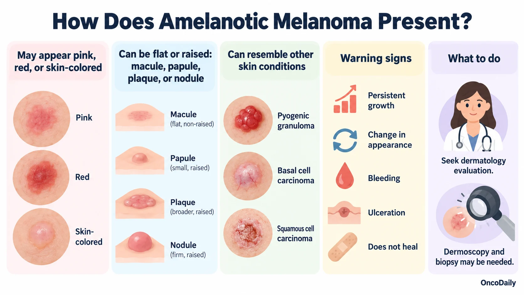
Amelanotic melanoma can appear as a pink, red, or skin-colored macule, papule, plaque, or nodule. Its appearance varies depending on the tumor’s growth pattern, location, and degree of pigmentation.
Clinically, these lesions may resemble several benign and malignant skin conditions, including pyogenic granuloma, basal cell carcinoma, squamous cell carcinoma, and inflammatory dermatoses. This overlap can make early recognition challenging, particularly when the lesion lacks the typical pigmentation associated with melanoma.
Persistent growth, changes in appearance, spontaneous bleeding, ulceration, or a lesion that fails to heal should prompt further dermatological evaluation.
Careful clinical examination and dermoscopy are important in assessing suspicious nonpigmented lesions. When melanoma is suspected, histopathological examination of a biopsy is required to establish the diagnosis (Jiang et al., 2025).
What Can Dermoscopy Show?
Amelanotic and hypomelanotic melanomas present a diagnostic challenge because the absence of visible pigmentation can make them difficult to distinguish from other skin lesions.
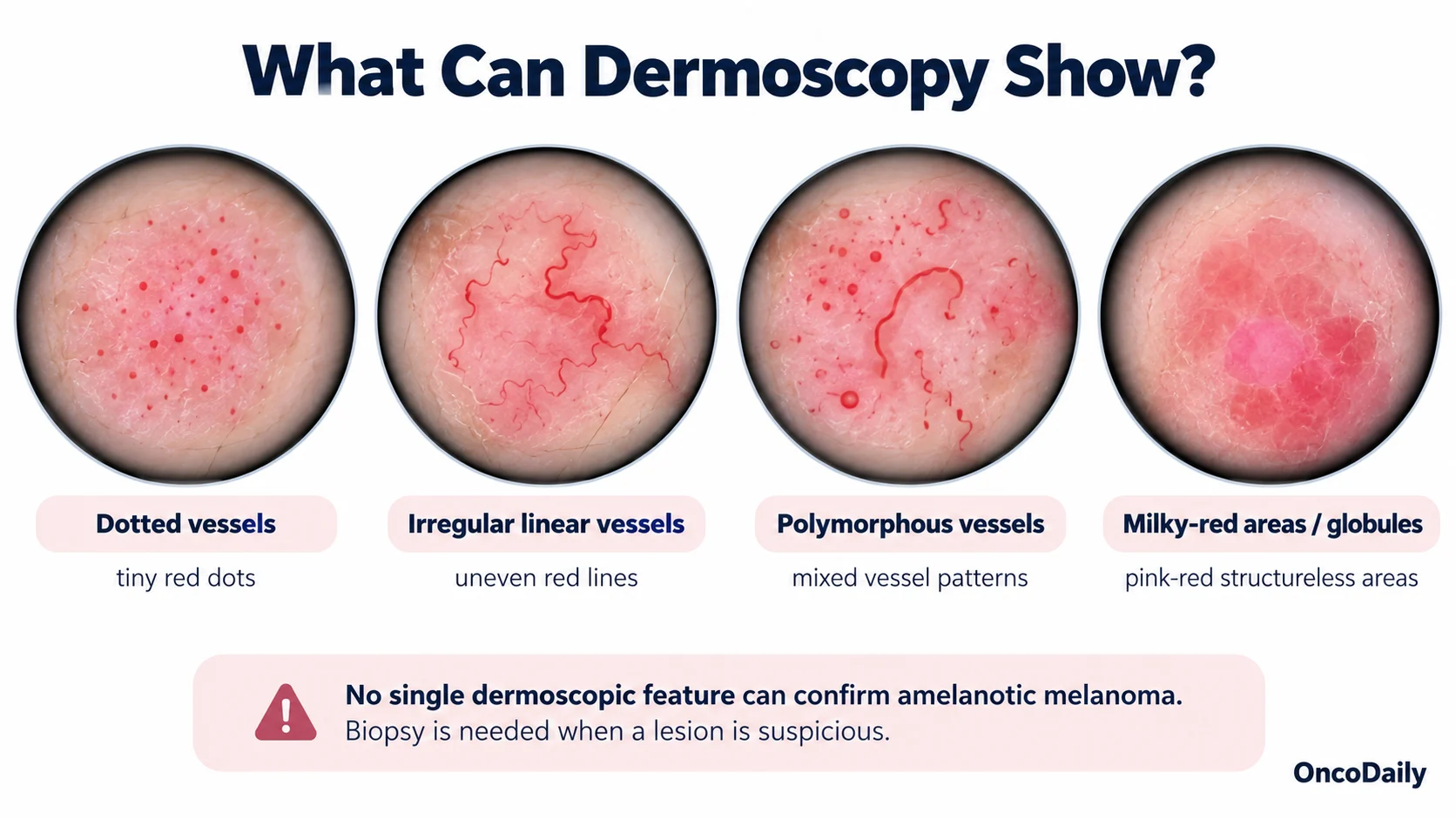
Dermoscopy is a valuable noninvasive examination technique that allows clinicians to identify structures that may not be apparent to the naked eye. In amelanotic melanoma, vascular patterns are particularly important.
Common dermoscopic findings include dotted vessels, irregular linear vessels, polymorphous vascular patterns, and milky-red areas or globules. These features can provide important diagnostic clues, although their appearance may vary between flat and raised lesions (Stojkovic-Filipovic et al., 2014).
However, no single dermoscopic feature is specific enough to confirm amelanotic melanoma. Findings must be interpreted alongside the patient’s clinical history, changes in the lesion over time, and the overall clinical appearance.
When a suspicious lesion cannot be confidently classified as benign, biopsy and histopathological examination remain essential for accurate diagnosis.
Histopathological Features
Amelanotic melanoma is not limited to a single histopathological subtype. It may occur in superficial spreading, nodular, lentigo maligna, desmoplastic, spindle cell, nevoid, and Spitz-like melanomas. Its defining characteristic is reduced or absent melanin pigmentation rather than a distinct microscopic growth pattern.
Certain melanoma subtypes, including desmoplastic and nodular melanoma, may present with reduced pigmentation. However, the frequency of amelanotic features varies across studies and patient populations.
Histopathological assessment is essential because the absence of pigment can complicate the distinction between melanoma and other malignancies. Examination of cellular morphology and tissue architecture, supported by immunohistochemical markers such as S100, SOX10, Melan-A, and HMB-45 when appropriate, helps confirm melanocytic differentiation.
Pathological examination also provides important information for staging and treatment planning, including Breslow thickness, ulceration, and other relevant tumor characteristics (Jiang et al., 2025).
What Conditions Can Mimic Amelanotic Melanoma?
The differential diagnosis of amelanotic melanoma is broad because its lack of pigmentation can resemble both benign and malignant skin lesions.
Important conditions include basal cell carcinoma, squamous cell carcinoma, Spitz nevus, seborrheic keratosis, actinic keratosis, pyogenic granuloma, dermatofibroma, keratoacanthoma, Merkel cell carcinoma, cutaneous lymphoma, and cutaneous metastases. Certain inflammatory skin conditions may also have a similar appearance.
Although dermoscopy can help distinguish these lesions, overlapping clinical and vascular features may complicate diagnosis. Therefore, histopathological examination is necessary when amelanotic melanoma is suspected (DermNet, 2017).
How Is Amelanotic Melanoma Treated?
Amelanotic melanoma is treated according to the same principles as pigmented melanoma. Treatment decisions are guided by Breslow thickness, ulceration, disease stage, and the presence of regional or distant metastases.
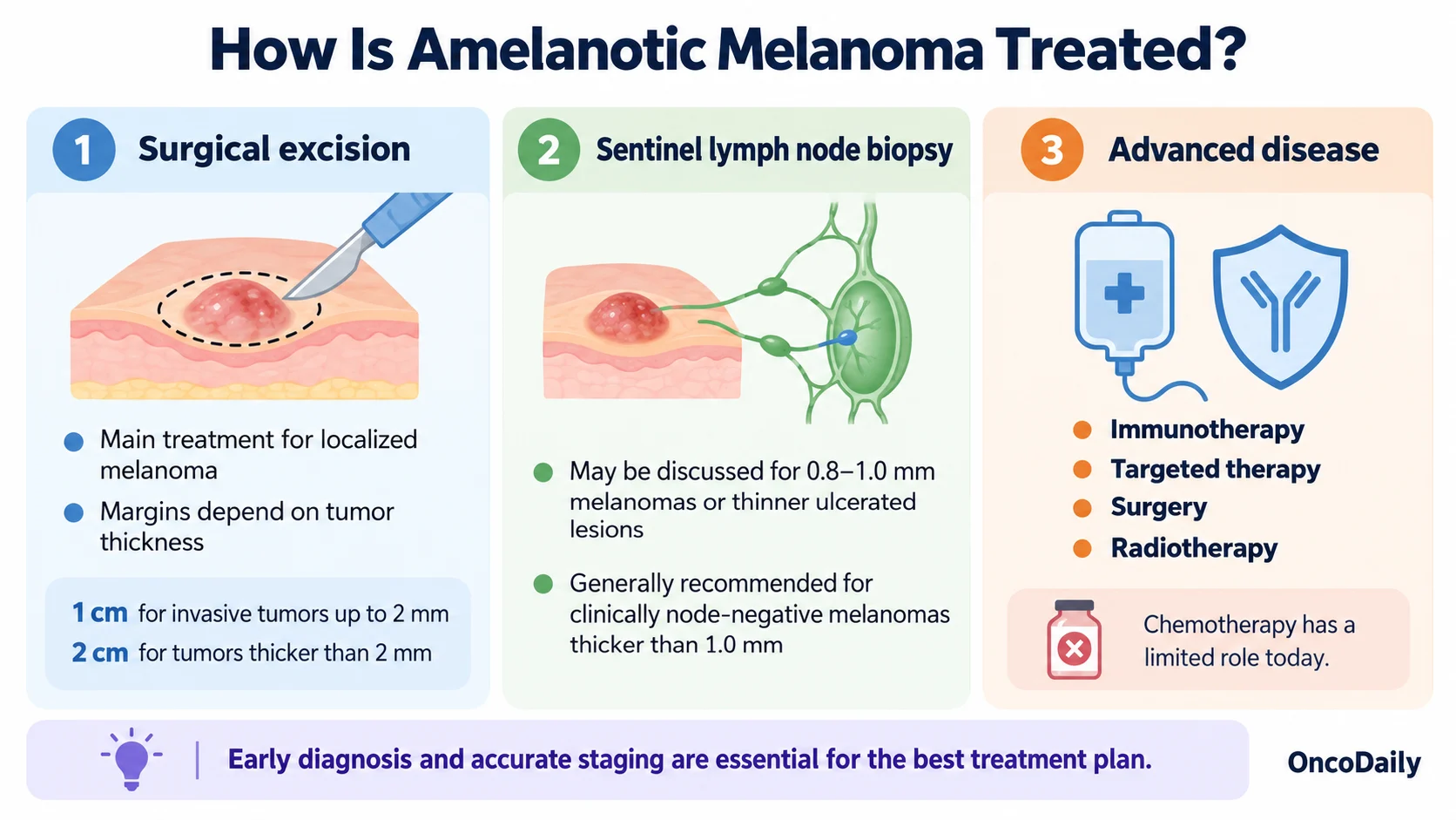
Surgical Excision: Wide local excision is the primary treatment for localized melanoma. Recommended surgical margins depend on tumor thickness, generally ranging from 1 cm for invasive melanomas measuring up to 2 mm in thickness to 2 cm for tumors thicker than 2 mm. Histopathological assessment is used to evaluate the excised tissue and confirm surgical margins.
Sentinel Lymph Node Biopsy: Sentinel lymph node biopsy may be discussed for melanomas measuring 0.8–1.0 mm in thickness or thinner lesions with ulceration. It is generally recommended for clinically node-negative melanomas thicker than 1.0 mm to assess regional lymph node involvement and guide staging.
Advanced Disease: Treatment may include immunotherapy, targeted therapy for tumors with actionable mutations, surgery, or radiotherapy, depending on disease extent and individual patient characteristics. Conventional chemotherapy has a more limited role in modern melanoma management.
Early recognition, accurate pathological diagnosis, and appropriate staging are essential for effective treatment. Although amelanotic melanoma may be more difficult to identify because of its unusual appearance, its management follows established melanoma treatment guidelines, emphasizing the importance of timely diagnosis and individualized care.
Written By Maria Asadour, MD
FAQ
What does amelanotic melanoma look like?
Amelanotic melanoma often appears as a pink, red, or skin-colored spot or bump rather than a dark mole. It may be flat or raised and can resemble a harmless skin lesion.
Is amelanotic melanoma dangerous?
Yes. Like other melanomas, it can invade surrounding tissue and spread to other organs. Its unusual appearance may delay diagnosis, making early detection particularly important.
How quickly does amelanotic melanoma grow?
Growth varies depending on the melanoma subtype. Some lesions develop slowly, while others, particularly nodular melanomas, may grow rapidly over weeks or months.
How is amelanotic melanoma diagnosed?
Diagnosis involves clinical examination, dermoscopy, and a skin biopsy. Histopathological examination confirms melanoma and provides information about tumor thickness and other important features.
Can amelanotic melanoma be cured?
Yes. Early-stage amelanotic melanoma can often be cured with complete surgical excision. Advanced cases may require immunotherapy, targeted therapy, or other treatments depending on disease stage.