Key takeaways
- Basal cell carcinoma (BCC) is the most common skin cancer, usually developing from basal cells in the epidermis and strongly linked to chronic ultraviolet (UV) radiation exposure.
- The main risk factors for BCC include fair skin, cumulative sun exposure, immunosuppression, genetic susceptibility, previous radiation exposure, and a history of prior BCC.
- Most BCCs are slow-growing and rarely metastasize, but untreated tumors can cause significant local tissue destruction and invade surrounding structures.
- Diagnosis requires skin biopsy with histopathologic examination, which remains the gold standard for confirming BCC and identifying the tumor subtype.
- Prevention and early detection are essential such as limiting UV exposure, using sunscreen, avoiding tanning beds, protecting the skin, and monitoring suspicious lesions can reduce risk and improve outcomes.
Basal cell carcinoma (BCC) is the most common type of skin cancer, originating from the basal cells located in the deepest layer of the epidermis. Although BCC is generally characterized by slow growth and a very low risk of distant metastasis, it can cause extensive local tissue destruction when diagnosis and treatment are delayed. The development of BCC is strongly associated with cumulative ultraviolet (UV) radiation exposure, with additional contributions from genetic susceptibility, immune status, environmental exposures, and individual skin characteristics.
What Is Basal Cell Carcinoma?
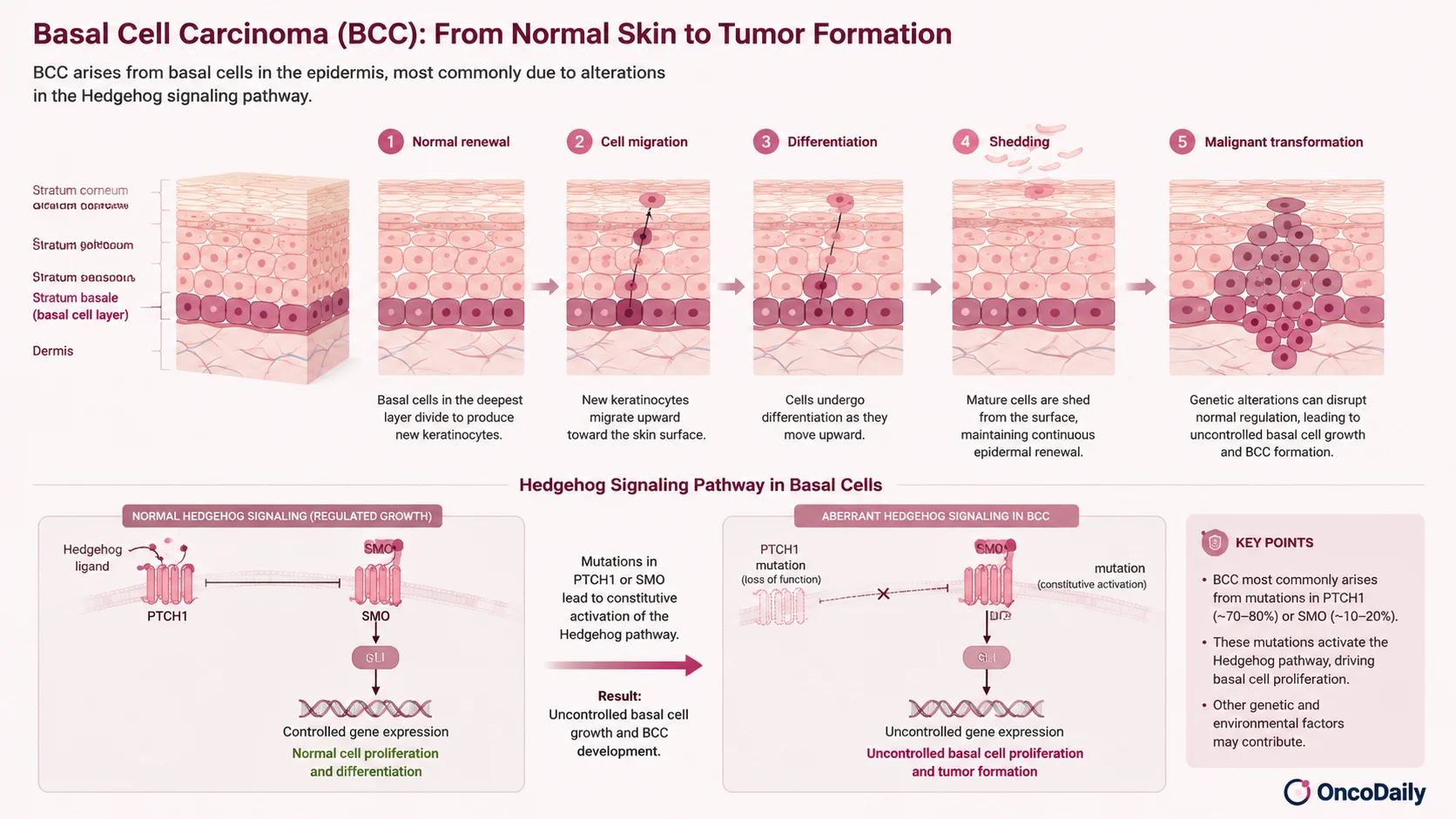
The epidermis is continuously renewed through the activity of basal keratinocytes, which are located in its deepest layer. These cells divide to generate new keratinocytes that gradually migrate toward the skin surface, undergoing differentiation before being shed.
When genetic alterations disrupt the normal regulation of basal cell growth, malignant transformation may occur, leading to the development of basal cell carcinoma.
The majority of BCCs are driven by abnormal activation of the Hedgehog signaling pathway, a pathway involved in regulating cell proliferation and differentiation. The most common molecular alterations include loss-of-function mutations in PTCH1 or activating mutations in SMO, resulting in uncontrolled pathway activation and abnormal basal cell proliferation. (Rubin et al., 2005)
Who Is at Higher Risk of Developing Basal Cell Carcinoma?
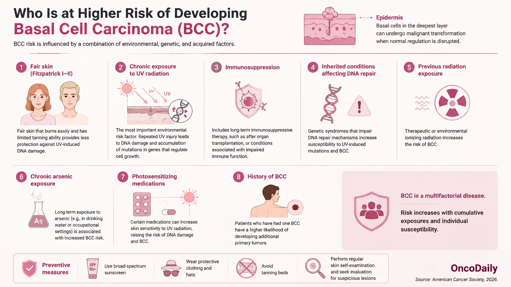
The risk of developing BCC is influenced by a combination of environmental, genetic, and acquired factors. Individuals with Fitzpatrick skin types I and II, characterized by fair skin that burns easily and has limited tanning ability, are at increased risk because of reduced protection against UV-induced DNA damage.
Chronic exposure to solar UV radiation remains the most important environmental risk factor. Repeated UV-induced DNA injury can lead to accumulation of mutations in tumor suppressor genes and signaling pathways involved in skin cell regulation.
Other factors associated with increased BCC risk include immunosuppression, such as long-term therapy after organ transplantation, inherited conditions affecting DNA repair, previous radiation exposure, chronic arsenic exposure, and certain photosensitizing medications. Previous development of BCC is also a significant risk factor, as patients with one BCC have a higher likelihood of developing additional primary tumors. (American Cancer Society, 2026)
How Does Basal Cell Carcinoma Appear on the Skin and Under the Microscope?
Basal cell carcinoma most commonly develops on sun-exposed areas of the body, particularly the head and neck region. The nose, ears, eyelids, and central face are among the most frequently affected sites, although tumors can also occur on the trunk and extremities.
The clinical appearance of BCC varies depending on the subtype. Nodular BCC, the most common form, typically presents as a pearly or translucent papule or nodule with visible small blood vessels (telangiectasia) and may develop crusting or ulceration.
Superficial BCC often appears as a red, scaly patch or plaque, while morpheaform or infiltrative BCC may resemble a scar-like lesion with poorly defined borders. Pigmented BCC may contain brown, blue, or black pigmentation and can sometimes mimic melanoma clinically.
Histologically, BCC is characterized by nests or cords of basaloid cells showing peripheral palisading and stromal retraction. Common subtypes include nodular, superficial, micronodular, infiltrative, morpheaform, and basosquamous variants.
Aggressive subtypes, particularly infiltrative, micronodular, morpheaform, and basosquamous BCC, are associated with greater local invasion and higher recurrence risk compared with nodular and superficial forms. (Rubin et al., 2005)
You can also read Melanoma (Skin Cancer): Symptoms, Causes, Stages, Diagnosis and Treatment on OncoDaily.
Can Basal Cell Carcinoma Spread or Come Back?
Although metastatic spread of BCC is extremely uncommon, some tumors demonstrate aggressive behavior with extensive local invasion and an increased risk of recurrence. Features associated with higher recurrence risk include tumor size greater than 2 cm, location in high-risk areas such as the central face and ears, long-standing lesions, incomplete surgical removal, aggressive histologic subtype, perineural invasion, and poorly defined clinical borders.
When metastasis occurs, regional lymph nodes are the most frequently affected sites, followed by distant organs such as the lungs, bones, and liver. Metastatic BCC is rare but associated with a poor prognosis, emphasizing the importance of early diagnosis and complete treatment. (Rubin et al., 2005)
How Is Basal Cell Carcinoma Diagnosed?
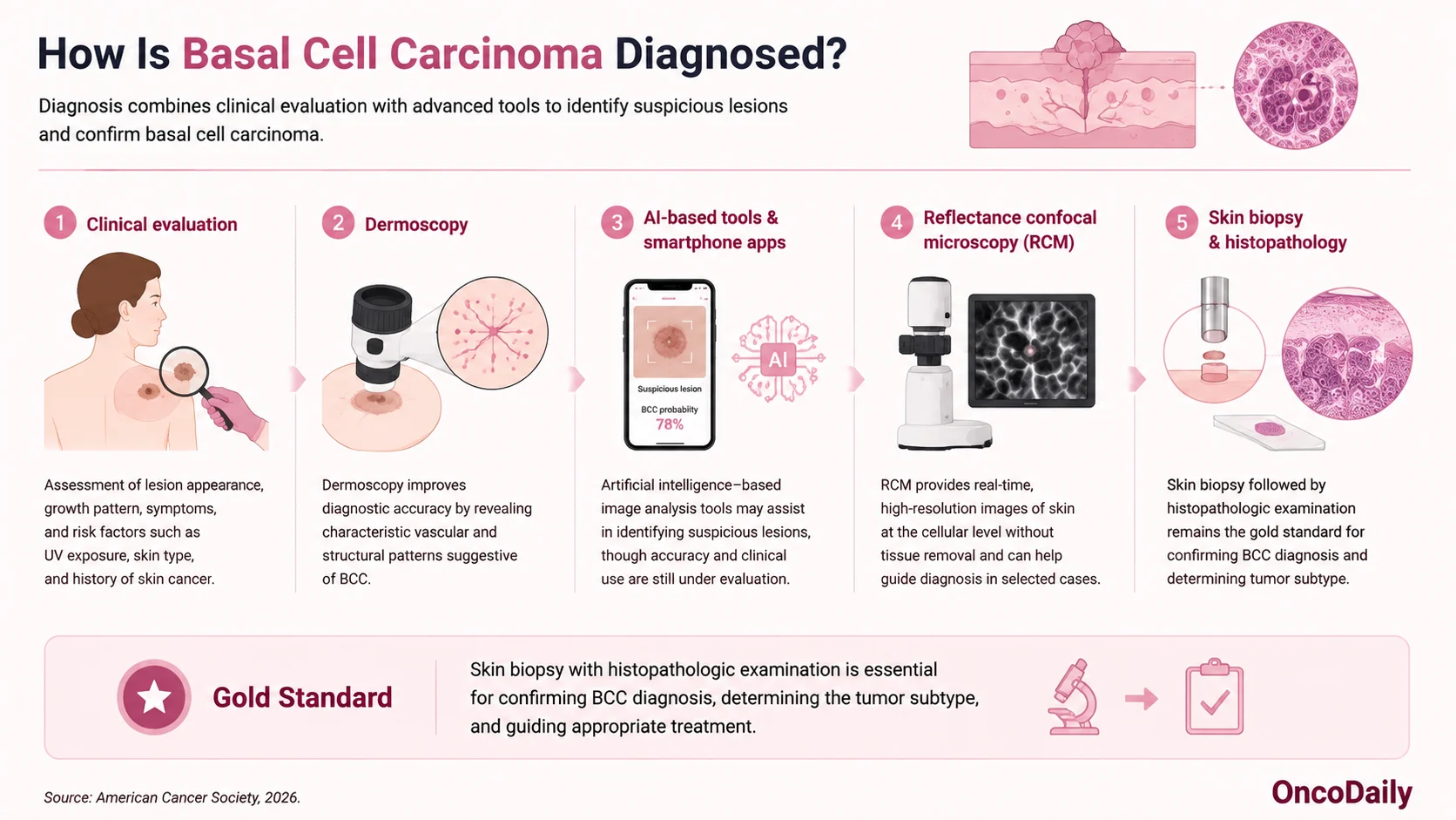
Diagnosis of BCC begins with clinical evaluation of suspicious skin lesions, including assessment of lesion appearance, growth pattern, and risk factors. Dermoscopy can improve diagnostic accuracy by identifying characteristic vascular and structural patterns.
Several emerging technologies are being investigated to support earlier detection. Artificial intelligence-based image analysis tools and smartphone applications may assist in identifying suspicious lesions, although their accuracy and clinical implementation continue to be evaluated. Reflectance confocal microscopy (RCM) provides high-resolution imaging of the skin at the cellular level without requiring tissue removal and may help guide diagnosis in selected cases.
Despite advances in noninvasive technologies, skin biopsy followed by histopathologic examination remains the gold standard for confirming BCC diagnosis and determining tumor subtype, which is essential for treatment planning. (American Cancer Society, 2026)
How Is Basal Cell Carcinoma Treated?
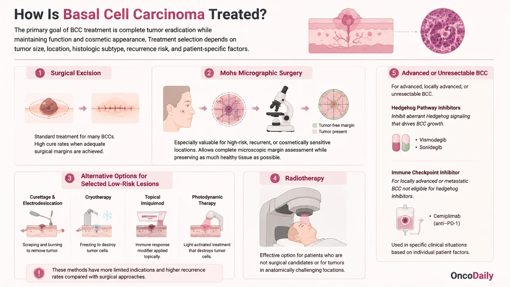
The primary goal of BCC treatment is complete tumor eradication while preserving function and cosmetic appearance. Treatment selection depends on tumor size, location, histologic subtype, recurrence risk, and patient-specific factors.
Surgical excision remains the standard treatment for many BCCs, providing high cure rates when adequate margins are achieved. Mohs micrographic surgery is particularly valuable for high-risk tumors, recurrent lesions, and tumors located in cosmetically or functionally sensitive areas because it allows complete microscopic margin assessment while preserving as much healthy tissue as possible.
For selected low-risk superficial tumors, alternative approaches may be considered. These include curettage and electrodesiccation, cryotherapy, topical imiquimod, and photodynamic therapy. However, these approaches generally have more limited indications and may have higher recurrence rates compared with surgical treatment.
Radiotherapy can be an effective option for patients who are not suitable surgical candidates or for tumors located in anatomically challenging areas where surgery may result in significant functional or cosmetic impairment.
Advanced, locally advanced, or unresectable BCC may require systemic therapy, including Hedgehog pathway inhibitors such as vismodegib and sonidegib. The immune checkpoint inhibitor cemiplimab, targeting PD-1, may be considered for selected patients with advanced BCC who are not candidates for Hedgehog inhibitor therapy.
How Can Basal Cell Carcinoma Be Prevented?
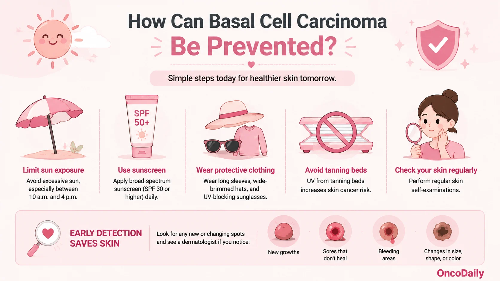
Prevention of BCC focuses primarily on reducing UV exposure and promoting early detection. Protective measures include avoiding excessive sun exposure, using broad-spectrum sunscreen, wearing protective clothing, avoiding tanning beds, and performing regular skin examinations.
Early recognition of suspicious lesions, particularly new growths, persistent sores, bleeding areas, or changing skin lesions, allows treatment at an earlier stage when cure rates are highest.
Can Chemoprevention Reduce the Risk?
Chemoprevention strategies may be considered for individuals with a significantly increased risk of developing multiple skin cancers, including patients with previous BCCs, genetic predisposition syndromes, or chronic immunosuppression.
Nicotinamide (vitamin B3) has shown potential in reducing the incidence of new basal and squamous cell carcinomas in high-risk individuals and is generally well tolerated. Retinoids may also reduce skin cancer risk in selected populations but are limited by potential toxicity. In rare hereditary conditions associated with multiple BCCs, such as basal cell nevus syndrome, hedgehog pathway inhibitors may reduce tumor development and treat existing disease. (American Cancer Society, 2026)
Written by Maria Asadour, MD
FAQ
What is basal cell carcinoma (BCC)?
BCC is the most common type of skin cancer, arising from basal cells in the epidermis.
What causes basal cell carcinoma?
The main cause is chronic ultraviolet (UV) radiation exposure, which can lead to DNA damage and abnormal cell growth.
Is basal cell carcinoma dangerous?
BCC rarely spreads to distant organs, but untreated tumors can invade and damage nearby tissues.
Who is at higher risk of developing BCC?
People with fair skin, high UV exposure, weakened immune systems, genetic risk factors, or a previous history of BCC have higher risk.
Where does BCC usually occur?
BCC most commonly develops on sun-exposed areas, especially the face, ears, scalp, and neck.
What does basal cell carcinoma look like?
It may appear as a pearly bump, a scaly red patch, a scar-like area, or a lesion that bleeds and does not heal.
How is BCC diagnosed?
Diagnosis is confirmed with a skin biopsy followed by microscopic examination of the tissue.
What is the main treatment for BCC?
Surgical removal is the standard treatment for many BCCs, while Mohs surgery is used for high-risk or sensitive areas.
Can basal cell carcinoma come back after treatment?
Yes. Recurrence risk is higher with aggressive subtypes, incomplete removal, large tumors, or high-risk locations.
How can BCC be prevented?
Reducing UV exposure, using sunscreen, avoiding tanning beds, wearing protective clothing, and checking the skin regularly can lower risk.