Central nervous system (CNS) lymphoma comes with a problem that extends beyond the disease itself: reaching it. Neurological symptoms can delay diagnosis, corticosteroids can shrink or obscure lesions before biopsy, the blood-brain barrier limits drug penetration, and treatment must balance disease control against the risk of long-term neurotoxicity. Nor is CNS lymphoma a single disease.
- Primary CNS lymphoma (PCNSL) arises within the CNS without systemic involvement at diagnosis and may affect the brain, leptomeninges, cerebrospinal fluid (CSF), spinal cord, or eyes.
- Primary vitreoretinal lymphoma (PVRL) is closely related to PCNSL but can present initially as isolated ocular disease.
- Secondary CNS lymphoma (SCNSL) occurs when systemic lymphoma spreads to the CNS, either at diagnosis or at relapse.
Each carries a different course, treatment approach, and in the case of SCNSL, disease outside the CNS that has to be managed as well.
What Is Primary CNS Lymphoma?
Most PCNSL cases are diffuse large B-cell lymphoma (DLBCL) by histology, now classified among large B-cell lymphomas arising in immune-privileged sites.
PCNSL has a distinctive molecular profile. MYD88 mutations (particularly L265P) and CD79B alterations are common and promote lymphoma-cell survival through B-cell receptor and Toll-like receptor signaling, with activation of BTK and NF-κB. Changes affecting antigen presentation and immune recognition also help lymphoma cells persist within the CNS environment.
These molecular features are already clinically relevant. Detection of MYD88 mutations in CSF or ocular samples can support diagnosis, and dependence on B-cell receptor signaling is the rationale behind using BTK inhibitors at relapse.
You can also read about
Why CNS Lymphoma Can Be Difficult to Recognize
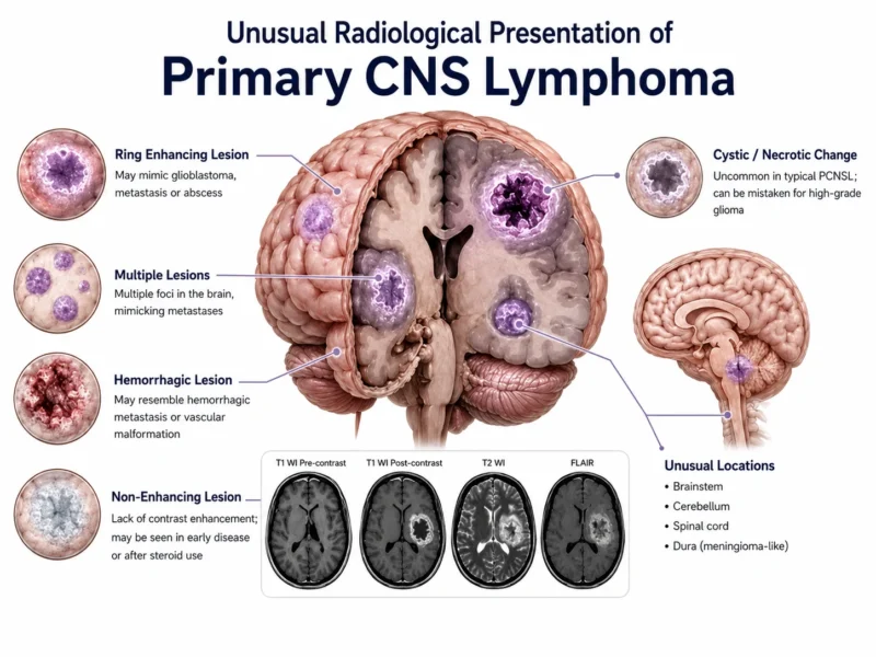
PCNSL can present with a wide range of neurological symptoms, including cognitive or behavioral changes, focal deficits, gait disturbance, headache, and seizures. Lesions often involve deep cerebral or periventricular structures and may be multifocal.
Contrast-enhanced brain MRI is the main imaging tool. PCNSL lesions typically show strong, relatively homogeneous enhancement and restricted diffusion, but these findings are not specific. Glioblastoma, metastases, demyelinating or inflammatory disorders, and infections can have a similar appearance.
Advanced imaging techniques, including diffusion-weighted imaging (DWI), perfusion and permeability imaging, magnetic resonance spectroscopy (MRS), and susceptibility-weighted imaging (SWI), can improve diagnostic accuracy and help distinguish from the mimics.
Stereotactic brain biopsy remains the diagnostic standard for most patients with parenchymal disease. CSF cytology and flow cytometry can identify lymphoma cells, though sensitivity is limited. Molecular testing adds another layer: MYD88 mutations, interleukin-10, and tumor-derived DNA in CSF.
Of these, CSF circulating tumor DNA (ctDNA) is drawing the most attention, since it may offer a molecular picture of the lymphoma without repeat biopsy. Its role is still evolving – beyond diagnosis, it’s also being studied for treatment-response monitoring and early relapse detection.
drdeepakaiims.com
The Steroid Dilemma: “Ghost Tumor” Phenomenon
Corticosteroids are lymphotoxic to PCNSL cells, and even a short course can shrink lesions enough to vanish from MRI entirely. The problem is timing: steroids are a default response to neurological symptoms like headache or focal deficits, often started in the emergency department.
The consequence is a diagnostic trap. A patient may present with a clear lesion, receive steroids for presumed edema or a presumed inflammatory process, and return for biopsy days later to find nothing to sample. Even when some lesion remains, steroid-induced changes can distort histology enough to obscure the diagnosis.
This is why current EHA-ESMO guidance is explicit: avoid corticosteroids before tissue diagnosis whenever the patient’s clinical condition allows. The instruction is straightforward, but adherence depends on lymphoma being on the differential early.
How Is CNS Lymphoma Staged?
There is no conventional stage I-IV system to guide treatment. The main goals are to determine which CNS compartments are involved and whether the lymphoma is truly confined to the CNS.
Systemic evaluation is needed to exclude secondary CNS lymphoma. FDG-PET/CT detects systemic disease in approximately 4%-12% of patients initially thought to have PCNSL; contrast-enhanced body CT can be used when PET/CT is unavailable.
Primary Vitreoretinal Lymphoma
PVRL often presents with blurred vision, floaters, eye pain, photophobia and can closely resemble chronic uveitis or other inflammatory eye disorders. Corticosteroids may temporarily improve symptoms and ocular findings, further delaying recognition.
Most PVRL is B-cell lymphoma and shares molecular features with PCNSL. Diagnosis usually relies on vitreous sampling, with cytology, flow cytometry, molecular testing, and intraocular interleukin-10 providing complementary information.
PVRL may occur alongside PCNSL or precede brain involvement, making CNS assessment important even when apparently confined to the eye. Treatment depends on the extent and may include intravitreal methotrexate or rituximab, ocular radiotherapy, or systemic CNS-penetrating therapy.
Why High-Dose Methotrexate Became the Backbone of PCNSL Treatment
Treating PCNSL depends heavily on whether a drug can reach effective concentrations within the CNS – which is why R-CHOP, despite its central role in systemic DLBCL, is not adequate treatment here.
High-dose methotrexate overcomes this problem and remains the backbone of first-line therapy. Its use requires careful attention to renal function, drug interactions, hydration, urinary alkalinization, methotrexate clearance, and leucovorin rescue.
In younger, fit patients, methotrexate is commonly combined with other CNS-active agents. MATRix (high-dose methotrexate, cytarabine, thiotepa, and rituximab) is a major induction regimen through the IELSG32 trial and is followed by consolidation in eligible patients.
Elderly and Frail Patients Are the Majority, Not a Subgroup
PCNSL has a median age at diagnosis in the mid-60s, and a substantial share of patients are older than 70. Frailty assessment, not chronological age, increasingly guides this decision.
For patients who can’t tolerate full-intensity induction, options include reduced-dose or attenuated methotrexate schedules, and combination regimens such as R-MPV (rituximab, methotrexate, procarbazine, vincristine), which preserve CNS-penetrating chemo while lowering the toxicity burden.
Consolidation: Transplantation or Radiotherapy?
Whole-brain radiotherapy (WBRT) can provide effective control, but its role has narrowed because of delayed neurotoxicity, particularly in older patients, cognitive decline, gait impairment, and loss of independence can emerge months or years later.
For fit patients, consolidation has increasingly moved toward high-dose chemotherapy followed by autologous stem cell transplantation (ASCT), with thiotepa-based conditioning as the preferred approach.
You can also read about
Secondary CNS Lymphoma: Who Is at Risk?
SCNSL occurs when systemic lymphoma involves the CNS, most often in aggressive B-cell lymphomas. CNS involvement may be present at initial diagnosis or develop later as relapse, either alone or together with recurrent systemic disease. Treatment has to account for disease inside and outside the CNS.
CNS relapse occurs in approximately 5% of patients with DLBCL overall, but the risk concentrates in specific groups. The CNS International Prognostic Index (CNS-IPI) combines standard IPI factors with kidney or adrenal involvement to identify higher-risk patients, testicular and kidney/adrenal involvement are also associated with increased CNS risk.
Many CNS relapses occur during or relatively soon after frontline treatment, suggesting that some may reflect occult CNS involvement already present at diagnosis.
Why CNS Prophylaxis in DLBCL Remains Controversial
CNS prophylaxis remains one of the most debated areas of DLBCL management: who is likely to benefit, and which approach actually works. Intrathecal methotrexate delivers chemotherapy directly into the CSF but provides limited protection against parenchymal disease, where many CNS relapses occur.
Systemic high-dose methotrexate reaches the brain more effectively, but evidence for its preventive benefit remains inconsistent, large retrospective studies have questioned whether it meaningfully reduces CNS relapse, while also showing toxicity and disruption of systemic therapy.
No randomized trial has established the optimal prophylactic strategy. Current decisions therefore rely on individual CNS risk, extranodal disease pattern, patient fitness, and the potential toxicity of prophylaxis.
You can also read about

How Secondary CNS Lymphoma Is Treated
Treatment of SCNSL often has to control disease inside and outside the CNS at once. The prospective MARIETTA trial provided important evidence here: patients received 3 courses of MATRix followed by 3 courses of R-ICE, with responders proceeding to thiotepa-based high-dose therapy and ASCT. Among 75 patients with DLBCL and secondary CNS involvement, the ORR was 61%, including a 55% CR rate, and one-year PFS was 58%.
CAR T-cell therapy is also emerging as an option for relapsed SCNSL, with early studies showing responses without neurotoxicity proving prohibitive, consistent with the pattern seen in PCNSL relapse.
Immunodeficiency-Associated CNS Lymphoma
In patients with HIV or post-transplant immunosuppression, CNS lymphoma is driven almost entirely by Epstein-Barr virus (EBV), reflecting loss of the T-cell surveillance that normally keeps EBV-infected B cells in check.
In HIV-associated CNS lymphoma, EBV DNA in the CSF is a useful diagnostic aid, often combined with FDG-PET or thallium-201 SPECT to help distinguish lymphoma from toxoplasmosis, the main differential in this population and one that’s rarely a consideration in immunocompetent patients.
Antiretroviral therapy is central to management: immune reconstitution alone can improve outcomes, and untreated or poorly controlled HIV limits how much chemotherapy a patient can tolerate.
Post-transplant CNS lymphoma, a form of post-transplant lymphoproliferative disorder, follows a similar logic. Reducing immunosuppression is often the first intervention, sometimes alongside rituximab, before more intensive chemotherapy is considered.
You can also read about

How Is Treatment Response Assessed, and What Determines Prognosis?
Prognosis depends on both disease and patient factors. The IELSG prognostic score incorporates age, performance status, serum LDH, CSF protein, and involvement of deep brain structures, the Memorial Sloan Kettering model uses age and performance status alone.
The International Primary CNS Lymphoma Collaborative Group (IPCG) criteria combine contrast-enhanced brain MRI with CSF findings, ophthalmologic assessment when the eyes are involved, and corticosteroid use. Complete response requires resolution of enhancing brain lesions and clearance of lymphoma from other previously involved CNS compartments.
EHA-ESMO guidance recommends brain MRI during induction and after consolidation, with repeat CSF or ocular assessment when these sites were involved at diagnosis. MRI isn’t a direct measure of viable lymphoma, though: residual enhancement may reflect persistent disease or treatment-related change, and corticosteroids can alter enhancement independently of lymphoma control.
In PCNSL, response has to be weighed alongside neurocognitive outcomes and quality of life, which complement progression-free and overall survival as endpoints.
You can also read about

Where CNS Lymphoma Treatment Is Heading
Beyond ctDNA monitoring and the targeted agents already in use for relapse, bispecific antibodies are now being explored in CNS lymphoma, extending the same logic that’s driven progress in systemic B-cell lymphoma into a disease behind the blood-brain barrier.
The larger goal is durable remission with preserved cognition and independence, given how much both the lymphoma and its treatment can still cost the CNS.
FAQ
Can CNS lymphoma spread outside the brain?
Yes. Primary CNS lymphoma can involve several CNS compartments, including the brain, cerebrospinal fluid, leptomeninges, spinal cord, and eyes. Systemic lymphoma outside these sites at diagnosis generally points toward secondary CNS involvement rather than classic PCNSL.
Can CNS lymphoma affect the eyes before the brain?
Yes. Primary vitreoretinal lymphoma can initially appear confined to the eyes and may cause blurred vision, floaters, eye pain, or photophobia. Some patients later develop brain involvement, which is why CNS evaluation remains important even when lymphoma initially appears limited to the eye.
Why can steroids make CNS lymphoma harder to diagnose?
PCNSL cells are highly sensitive to corticosteroids. Even a short course can shrink lesions substantially or make them temporarily disappear on MRI, reducing the amount of diagnostic tissue available for biopsy. When clinically possible, corticosteroids are therefore avoided until tissue has been obtained.
Why doesn't R-CHOP work well for primary CNS lymphoma?
The problem is drug penetration. Several major components of R-CHOP do not reach adequate concentrations within the CNS. High-dose methotrexate can achieve therapeutic CNS exposure, which is why it remains the backbone of PCNSL treatment.
Can a blood test detect CNS lymphoma?
There is currently no routine blood test that can reliably diagnose PCNSL. CSF may be more informative because it is in closer contact with CNS disease. Researchers are studying circulating tumor DNA in CSF as a potential tool for diagnosis, molecular profiling, treatment monitoring, and earlier detection of relapse.
Who is most at risk of CNS relapse from DLBCL?
Overall, CNS relapse occurs in a minority of patients with DLBCL, but risk is higher in selected groups. The CNS-IPI helps estimate this risk, while involvement of certain extranodal sites—particularly the testes and kidney/adrenal glands—also raises concern.
Does CNS lymphoma rule out CAR T-cell therapy?
Not necessarily. Patients with CNS lymphoma were historically approached cautiously because CAR T-cell therapy can cause neurological toxicity. Emerging experience in relapsed PCNSL and secondary CNS lymphoma suggests that CNS involvement alone does not automatically exclude CAR T-cell therapy, although evidence remains less mature than for systemic large B-cell lymphoma.