


Sahar Mansour, Professor of Radiology at Kasr Al-Ainy Hospital, Cairo University, and Consultant of Breast Imaging and Intervention at Baheya Charity Women’s Cancer Hospital, shared a post on LinkedIn:
“Sometimes, it’s not what you see… but how clearly you see it.
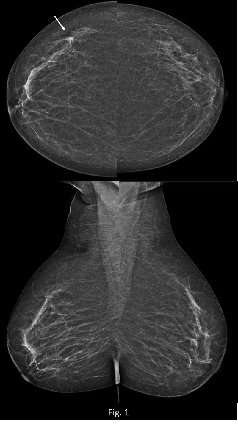
A routine case came in, nothing particularly alarming at first glance. Just a subtle unease… the kind that makes you look twice (fig.1).
On standard imaging, it could have easily passed as insignificant. But using high tech. mammogram something began to unfold; fine, delicate tentacles forming a pattern of architectural distortion (fig.2).
Was this early malignancy quietly emerging, or simply fibrous traction from benign proliferative changes?
In moments like these, technology sharpens judgment.
Initial assign was suspicious (BI-RADS 4) and a stereotactic vacuum-assisted_biopsy, guided precisely by tomosynthesis (fig.3), ensured that what we sampled truly represented what we saw.
And then pathology confirmed: No invasion, not a malignancy, but rather: Fibrocystic changes with apocrine metaplasia and focal atypia (B3).
Then the next step, does imaging and pathology are speaking the same language? YES. No need to re-biopsy, as confidence comes from correlation. While architectural distortion is present, high resolution mammography combined with wide angle tomosynthesis (fig.2) reveals reassuring features of preserved architecture, central fat lucency, and fine fibrous strands… findings that favor benignity rather than aggressive infiltration.
Early detection isn’t just about finding abnormalities…It’s about understanding them correctly.
Case Credit: Baheya Foundation
Imaging: Siemens Healthineers Mammomat B.brilliant mammography.”

More posts featuring Sahar Mansour.