Key takeaways
- Radiation necrosis is a delayed injury to normal brain tissue that can develop months or years after brain radiotherapy, especially after high-dose focal treatments such as SRS.
- It results from vascular injury, blood–brain barrier disruption, inflammation, hypoxia, and tissue breakdown, often with surrounding vasogenic edema.
- Symptoms vary by lesion location and size and may include headache, seizures, weakness, speech or visual changes, balance problems, or cognitive impairment.
- Radiation necrosis can closely mimic tumor recurrence on MRI, so diagnosis often relies on perfusion MRI, MR spectroscopy, amino-acid PET, serial imaging, and occasionally biopsy.
- - Management ranges from observation and corticosteroids to bevacizumab, surgery, or LITT, and many patients achieve radiographic or symptomatic improvement with appropriate treatment.
Radiation necrosis after brain radiation is a delayed injury to normal brain tissue that can develop months or years after radiotherapy. It is particularly relevant after high-dose focal techniques such as stereotactic radiosurgery (SRS), although it can also occur after conventionally fractionated brain radiation.
Radiation necrosis can cause headaches, seizures, cognitive changes, and focal neurological deficits, but some lesions are discovered incidentally on follow-up MRI. One of the major clinical challenges is that radiation necrosis can closely resemble recurrent brain tumor on conventional imaging. Diagnosis may therefore require advanced MRI, metabolic imaging, serial follow-up, and occasionally biopsy or surgical resection.
Treatment ranges from observation for stable, asymptomatic lesions to corticosteroids, bevacizumab, surgery, or laser interstitial thermal therapy (LITT) for symptomatic or progressive disease. The clinical course is variable, and some lesions stabilize or regress while others produce persistent neurological morbidity.
What Is Radiation Necrosis and Why Can It Happen After Brain Radiation?
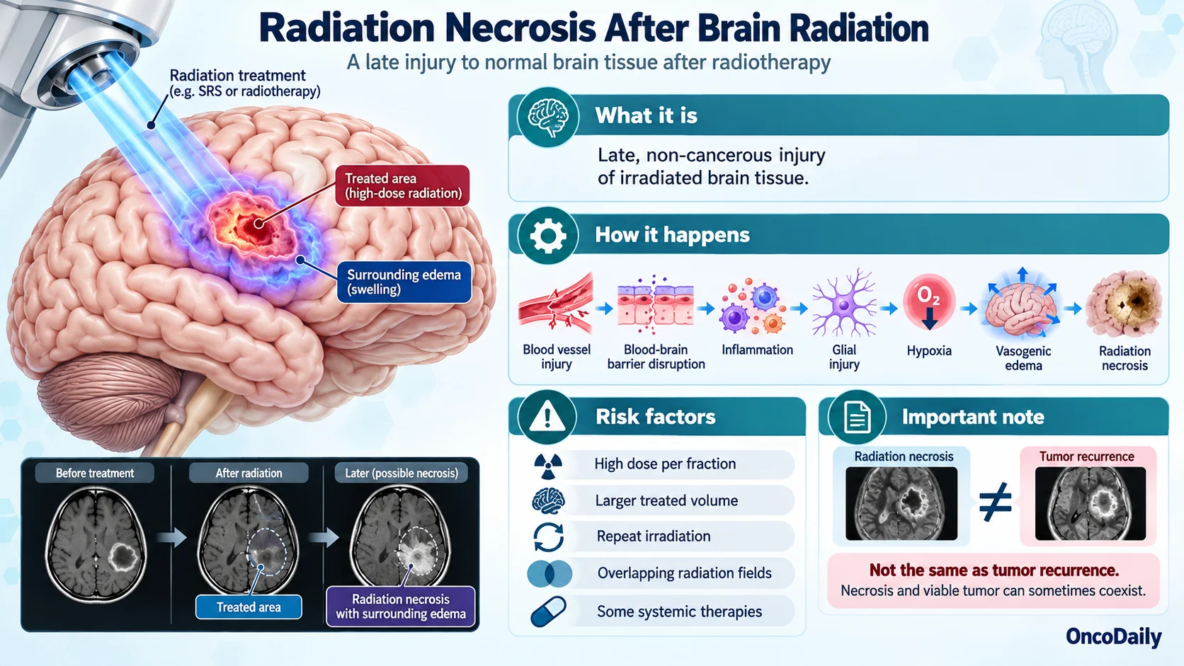
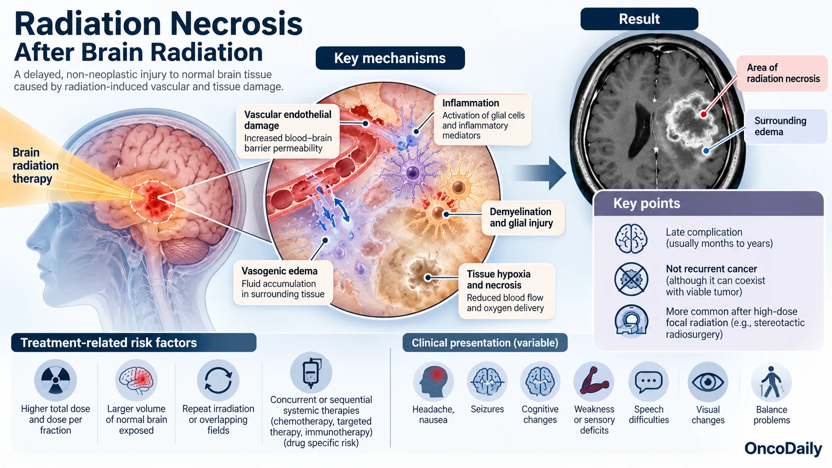
Radiation necrosis is a late, non-neoplastic injury of irradiated brain tissue. Its pathophysiology is multifactorial and involves vascular endothelial damage, blood–brain barrier disruption, glial injury, inflammation, demyelination, and tissue hypoxia. Increased vascular permeability can produce vasogenic edema and further contribute to local tissue injury (Rahmathulla et al., 2013; Katsura et al., 2021).
The risk depends strongly on radiation dose and the volume of normal brain exposed. Radiation necrosis is particularly associated with high-dose focal radiation such as SRS and stereotactic radiotherapy.
Important treatment-related risk factors include:
- higher dose per fraction
- larger high-dose treatment volumes
- repeat irradiation of the same region
- overlapping radiation fields
- selected systemic therapies given near radiation treatment
The interaction with systemic therapy is complex and drug specific; chemotherapy, targeted therapy, and immunotherapy should not be considered uniform risk factors (Mayo et al., 2024).
Radiation necrosis is not recurrent cancer, although necrosis and viable tumor can coexist within the same lesion. This overlap is one reason diagnosis can be difficult.
When Does Radiation Necrosis Develop After Radiation Therapy?
Radiation necrosis is classified as a late radiation effect. The 2026 European Association for Neuro-Oncology (EANO) consensus indicates that it most commonly appears approximately 6–24 months after radiotherapy, although the interval is variable and cases may occur considerably later (Duerinck et al., 2026).
A 2025 series of histologically confirmed radiation necrosis after treatment of primary brain tumors reported a latency of 3–40 months (Hazaymeh et al., 2025).
Timing can help frame the differential diagnosis:
- Days to weeks after radiation: acute treatment effects are more likely.
- Weeks to several months: early delayed changes and pseudoprogression become important considerations.
- Several months to years: radiation necrosis becomes increasingly relevant.
These intervals are not diagnostic boundaries. Tumor progression, pseudoprogression, and radiation necrosis can overlap temporally, particularly in patients who have received combined-modality treatment (Katsura et al., 2021).
What Are the Most Common Symptoms of Brain Radiation Necrosis?
The clinical presentation depends primarily on the location and volume of injured brain tissue and the extent of associated edema. Radiation necrosis may be completely asymptomatic and detected only during surveillance imaging.
When symptomatic, common manifestations include:
- headache, nausea, or somnolence
- seizures
- weakness or sensory loss
- speech or language disturbance
- visual abnormalities
- impaired balance or coordination
- memory, attention, or other cognitive changes
Larger lesions or substantial edema can produce mass effect and progressive neurological deterioration. Symptoms frequently resemble those caused by recurrent tumor, so the clinical presentation alone cannot reliably distinguish the two conditions (Mayo et al., 2024).

How Can Doctors Tell Radiation Necrosis From Tumor Recurrence?
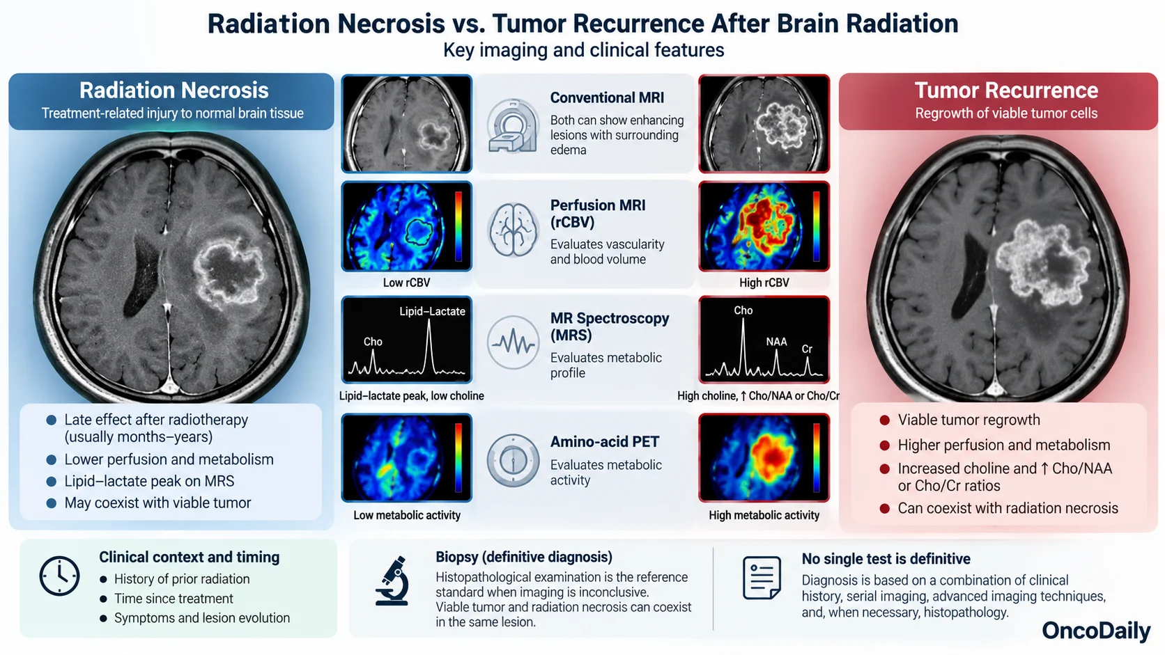
Distinguishing radiation necrosis from recurrent tumor remains one of the major diagnostic challenges after brain radiotherapy. Both may appear as new or enlarging contrast-enhancing lesions with surrounding T2/FLAIR abnormality and edema on conventional MRI (Verma et al., 2013).
Diagnosis therefore usually combines treatment history, lesion timing, serial imaging, and advanced imaging techniques.
Perfusion MRI evaluates tumor vascularity. Recurrent tumor generally shows increased perfusion and relative cerebral blood volume (rCBV), whereas radiation necrosis more often demonstrates lower perfusion. However, thresholds vary between studies, and mixed tumor–necrosis lesions can reduce diagnostic accuracy (Barajas et al., 2009).
MR spectroscopy provides metabolic information. Recurrent tumor commonly shows increased choline and higher choline-to-NAA or choline-to-creatine ratios, reflecting increased cellular turnover. Necrosis is more often associated with lipid–lactate peaks and lower choline levels (Weybright et al., 2005).
PET imaging can provide additional metabolic information. Current EANO guidance considers amino-acid PET, where available, one of the most useful imaging approaches alongside perfusion MRI for differentiating radiation necrosis from recurrent tumor (Duerinck et al., 2026).
No imaging modality provides perfect diagnostic certainty. Histopathological examination remains the reference standard when the diagnosis remains unresolved and would substantially change management. Even pathology can be challenging because viable tumor and treatment-related necrosis may coexist within the same lesion (Duerinck et al., 2026).
What Are the Treatment Options for Radiation Necrosis?
Management depends on symptoms, lesion size and location, surrounding edema, radiographic progression, diagnostic certainty, and the status of the underlying cancer.
Observation is appropriate for selected patients with small, asymptomatic, and radiographically stable lesions. These patients are usually followed with serial MRI and clinical assessment (Bernhardt et al., 2022).
Corticosteroids, most commonly dexamethasone, are generally used first for symptomatic edema. They reduce vascular permeability and can rapidly improve headache or focal neurological symptoms. Because prolonged corticosteroid exposure carries substantial toxicity, the lowest effective dose and appropriate taper are preferred (Bernhardt et al., 2022; Duerinck et al., 2026).
Bevacizumab, an anti-VEGF monoclonal antibody, is an important option for persistent or corticosteroid-refractory symptomatic radiation necrosis. By reducing abnormal vascular permeability, bevacizumab can markedly decrease contrast enhancement and FLAIR edema and improve neurological symptoms. A systematic review of 236 treated patients reported high radiographic response rates, although recurrence after treatment can occur (Liao et al., 2021).
Surgical resection can be considered for accessible lesions causing major mass effect, progressive neurological deterioration, or persistent diagnostic uncertainty. Surgery provides both decompression and tissue for histopathological examination (Duerinck et al., 2026).
Laser interstitial thermal therapy (LITT) is a minimally invasive option for selected patients with treatment-resistant radiation necrosis, particularly when conventional resection is less suitable. Evidence is still more limited than for corticosteroids, bevacizumab, and surgery (Vellayappan et al., 2024).
Other treatments, including hyperbaric oxygen therapy, have been reported, but the supporting evidence remains comparatively limited and they are not considered core treatment options in most contemporary management pathways.
Can Radiation Necrosis Improve Over Time, and What Is the Outlook?
Radiation necrosis does not follow a single clinical course. Some asymptomatic lesions remain stable or regress on serial imaging, while symptomatic disease may improve when edema and vascular permeability are controlled.
Radiographic improvement does not always parallel neurological recovery. Outcome depends on the size and anatomical location of the lesion, degree of irreversible tissue injury, severity of edema, prior radiation exposure, response to treatment, and control of the underlying malignancy.
For corticosteroid-refractory disease, bevacizumab can produce substantial symptomatic and radiographic improvement, although recurrence after discontinuation has been reported (Liao et al., 2021). Surgically accessible, treatment-refractory lesions may also improve after resection; retrospective studies have reported reductions in edema and meaningful neurological improvement following surgery (Shah et al., 2020; Newman et al., 2021).
Radiation necrosis can also emerge years after SRS. Long-term follow-up studies demonstrate that cumulative risk continues beyond the first year after treatment, particularly among patients surviving long enough for late radiation effects to become apparent (Kohutek et al., 2015).
The prognosis therefore depends on both the radiation injury and the underlying cancer. Radiation necrosis can stabilize or improve, but severe lesions may leave persistent neurological deficits, particularly when extensive damage involves functionally important brain regions.
FAQ
What is radiation necrosis after brain radiation?
Radiation necrosis is a delayed injury to normal brain tissue caused by radiation-related vascular and tissue damage.
How long after radiation therapy can radiation necrosis occur?
It most often develops several months to a few years after treatment, but later cases can also occur.
What are the symptoms of brain radiation necrosis?
Symptoms may include headache, seizures, weakness, speech or visual changes, balance problems, and cognitive difficulties. Some patients remain asymptomatic.
How is radiation necrosis distinguished from tumor recurrence?
Doctors use clinical history, serial MRI, perfusion MRI, MR spectroscopy, amino-acid PET, and sometimes biopsy because no single imaging test is completely definitive.
Can radiation necrosis be treated successfully?
Yes. Depending on severity, treatment may include observation, corticosteroids, bevacizumab, LITT, or surgery, and many patients achieve symptom control or radiographic improvement.