





Authors: Pasquale Lombardi, Erik Ramon-Gil, Rabial Q. Raja, Leonardo Brunetti, Giulia Francesca Manfredi, Zhongguo Zhou, George Merces, Sarah Cappuyns, Claudia Angela Maria Fulgenzi, Antonio D’Alessio, Aria Torkpour, Ciro Celsa, Bernardo Stefanini, Hannah Yang, Fionnuala Crowley, Thomas U. Marron, Anwaar Saeed, Matthias Pinter, Bernhard Scheiner, Yi-Hsiang Huang, Pei-Chang Lee, Naoshi Nishida, Ryan Po-Ting Lin, Andrea Dalbeni, Caterina Vivaldi, Gianluca Masi, Natascha Rohlen, Johann von Felden, Ahmed Kaseb, Peter R. Galle, Masatoshi Kudo, Wei-Fan Hsu, Lorenza Rimassa, Alessandro Parisi, Robin Kate Kelley, Hidenori Toyoda, Mario Pirisi, Falah Jabar, Mehrdad Rakaee, Giuseppe Cabibbo, Calogero Cammà, Fabio Piscaglia, Sohyun Hwang, Dong Jun Shin, Michael Li, Jeroen Dekervel, Nadia Guerra, Tim Meyer, Helen L. Reeves, Bertram Bengsch, Gennaro Daniele, Derek A. Mann, Hong Jae Chon, Jack Leslie, and David J. Pinato.
The combination of atezolizumab plus bevacizumab has transformed first-line treatment for advanced hepatocellular carcinoma (HCC), extending survival well beyond what was previously achievable with tyrosine kinase inhibitors. However, not every patient benefits from this regimen. Nearly half of patients experience primary refractoriness, meaning their disease progresses rapidly or achieves only short-lived disease stabilization despite treatment. Until now, the biological basis of this early resistance has remained poorly understood.
In this landmark translational study, Lombardi and colleagues sought to determine why some tumors fail to respond from the very beginning. Rather than simply evaluating clinical outcomes, the investigators integrated data from large clinical cohorts with cutting-edge technologies, including imaging mass cytometry, bulk and single-cell RNA sequencing, machine-learning–based tissue analysis, and spatial immune profiling. Their goal was to characterize the tumor microenvironment associated with primary resistance and identify biomarkers that could predict which patients are unlikely to benefit from atezolizumab plus bevacizumab.
Study Design
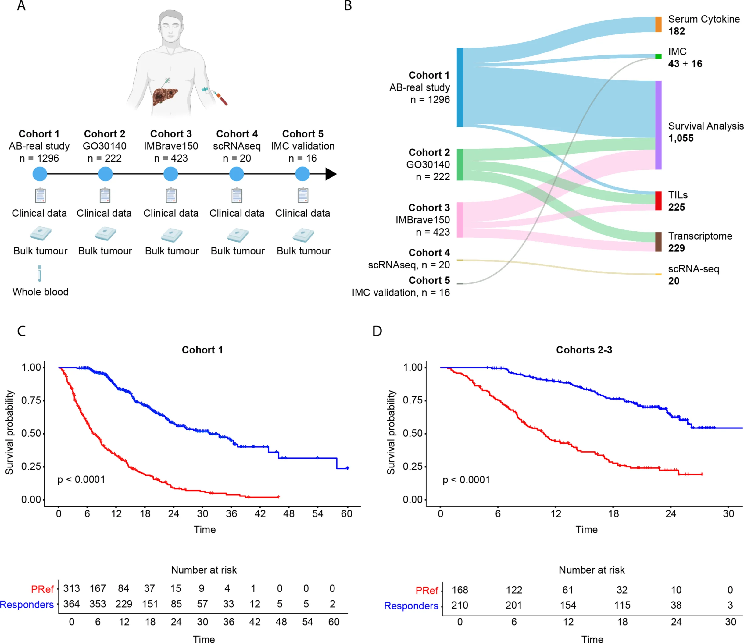
The investigators analyzed data from 1,296 patients treated in a prospective real-world cohort and validated their findings using 645 additional patients enrolled in the landmark IMbrave150 and GO30140 clinical trials. Multiple independent translational cohorts were also included for imaging mass cytometry, single-cell transcriptomics, serum cytokine analysis, and gene-expression profiling, making this one of the most comprehensive biological studies of immunotherapy resistance in hepatocellular carcinoma performed to date.
Primary refractoriness was defined according to the Society for Immunotherapy of Cancer (SITC) criteria as either progressive disease as the best response or stable disease lasting less than six months after initiation of atezolizumab plus bevacizumab.
Clinical Outcomes
The first important observation was that primary resistance is remarkably common. Approximately 45% of patients met the criteria for primary refractoriness across both the real-world and clinical trial populations, confirming that early resistance represents a major clinical challenge rather than an uncommon phenomenon.
Patients with primary refractory disease experienced dramatically worse outcomes than those who responded to treatment. Overall survival was reduced by more than threefold, establishing primary refractoriness as one of the strongest adverse prognostic factors in advanced hepatocellular carcinoma.
- Approximately 46% of patients in the real-world cohort and 44% in the validation cohorts demonstrated primary refractoriness.
- Median overall survival was 31.5 months in responders versus 7.3 months in patients with primary refractoriness in the real-world cohort.
- In the validation cohorts, median overall survival was 10.8 months for primary refractory patients, while it was not reached among responders.
The Tumor Microenvironment Behind Primary Resistance
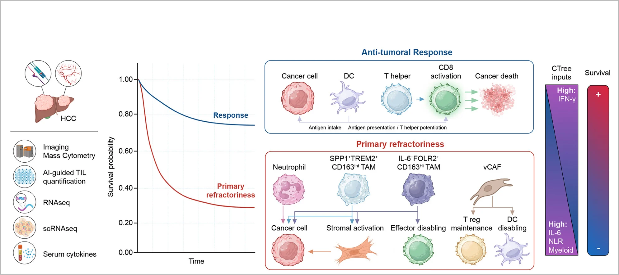
One of the most striking findings was that resistant tumors were not simply “immune-cold.” Machine-learning analysis showed that the overall density of tumor-infiltrating lymphocytes was similar between responders and non-responders. Instead, the decisive difference lay in the composition and organization of the immune microenvironment.
Using imaging mass cytometry, the investigators demonstrated that primary refractory tumors were heavily infiltrated by CD163-positive tumor-associated macrophages, vascular cancer-associated fibroblasts, regulatory T cells, and neutrophils. In contrast, tumors that responded to therapy contained greater numbers of cytotoxic CD8⁺ T cells, dendritic cells, helper T cells, and activated natural killer cells. These findings suggest that primary resistance is driven by an immune ecosystem dominated by suppressive myeloid cells rather than by an absence of immune infiltration.
Spatial analysis further revealed that these suppressive immune cells did not act independently. Instead, macrophages, fibroblasts, neutrophils, and regulatory T cells formed interconnected cellular networks surrounding tumor cells, creating a highly immunosuppressive microenvironment. Responding tumors displayed the opposite architecture, characterized by interactions between dendritic cells, cytotoxic T cells, helper T cells, and cancer cells that are consistent with active antitumor immunity.
Gene Expression Reveals an Immune-Suppressed Phenotype
Transcriptomic analysis provided additional evidence that resistant tumors possess a fundamentally different biology. Primary refractory tumors showed marked suppression of interferon-γ signaling together with reduced expression of genes associated with cytotoxic T-cell and natural killer cell activity. At the same time, genes involved in myeloid-cell recruitment, IL-6 signaling, TGF-β activation, hypoxia, angiogenesis, and stromal remodeling were significantly upregulated. Together, these findings indicate that resistant tumors exist in a profoundly immunosuppressive state before treatment even begins.
- Reduced IFN-γ, cytotoxic T-cell, and NK-cell signatures.
- Lower expression of immune effector genes including CD8A, GZMB, FASLG, and IFNG.
- Increased expression of genes associated with myeloid infiltration, IL-6 signaling, TGF-β, hypoxia, angiogenesis, epithelial-mesenchymal transition, and immune suppression.
Systemic Inflammation Reflects Tumor Biology
An important clinical observation was that the biology of the tumor could also be detected in peripheral blood. Patients with an elevated neutrophil-to-lymphocyte ratio (NLR ≥3) exhibited tumors enriched with immunosuppressive macrophages, neutrophils, and inflammatory cytokines such as IL-6. This suggests that a simple blood test may serve as a surrogate marker for the underlying immune landscape of the tumor and help identify patients at increased risk of primary resistance.
A Predictive Model for Primary Resistance
Finally, the investigators combined clinical variables with transcriptomic biomarkers using a machine-learning conditional inference tree. They found that suppressed IFN-γ signaling was the strongest determinant of primary resistance. When low IFN-γ activity was combined with an elevated neutrophil-to-lymphocyte ratio, the probability of primary refractoriness increased to nearly 74%. Even among tumors with preserved IFN-γ signaling, a high ratio of myeloid cells to effector T cells substantially increased the likelihood of treatment failure, emphasizing that the balance between immune activation and immune suppression is a critical determinant of response.
- Low IFN-γ signaling emerged as the strongest predictor of primary resistance.
- IFN-γ-low + NLR ≥3 identified patients with approximately 74% probability of primary refractoriness.
- A high myeloid-to-effector T-cell ratio further increased the risk of resistance, even in tumors with preserved IFN-γ activity.
Clinical Implications
This study fundamentally changes our understanding of why atezolizumab plus bevacizumab fails in a substantial proportion of patients with hepatocellular carcinoma. Rather than being driven simply by insufficient immune-cell infiltration, primary resistance appears to result from a highly organized myeloid-dominant immunosuppressive tumor microenvironment that prevents effective antitumor immunity. These findings provide a strong biological rationale for combining immune checkpoint inhibitors with therapies targeting tumor-associated macrophages, neutrophils, inflammatory cytokines, and other components of the myeloid compartment.
You Can Read Full Article Here