
Bispecific antibodies and other immune-based therapies have rapidly expanded treatment options for relapsed or refractory B-cell lymphoma, but predicting who will respond has not kept pace. Differences in outcomes cannot be explained by tumor genetics alone, as immunotherapy response also depends on the functional relationship between malignant B cells and the patient’s immune microenvironment.
Standard lymphoma cell lines lose much of the heterogeneity of the original disease. 2D cultures can’t reproduce the spatial interactions. More sophisticated 3D systems address some of this, but keeping malignant cells alive alongside functional autologous immune cells has been difficult, and for therapies built on T-cell engagement, that missing immune context is not a minor gap.
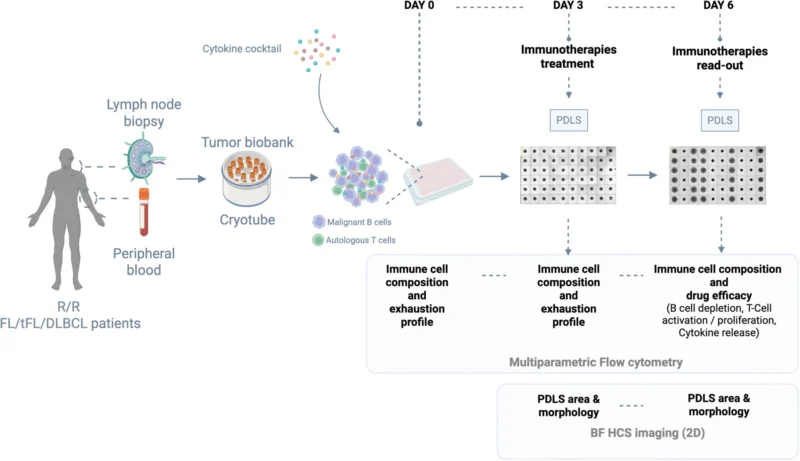
In a study published in HemaSphere, researchers developed patient-derived lymphoma spheroids (PDLS), a 3D ex vivo platform grown directly from lymph node or peripheral blood samples. The model keeps the patient’s lymphoma and immune cells together, allowing immunotherapies to be tested within a patient-specific tumor-immune environment.
Building Patient-Derived Lymphoma Spheroids
The study included 45 samples from patients with follicular lymphoma (FL), transformed FL, or diffuse large B-cell lymphoma (DLBCL), obtained from peripheral blood or lymph nodes. For treatment experiments, 33 PDLS samples were exposed to a broad panel of therapies: glofitamab, checkpoint-directed agents, CD19-targeted costimulatory antibodies, and polatuzumab vedotin, alone or in combination.
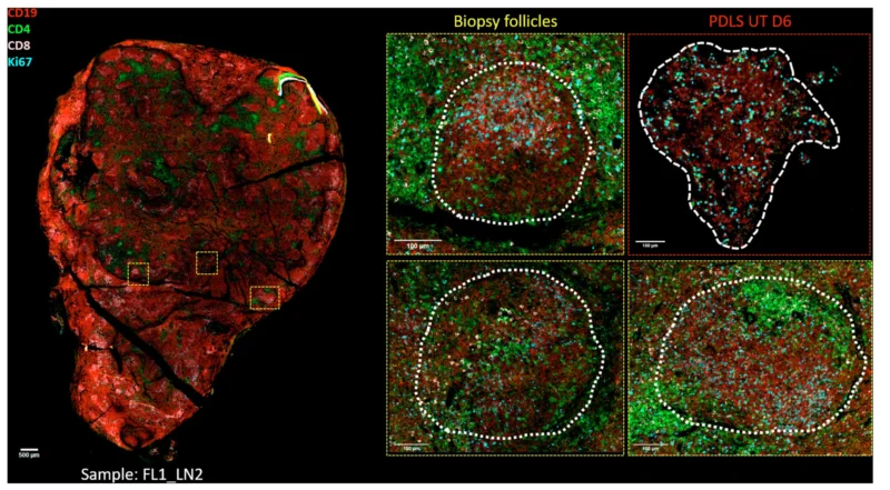
Response was assessed through B-cell depletion, T-cell activation, cytokine release, and imaging. The workflow moves from establishing the spheroids to treatment assessment within six days. PDLS formed from both blood and lymph-node specimens, remained viable through the experimental window, and preserved major immune populations. Spatial profiling in an FL sample showed preservation of follicular organization, with malignant B cells surrounded by CD4+ and CD8+ T cells.
Preserving immune-cell numbers would not be enough for immunotherapy testing if those cells lost their functional state in culture. Expression of costimulatory molecules and immune checkpoints, including PD-1, LAG-3, TIGIT, and TIM-3, remained correlated between the original samples and their PDLS counterparts.
Glofitamab Produced the Strongest Single-Agent Response
Several single agents produced significant B-cell depletion, including the CD19-CD28 costimulatory bispecific, polatuzumab vedotin, and a PD-1/TIM-3 bispecific antibody. Glofitamab produced the strongest effect, with dose-dependent B-cell depletion of approximately 39% to 51%.
The model captured more than disappearance of malignant B cells. Glofitamab increased activation of both CD4+ and CD8+ T cells and produced dose-dependent granzyme B release. TNFα, IFNγ, and IL-10 also increased, while IL-6 and IL-8 remained near baseline. Imaging showed proliferating, granzyme B-positive T cells near malignant B cells and dying cells, and another FL sample showed increased cleaved caspase-3 in B cells after treatment.
Hierarchical clustering separated samples into 14 low responders across treatment classes, 11 intermediate responders mainly to glofitamab and antibody-drug conjugates, and six high responders to glofitamab. Low responders contained substantially more exhausted CD4+ T cells than high responders, 43.3% versus 19.3%, and fewer non-exhausted CD8+ T cells, 16.8% versus 29.3%. Intermediate responders fell in between.
Resistance Was Not Explained by One Mechanism
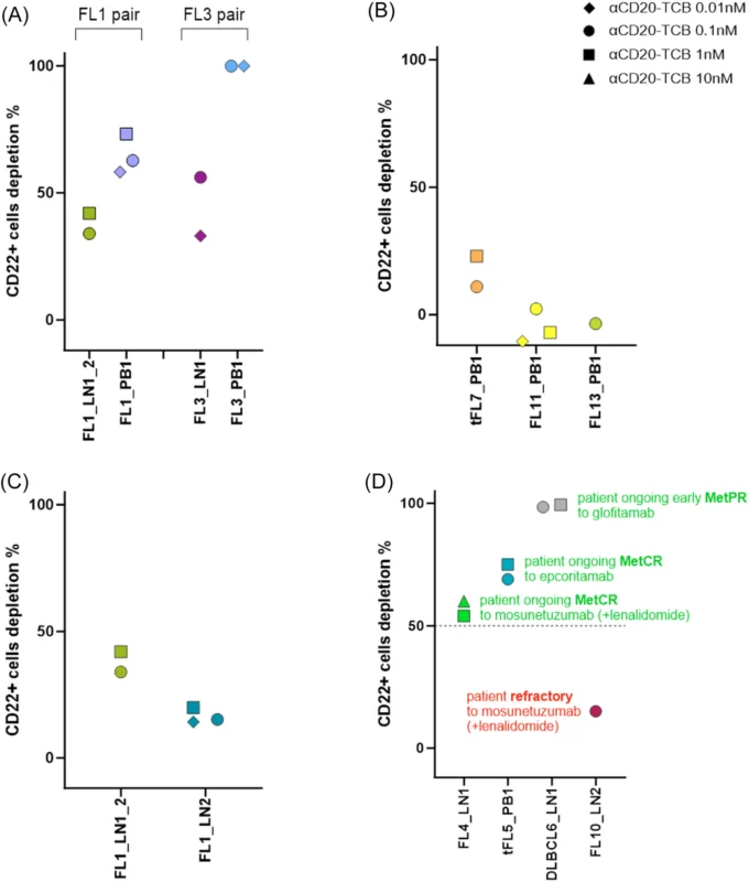
Three of 34 samples tested with glofitamab had very low or absent CD20 expression and responded poorly at every tested concentration. Paired lymph-node and peripheral-blood samples from the same patients could also differ in sensitivity. In one patient, a lymph-node sample collected after second-line treatment was less sensitive to glofitamab than an earlier sample from the same individual.
In some tumors the therapeutic target may be absent or reduced, in others the immune compartment may be too dysfunctional. PDLS may help distinguish tumor-related from immune-related mechanisms of resistance.
Ex Vivo Response Matched Clinical Response, but in Only Four Patients
The most clinically provocative part of the study involved four patients who had received a CD20×CD3 bispecific antibody, glofitamab, epcoritamab, or mosunetuzumab, and whose PDLS were tested ex vivo with glofitamab.
All three patients who achieved an early partial or complete metabolic response clinically also showed strong ex vivo sensitivity, with 50% to 98% B-cell depletion. The fourth patient was clinically refractory to bispecific-antibody therapy and showed little B-cell depletion.
The authors note the observation is consistent with a separate nine-patient cohort from their group, but the platform still requires formal prospective validation in larger populations, testing samples before therapy, scoring the ex vivo response blind to outcome, and then checking if it matches the clinical response.
Can the Platform Find a Way Around Resistance?
The researchers tested up to 60 combinations per sample to see if functional screening could rescue weak responses. Several checkpoint-based combinations increased B-cell depletion compared with untreated controls, particularly regimens combining glofitamab with TIGIT or PD-1/TIM-3 targeting.
Checkpoint inhibition alone generally had limited direct activity. When samples were separated by response class, none of the combinations significantly improved B-cell depletion in the low-responder group.
One transformed FL sample with less than 50% B-cell depletion on glofitamab alone reached ~65% – 70% depletion with several combinations. A combination that showed little benefit across a cohort may still help a particular tumor.
Important Clinical Limitations
The model does not preserve the complete tumor microenvironment. Mechanical dissociation and cryopreservation can remove stromal populations, so PDLS model autologous immune-tumor interactions. More complex systems: tumor-fragment cultures, lymphoma organoids, lymphoma-on-chip, and microfluidic models, may retain components that PDLS lose.
Comprehensive screening currently requires up to 50,000 cells per well and approximately 30 million cells for a workflow testing 60 to 80 conditions, difficult to obtain from small biopsies. Relapsed or refractory ABC-type DLBCL samples were relatively fragile in culture, and peripheral-blood PDLS require adequate circulating tumor B cells. Miniaturization will be important if the approach is to become practical.
The six-day assay is also a trade-off: rapid, but unable to reproduce longer-term processes such as chronic immune remodeling or acquired resistance. Further study will be needed to characterize effects on proliferation and mechanisms of cell death.
The workflow could potentially run in cytometry-equipped laboratories and be incorporated into multicenter studies, but this requires reproducibility across sites with minimal batch effects. Integration with spatial profiling and single-cell RNA sequencing could add deeper biological information.
Can PDLS Inform Treatment Decisions?
This study does not show that clinicians can send a lymphoma biopsy to the laboratory and receive a validated answer about which immunotherapy to prescribe. It shows that such a strategy is experimentally plausible.
PDLS preserved enough patient-specific biology to reproduce differences in immune state, antigen expression, treatment sensitivity, and resistance, screened multiple therapies within days, distinguished responder phenotypes, and, in a very small clinical comparison, showed ex vivo bispecific-antibody sensitivity aligned with outcomes.
Next is prospective validation in larger cohorts, with predefined ex vivo response criteria compared against clinical outcomes. PDLS could add something genuinely new to precision lymphoma therapy: a functional test of how that patient’s lymphoma and immune cells respond when actually exposed to treatment.
Can ctDNA Predict Both Benefit and Toxicity From Immunotherapy?