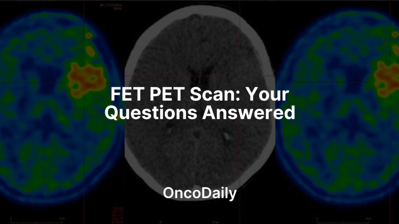
A FET PET scan is an advanced medical imaging technique, frequently combined with a CT scan, specifically designed to visualize and assess brain tumors, whether they are primary or have spread from other parts of the body. This examination uniquely focuses on the amino acid metabolism within cells, leveraging the fact that diseased cells, particularly tumor tissue, often exhibit heightened metabolic activity compared to healthy brain tissue. By injecting a special radioactively labeled amino acid tracer into the bloodstream, areas with elevated metabolic activity light up, providing crucial information to physicians.
This detailed insight into the location and extent of tumors is invaluable for determining biopsy sites, planning precise surgical resections or radiation therapy, and effectively monitoring a patient’s response to treatment.
How Does FET PET Scan Work?
A FET PET scan, often performed in conjunction with a CT scan, operates by examining the amino acid processes within cells, which can indicate disease. Initially, a small amount of a weakly radioactive amino acid solution, known as a tracer, is introduced into a vein in the arm. This tracer circulates through the body, and unlike healthy brain tissue, cells with elevated amino acid metabolic activity, such as those found in tumors, readily absorb and bind to it. As the tracer is taken up, it emits a radioactive signal.
After the injection, there’s a necessary waiting period, typically between 60 to 90 minutes, allowing the tracer sufficient time to accumulate in the target areas. During this phase, the patient is encouraged to relax. Once the uptake period concludes, the patient is positioned comfortably on an examination table that slides into the PET/CT scanner.
Within the scanner, two distinct imaging processes occur. The PET component is responsible for detecting the radiation signals released by the accumulated tracer, pinpointing its quantity and exact location within the brain. Simultaneously, the CT component captures detailed anatomical images, revealing the structure of the brain and any changes, such as tumor growth or other disease-related alterations. It is crucial for the patient to remain perfectly still throughout this 15-20 minute scanning period to ensure high-quality images.
The collected PET and CT images are then combined. This comprehensive data provides valuable insight into the tumor’s position and size. This information is then sent to a specialist physician who interprets the findings, providing a report to the referring doctor. This detailed metabolic and structural information is critical for decisions regarding biopsy sites, planning precise surgical removal, guiding radiation therapy, or differentiating between actual tumor growth and post-treatment effects like swelling or changes from radiation.
What to Do Before Your PET PET Scan?
Before your FET PET scan, certain preparations are advised to ensure optimal imaging and your comfort during the procedure.
One important aspect mentioned in the provided information is hydration; it is crucial to be well hydrated prior to your scan. Regarding food intake, the texts present slightly differing advice: one indicates there is no requirement to fast for a FET PET scan, while another states it is essential to fast for at least four hours before the examination. However, both agree that you can continue to drink water and take your regular medications as you normally would.
Upon your arrival at the clinic, you will be requested to complete a questionnaire and sign a consent form as part of the initial check-in process. You will also need to change into a gown.
Should you experience claustrophobia, you have the option to request a sedative upon arrival at the clinic to help you relax during the PET/CT scan. If you choose to take this medication, it is important to arrange for someone to accompany you and drive you home afterwards, as you should not drive for the remainder of the day. Ideally, you should have someone available to pick you up following the examination.
FET PET-Scan vs Other Imaging
FET PET-CT offers significant advantages over other common imaging techniques like FDG PET, CT, and MRI, particularly in the context of brain tumors.
One key benefit is its superior ability to distinguish viable tumor tissue from non-malignant changes that can result from treatment, such as post-operative effects, swelling, or radiation damage (radionecrosis). While conventional imaging might show ambiguous findings in these situations, FET PET’s focus on amino acid metabolism allows for a clearer differentiation. It is also noted to be more effective than FDG PET in identifying low-grade tumor recurrence.
Furthermore, FET PET provides more precise tumor delineation, meaning it offers a better estimate of the actual spread of the tumor in both low and high-grade gliomas. This enhanced clarity is crucial for selecting optimal biopsy sites, guiding stereotactic biopsies to ensure accurate classification and grading of gliomas.
The scan also aids in non-invasive tumor grading, potentially helping to differentiate between high-grade gliomas and benign or non-cancerous brain lesions. This is particularly valuable for therapy planning, as FET PET, when used alongside anatomical imaging, can more accurately define the tumor volumes for surgical removal or radiation therapy.
Lastly, FET PET is beneficial for monitoring a tumor’s response to treatment, including chemotherapy and radiation therapy. It enables earlier detection of any remaining tumor tissue after surgery, helping physicians assess the effectiveness of interventions and adjust treatment strategies as needed. Overall, FET PET’s distinct capability to visualize cellular metabolic activity provides a more specific and sensitive assessment of brain tumors compared to methods that primarily focus on anatomy or general glucose uptake.

Read OncoDaily’s Special Article About PET Scan
Adverse Effects
While the substances utilized in the FET PET examination are not known to cause any direct side effects or allergic reactions, and the procedure is even deemed suitable for children, there are important considerations related to the temporary presence of the radioactive tracer in your system.
Following the scan, the small amount of radioactive material remains in your body for a short duration. Consequently, it is advised to limit prolonged close contact with children, particularly those under 16 years of age, and pregnant women. One text suggests this precaution for a few hours, while another specifies a 12-hour period on the day of the examination. For breastfeeding women, it is recommended to refrain from breastfeeding for 24 hours after the procedure.
To help the radioactive substances exit your body more quickly and reduce overall radiation exposure, it is encouraged to drink plenty of fluids and urinate frequently after the examination.
Additionally, if a sedative is administered to help with claustrophobia during the scan, you should not drive a car for the rest of that day. In such cases, it is ideal to have someone accompany you to pick you up following your appointment. Aside from these specific considerations, you are generally free to resume all other normal activities without restriction after the examination.
Written By Aren Karapetyan, MD
FAQ
What is a FET PET scan?
A FET PET scan is an advanced medical imaging test, often combined with a CT, used to visualize and evaluate brain tumors by examining their amino acid metabolism.
How does a FET PET scan work?
A radioactive amino acid tracer is injected, which accumulates in tumor cells due to their elevated metabolic activity. A scanner then detects the emitted signals to pinpoint the tumor's location and extent.
How does FET PET compare to other brain imaging methods?
FET PET is considered superior to other methods like FDG PET, CT, and MRI for distinguishing viable tumor tissue from treatment-related changes, delineating tumor boundaries, and guiding biopsies.
How should I prepare for a FET PET scan?
You should be well-hydrated. Regarding food, one text indicates no fasting is required, while another states essential fasting for at least 4 hours. You can generally take medications with water.
Are there any side effects or post-scan precautions for FET PET?
The substances used for the scan are not known to cause side effects or allergic reactions. However, due to the temporary radioactivity, it's advised to limit close contact with children and pregnant women for several hours after the scan.


