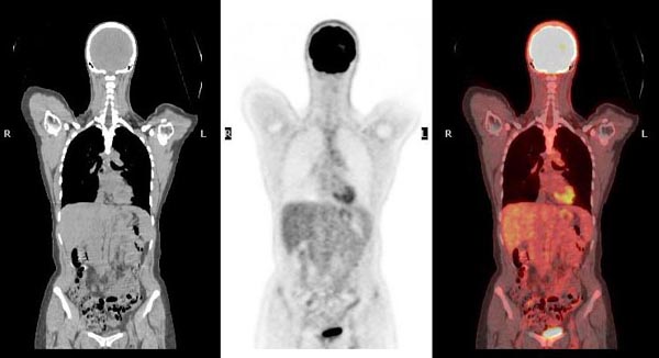
A Positron Emission Tomography (PET) scan is an advanced medical imaging test that provides a unique view into how your body’s organs and tissues are actively functioning at a cellular level. Unlike traditional imaging methods that show physical structures, a PET scan utilizes a small amount of a safe, injectable radioactive substance, or “tracer,” which is absorbed differently by healthy and diseased cells.
By detecting where this tracer accumulates, particularly in areas of higher metabolic activity, often seen in illness, PET scans can identify potential health problems, such as cancer, heart conditions, and brain disorders, often much earlier than other scans. Frequently combined with CT or MRI for a comprehensive view, PET scans play a crucial role in diagnosing conditions, planning effective treatments, and monitoring their progress.
How does a PET Scan work?
A PET scan is a type of nuclear medicine imaging that reveals the body’s internal processes and molecular activity, enabling the detection of diseases in their earliest stages.
Tracer Administration
A small, safe amount of a radioactive chemical, known as a radiotracer (commonly FDG), is introduced into your body. This is usually given intravenously through an IV in your arm or hand, but can sometimes be swallowed or inhaled, depending on the examined area.
Targeted Absorption
The tracer travels through the bloodstream and is absorbed by cells and tissues. Diseased cells, such as cancer cells, exhibit a higher metabolic rate, leading them to absorb more radiotracer than healthy cells. The tracer’s radioactive component highlights these areas of differing uptake, making them visible.
Rest and Absorption Period
After the injection, you’ll rest quietly for approximately 45 to 60 minutes. This allows sufficient time for the tracer to distribute and accumulate in the target organs and tissues. Limiting movement during this phase ensures the tracer goes to the intended areas. Minimizing mental activity (like reading or listening to music) may also be requested for brain scans.
Scanning Process
You then lie on a narrow table that slides into a doughnut-shaped PET scanner. The scanner detects the radiation emitted by the concentrated tracer within your body. These areas of increased tracer uptake, often referred to as “hot spots,” indicate elevated chemical activity and potential health issues.
Image Generation
A computer processes the signals from the scanner, transforming them into detailed 2D and 3D images. These images provide insights into crucial bodily functions such as blood flow, oxygen utilization, and glucose metabolism.
Integrated Imaging (PET/CT or PET/MRI)
Most PET scans are now combined with a CT scan (PET/CT) or sometimes an MRI scan (PET/MRI). The CT provides detailed structural X-ray images, while MRI offers images based on magnetic fields and radio waves. The fusion of these structural images with the functional PET images creates a highly comprehensive 3D picture, allowing for the precise anatomical localization of abnormalities and a more accurate diagnosis.
What to do before your FDG PET-CT scan?
To prepare for your FDG PET-CT scan, begin by avoiding strenuous physical activity and high-carbohydrate, sugary foods for 24 hours prior. Throughout this period and on scan day, stay warm, as cold can affect image clarity.
On the day of the scan, fast from food for 4-6 hours (only plain water allowed; no gum or candies), and take medications only with water. Diabetics will receive specific instructions regarding medication and meals to manage blood sugar. Remove any wearable medical devices like CGM or insulin pumps as advised.
For the appointment, wear comfortable, metal-free, warm clothing and remove all jewelry. Bring 1 liter of non-carbonated water, all relevant medical records, and identification. If you are claustrophobic, inform your provider to discuss relaxation options.
PET-Scan vs Other Imaging
PET scans differ fundamentally from other common imaging techniques like CT and MRI by focusing on the function and metabolic activity of organs and tissues at a cellular level, rather than just their physical structure.
While Computed Tomography (CT) scans utilize X-rays to generate still images depicting the form and blood flow to organs, and Magnetic Resonance Imaging (MRI) scans employ magnets and radio waves to create static pictures of body structures, both primarily reveal anatomical details. These structural imaging methods can only detect changes in organs and tissues once a disease has caused physical alterations.
In contrast, a PET scan uses a radioactive tracer that is absorbed by cells. Because diseased cells often exhibit higher metabolic rates, they accumulate more of this tracer, appearing as “bright spots.” This unique capability allows PET scans to show how an organ is working in real-time and to identify cellular changes much earlier than CT or MRI scans, often before any structural abnormalities are visible. This early detection ability provides doctors with a superior view of complex systemic diseases, such as coronary artery disease, brain tumors, and seizure disorders.
To combine the strengths of both approaches, PET scans are very frequently performed simultaneously with a CT scan, creating a PET/CT scan. This integrated technique merges the functional information from the PET scan with the detailed anatomical images from the CT scan. The result is a comprehensive, three-dimensional picture that allows for a more accurate and precise diagnosis by pinpointing the exact location of the metabolic abnormalities within the body’s structure.
Some advanced centers also use a hybrid PET/MRI scan for extremely high-contrast images, particularly beneficial for soft tissue cancers, combining the functional data of PET with the superior soft tissue detail of MRI while reducing radiation exposure compared to PET/CT. This fusion of functional and anatomical data provides a complete picture that significantly aids in diagnosing diseases, planning treatments, and monitoring their effectiveness.

source: www.brighamandwomens.org
PET Scan vs PET-CT
A PET scan is an advanced imaging test that visualizes your body’s functional activity using a small amount of a radioactive tracer. This tracer accumulates in areas of higher chemical activity, like diseased cells, appearing as bright spots. Unlike CT or MRI, which show structure, PET scans detect cellular changes earlier, measuring blood flow, oxygen use, and sugar metabolism. Often combined with CT (PET-CT) or MRI (PET-MRI) for precise anatomical localization, it aids in diagnosing and managing conditions like cancer, heart problems, and brain disorders.
The scan helps detect cancer’s presence, spread, and treatment response due to cancer cells’ higher metabolic rate. For heart issues, it shows areas of decreased blood flow, and for brain disorders, it indicates glucose usage patterns. PET scans’ ability to reveal cellular-level changes provides a superior view of complex systemic diseases, especially when fused with structural images.
While involving minimal radiation, PET scans are generally safe, with tracers quickly eliminated. Risks are low but include rare allergic reactions or issues for pregnant individuals, those breastfeeding, or with certain health conditions. Preparation involves dietary restrictions, avoiding strenuous activity, and keeping warm, along with informing staff about medical conditions or claustrophobia.
The procedure involves a tracer injection, a 45-60 minute absorption period where you rest quietly, followed by a 30-45 minute scan in a doughnut-shaped machine. Post-scan, you can resume normal activities, drinking plenty of fluids to flush the tracer. Contact with pregnant women and young children is advised to be limited for a few hours. Results are interpreted by a radiologist and sent to your doctor, typically within a few business days. Monitor the injection site for any unusual changes.
Adverse Effects
While PET scans are generally considered safe and complications are rare, it’s important to be aware of potential adverse effects. The primary concern stems from the radiation exposure. Although the radioactive tracer used contains a very small amount of radiation that quickly leaves the body, typically within 2 to 10 hours, any radiation exposure carries a minimal, slight risk of potentially increasing the risk of cancer over a long period. This risk is compounded if a PET-CT scan is performed due to the additional X-ray radiation from the CT component. Healthcare providers carefully weigh these slight risks against the significant benefits of obtaining a correct diagnosis for serious medical conditions.
Specific situations or conditions can also introduce or heighten risks. Pregnancy is a contraindication for PET scans, as the radiation may be harmful to a developing fetus; similarly, breastfeeding mothers are advised to temporarily stop breastfeeding for a period (e.g., 4 to 12 hours) after the scan to prevent the tracer from passing to the infant through breast milk.
Allergic reactions to the radioactive tracer or, in the case of PET/CT scans, to the oral or intravenous contrast dyes (which may contain iodine or barium), are possible but extremely rare and usually mild. However, individuals with a history of allergies, asthma, heart disease, dehydration, certain blood disorders, kidney disease, or specific medication regimens should inform their healthcare provider, as they might be at a slightly higher risk for such reactions. Medical staff are prepared to quickly manage any allergic responses that may occur.
For individuals with diabetes, their blood sugar or insulin levels can affect how the tracer is absorbed, potentially leading to inaccurate scan results. Special dietary and medication instructions are provided to manage this and ensure accurate readings.
Other potential discomforts or minor issues include a sharp sting when the IV with the tracer is inserted, or localized pain, redness, or swelling at the injection site, which typically resolves quickly. For those who experience claustrophobia (fear of enclosed spaces), the tunnel-shaped scanner might cause anxiety, but medication can be offered to help them relax. Lastly, some people might experience discomfort if they are uncomfortable with needles. Patients should always inform their healthcare team about any concerns or symptoms they experience during or after the scan.
Written By Aren Karapetyan, MD
FAQ
What is a PET scan?
A PET scan is an imaging test that shows how your organs and tissues are functioning at a cellular level, using a radioactive tracer to detect areas of high metabolic activity, often indicating disease.
How is a PET scan different from a CT or MRI?
Unlike CT and MRI scans which show physical structures, a PET scan reveals how organs are functioning in real-time and can detect cellular changes much earlier.
Why would my doctor order a PET scan?
Doctors commonly order PET scans to detect and assess cancer, evaluate heart problems (like blood flow), and diagnose brain disorders (such as tumors, epilepsy, or dementia).
What is a PET-CT scan?
A PET-CT scan combines the functional images from a PET scan with the detailed anatomical images from a CT scan into one procedure, providing a more precise 3D picture of any abnormalities.
How is the radioactive tracer given?
The tracer is typically injected into a vein in your arm or hand, but can sometimes be swallowed or inhaled depending on the area being examined.
How do I prepare for a PET scan?
Preparation usually involves fasting for 4-6 hours (only water), avoiding strenuous exercise and high-carb foods for 24 hours prior, staying warm, and removing all metal items. Diabetics receive special instructions.
How long does a PET scan take?
The entire process, including tracer absorption and the scan itself, usually takes about 2 to 4.5 hours, depending on the area being scanned and if a PET/CT is performed.
Are there any risks to a PET scan?
Risks are generally low, as the radiation dose is minimal and short-lived. However, it's not recommended for pregnant women, and breastfeeding mothers need to take precautions. Rare allergic reactions can occur.
What should I do after the scan?
Drink plenty of fluids to help flush the tracer from your body. You can usually resume normal activities immediately, but may need to limit close contact with pregnant women and young children for a few hours.
When will I get my PET scan results?
Results are typically available within 1 to 2 business days, interpreted by a radiologist and sent to your referring doctor, who will discuss them with you.


