Key takeaways
- CT, MRI, and PET/CT serve different purposes in cancer diagnosis, staging, and treatment monitoring.
- CT is widely used to detect tumors, measure their size, and assess cancer spread, particularly in the chest and abdomen.
- MRI provides detailed soft-tissue images, making it especially useful for brain, prostate, liver, and pelvic cancers.
- PET/CT evaluates metabolic activity, helping detect selected cancers, assess metastases, and monitor treatment response.
- No single scan is best for every cancer. Doctors choose imaging based on cancer type, clinical needs, and patient safety.
CT, MRI, and PET/CT are among the most commonly used imaging tests in cancer care. They help doctors detect suspicious tumors, determine how far cancer has spread, and evaluate how well treatment is working.
Although these scans may be used for similar purposes, they provide different information. CT and MRI show the structure of organs and tissues, while PET/CT combines anatomical imaging with information about metabolic activity.
No single imaging technique is suitable for every cancer. The choice depends on the tumor’s location, the type of cancer suspected, and the information needed to guide diagnosis or treatment.
What Is the Difference Between CT, MRI, and PET/CT Scans?
CT, MRI, and PET/CT differ mainly in the technology they use and the type of information they provide about tumors.
Computed tomography (CT) uses X-rays to produce detailed cross-sectional images of the body. It allows doctors to examine organs, identify abnormal masses, measure tumors, and assess their relationship with nearby structures. CT is relatively fast and widely available, making it a routine examination in cancer diagnosis and staging.
Magnetic resonance imaging (MRI) uses strong magnetic fields and radio waves rather than X-rays. It provides greater detail of many soft tissues, including the brain, spinal cord, prostate, and pelvic organs. MRI can also use specialized techniques, such as diffusion-weighted imaging, to evaluate tissue characteristics.
Positron emission tomography combined with computed tomography (PET/CT) works differently. PET uses a small amount of radioactive tracer to show biological activity within tissues, while CT provides the anatomical images needed to locate these findings.
The most commonly used PET tracer is fluorodeoxyglucose (FDG), a glucose-like substance that accumulates in many cancers because of their increased glucose metabolism. However, not all tumors absorb FDG to the same degree.
These differences explain why doctors may recommend more than one imaging examination. CT can show the size and location of a tumor, MRI may provide greater detail about its relationship with nearby tissues, and PET/CT can help assess metabolic activity when clinically indicated (National Cancer Institute, n.d.; Ding et al., 2026).

How Are CT, MRI, and PET/CT Used in Cancer Diagnosis?
Imaging tests are often performed when symptoms, physical examination findings, or laboratory results raise suspicion of cancer. They help doctors identify abnormalities and decide whether further investigations are necessary.
CT is frequently used to examine the chest, abdomen, and pelvis. It can reveal suspicious masses, enlarged lymph nodes, or changes in organs such as the lungs, liver, and pancreas. CT also helps guide needle biopsies, allowing tissue samples to be collected from areas that may be difficult to reach.
MRI is particularly useful when doctors need a detailed view of soft tissues. For example, prostate MRI can identify suspicious lesions before biopsy, while brain MRI helps evaluate the location and extent of tumors within the brain. In rectal cancer, MRI is used to assess how deeply a tumor extends into surrounding tissues.
PET/CT is generally requested for more specific clinical questions. It may help identify metabolically active disease, clarify findings from other scans, or assess whether cancer has spread beyond its original location.
However, an abnormal imaging result does not necessarily mean that cancer is present. Benign tumors, inflammation, and infections can sometimes resemble malignancy on CT or MRI. Similarly, increased FDG uptake on PET/CT may occur in inflammatory or infectious conditions.
When imaging identifies a suspicious lesion, biopsy is often needed to confirm whether cancer is present and determine its histological type. Imaging helps locate and characterize abnormalities, but it cannot replace microscopic examination of tissue in most cancer diagnoses (American Cancer Society, n.d.; RadiologyInfo.org, n.d.).
Which Cancers Are Best Evaluated With CT, MRI, or PET/CT?
Different cancers require different imaging approaches. The examination selected depends not only on the organ involved but also on whether doctors are investigating a new diagnosis, determining cancer stage, or looking for recurrence.
CT is commonly used in lung, pancreatic, colorectal, and several other abdominal cancers. It provides detailed images of the lungs and abdominal organs, making it useful for measuring tumors and assessing possible spread to lymph nodes or distant organs.
MRI is particularly important for cancers involving the brain, prostate, rectum, liver, and certain pelvic structures. Its high soft-tissue contrast allows doctors to evaluate the relationship between tumors and nearby muscles, nerves, blood vessels, or other organs.
In rectal cancer, for example, pelvic MRI is central to local staging because it helps assess tumor extension into surrounding tissues and the relationship to the mesorectal fascia. In prostate cancer, multiparametric MRI can identify suspicious lesions and guide biopsy decisions.
PET/CT is commonly used in selected patients with lymphoma, lung cancer, melanoma, and head and neck cancers. It can help identify metabolically active disease and assess whether cancer is present in other parts of the body.
The tracer used during PET/CT also matters. While FDG is suitable for many malignancies, other tracers are more useful for particular cancer types. For example, prostate-specific membrane antigen (PSMA) PET/CT is used in selected patients with prostate cancer for staging or recurrence assessment.
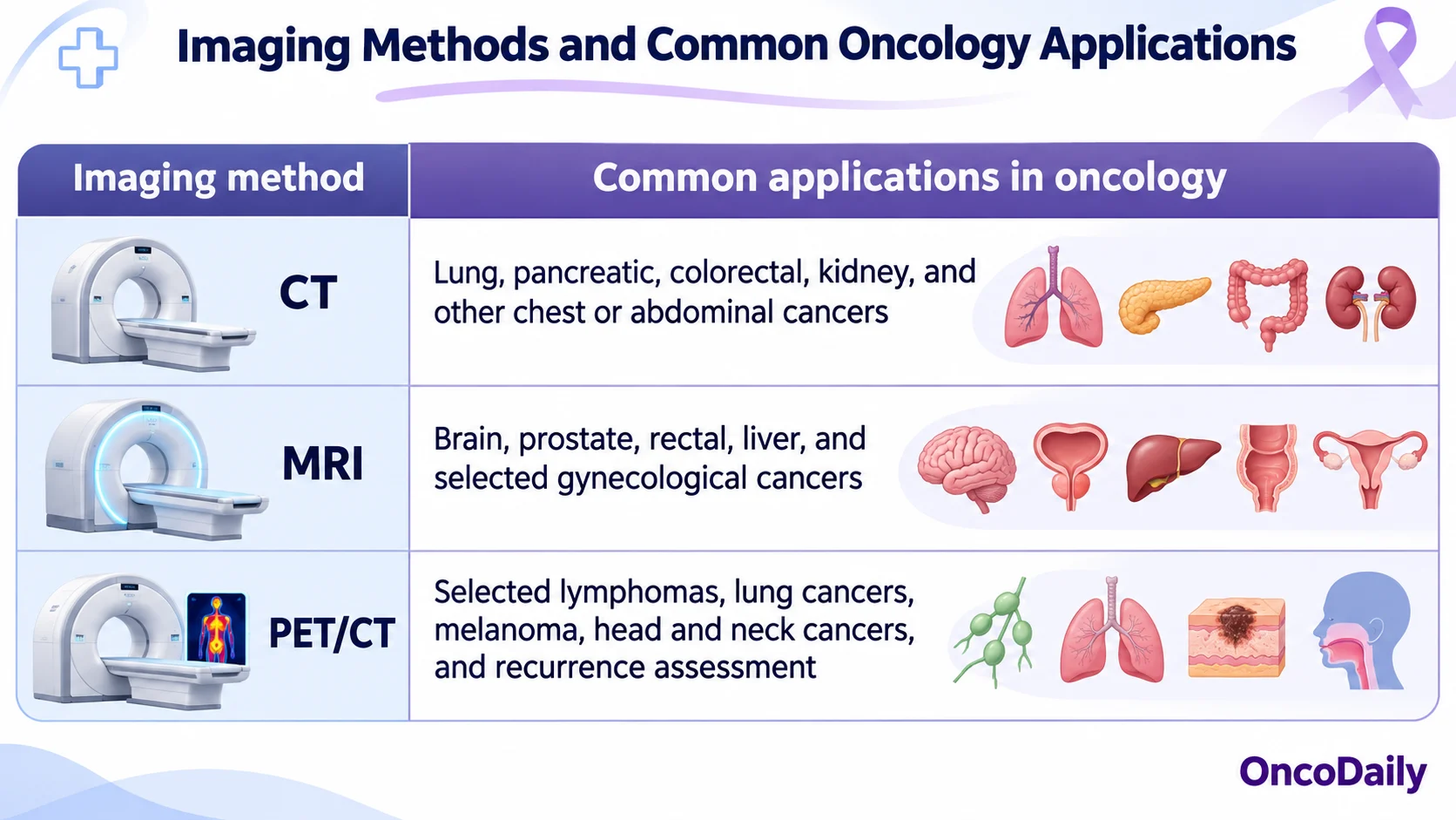
These are common applications rather than strict rules. Some cancers require several imaging methods during diagnosis and treatment, while others can be adequately assessed using one technique (National Cancer Institute, n.d.; Ding et al., 2026).
How Accurate Are CT, MRI, and PET/CT in Detecting Cancer?
The accuracy of cancer imaging depends on the tumor’s size, location, biological characteristics, and the technique being used. No scan can detect every cancer, and a normal result does not completely exclude malignancy.
CT can identify many tumors and structural abnormalities, particularly in the chest and abdomen. However, very small lesions may be difficult to detect, and some tumors have an appearance similar to surrounding normal tissue.
MRI provides excellent soft-tissue contrast and may detect abnormalities that are less visible on CT. This is particularly useful in the brain, liver, and pelvis. Nevertheless, MRI findings are not always specific for cancer, and benign changes can occasionally appear suspicious.
PET/CT provides additional information by measuring tracer uptake. Some cancers show increased metabolic activity before substantial changes in tumor size become visible. However, FDG PET/CT may miss tumors that are very small or have low glucose metabolism. Inflammation and infection can also produce increased tracer uptake, leading to false-positive results.
Two important measures of diagnostic accuracy are sensitivity and specificity. Sensitivity describes how often a test correctly identifies disease when it is present, while specificity describes how often it correctly identifies people without the disease.
These values vary considerably between cancer types and clinical situations, so it would be misleading to assign one overall accuracy percentage to CT, MRI, or PET/CT.
A 2016 review by Spick and colleagues, which examined studies involving more than 2,300 patients, found that FDG PET/CT and PET/MRI had broadly comparable diagnostic performance in many oncological applications. Differences were observed in particular anatomical regions and clinical settings (Spick et al., 2016).
Rather than choosing a scan based on its perceived accuracy alone, doctors consider which technique is most likely to answer the specific diagnostic question.
Can CT, MRI, and PET/CT Detect Cancer Metastases?
CT, MRI, and PET/CT can all help detect metastatic cancer, but their usefulness varies depending on where the disease has spread.
Metastasis occurs when cancer cells spread from the original tumor to other parts of the body. The most common sites vary by cancer type and may include the liver, lungs, bones, brain, and lymph nodes.
CT is routinely used to investigate possible metastases in the chest, abdomen, and pelvis. It can identify suspicious lung nodules, liver lesions, enlarged lymph nodes, and abnormalities in other organs.
MRI is especially useful when more detailed imaging is required. Brain MRI is generally preferred when brain metastases are suspected, while liver MRI may help characterize small lesions that remain uncertain on CT. MRI is also valuable for evaluating the spinal cord and surrounding tissues.
PET/CT can identify abnormal tracer uptake in different parts of the body, sometimes revealing disease that was not clearly visible on conventional imaging. This makes it particularly useful for staging certain cancers and investigating suspected recurrence.
However, PET/CT does not detect every metastatic lesion. Small deposits may be missed because of limited spatial resolution, while some cancers have insufficient FDG uptake to be clearly identified. The usefulness of PET/CT also depends on whether the tracer is appropriate for the suspected cancer.
A patient may therefore undergo CT followed by MRI or PET/CT when the initial examination does not provide enough information.
Imaging can suggest metastatic disease, but biopsy may be necessary when the findings are uncertain or when confirmation would change treatment decisions (National Cancer Institute, n.d.; RadiologyInfo.org, n.d.).
How Are CT, MRI, and PET/CT Used to Monitor Cancer Treatment?
Imaging allows doctors to assess whether cancer is responding to treatment, remaining stable, or progressing. The timing and type of examination depend on the cancer diagnosis and therapy received.
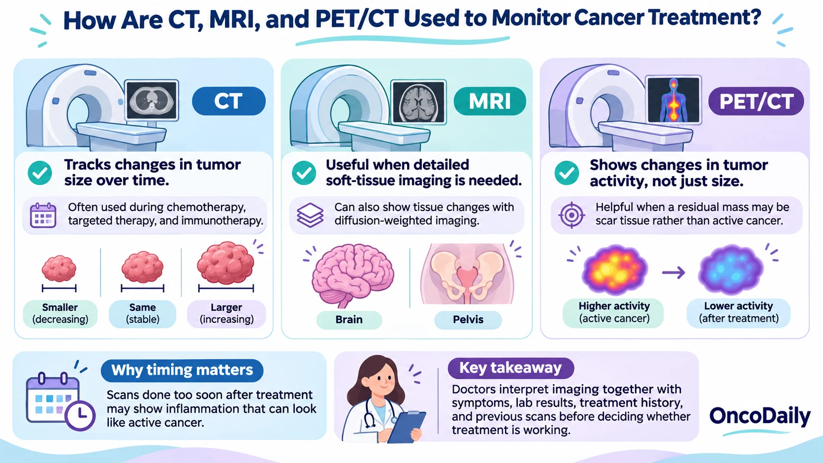
CT is widely used to monitor changes in tumor size during chemotherapy, targeted therapy, and immunotherapy. By comparing scans obtained at different times, doctors can evaluate whether measurable lesions have decreased, increased, or remained stable.
In many solid tumors, treatment response is assessed using standardized criteria such as RECIST (Response Evaluation Criteria in Solid Tumors). These criteria help ensure that changes in tumor measurements are interpreted consistently.
MRI is used for similar purposes when tumors are located in areas where detailed soft-tissue imaging is important. It can also provide information about tissue changes through techniques such as diffusion-weighted imaging, which measures the movement of water molecules within tissues.
PET/CT assesses treatment response through changes in metabolic activity rather than tumor size alone. A decrease in FDG uptake may indicate reduced tumor activity, even when a mass remains visible on CT.
This distinction is particularly useful in selected lymphomas, where a residual mass after treatment may contain scar tissue rather than active cancer.
However, treatment-related inflammation can complicate PET/CT interpretation. Radiation therapy, surgery, infection, and immune responses may cause increased FDG uptake without indicating persistent malignancy.
The timing of imaging is therefore important. Scans performed too soon after certain treatments may show inflammatory changes that are difficult to distinguish from active cancer.
In a 2026 review, Fu and colleagues discussed the clinical applications of PET/CT and PET/MRI in radiotherapy, including tumor localization, treatment planning, and response assessment (Fu et al., 2026).
Doctors interpret imaging changes alongside symptoms, laboratory findings, treatment history, and previous examinations before deciding whether treatment should continue or change.
What Are the Risks and Side Effects of CT, MRI, and PET/CT?
All three imaging techniques are generally safe when used appropriately, although they involve different precautions.
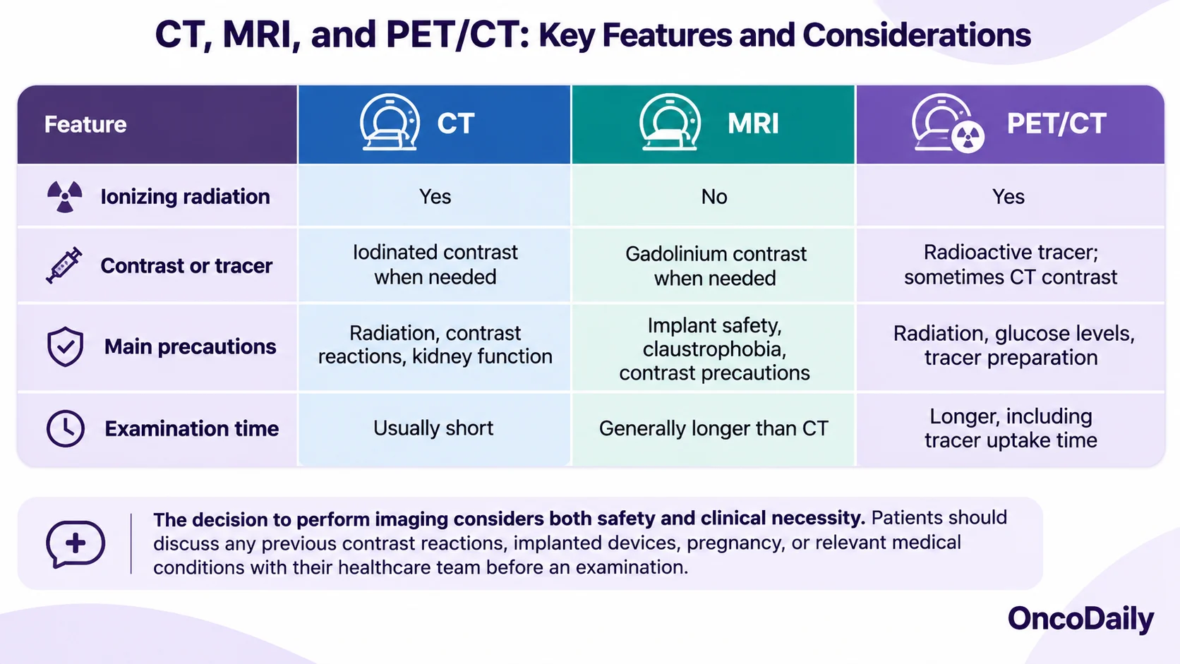
CT uses ionizing radiation, which can contribute to cumulative radiation exposure when examinations are repeated. The risk from a medically necessary scan is generally small, and the diagnostic benefits usually outweigh the potential harm.
Some CT examinations require intravenous iodinated contrast to improve visualization of tumors, blood vessels, and organs. Patients should inform their healthcare team about previous contrast reactions or kidney problems, as additional precautions may be necessary.
MRI does not use ionizing radiation. However, its strong magnetic field can be unsafe for certain metallic implants or foreign objects. Many modern medical devices can be scanned under specific MRI conditions, but their compatibility must be confirmed beforehand.
MRI examinations also tend to take longer than CT scans. Patients with claustrophobia may find the enclosed scanner uncomfortable, while those receiving gadolinium-based contrast may require additional assessment if they have severe kidney disease.
PET/CT involves radiation exposure from both the radioactive tracer and the CT examination. FDG is usually injected into a vein, followed by a waiting period before scanning to allow the tracer to distribute throughout the body.
Patients are generally instructed to fast before FDG PET/CT because blood glucose levels can affect tracer uptake. Those with diabetes may need specific instructions regarding meals, insulin, and other medications.
How Do Doctors Decide Which Cancer Imaging Scan to Recommend?
Doctors select cancer imaging tests according to the suspected diagnosis, the location of the abnormality, and the information needed to guide treatment.
For example, CT may be recommended when evaluating a suspected lung tumor or investigating possible disease in the abdomen. MRI is often preferred for assessing tumors in the brain, prostate, or rectum, where detailed visualization of surrounding tissues is essential.
PET/CT may be useful when doctors need to assess metabolic activity, investigate suspected recurrence, or determine whether cancer has spread to distant sites. However, it is not routinely necessary for every patient with cancer.
Previous imaging results also influence the decision. A suspicious liver lesion found on CT may require MRI for further characterization, while PET/CT may be considered when conventional imaging cannot fully explain suspected disease in a cancer for which PET is useful.
Patient-related factors, including kidney function, implanted medical devices, pregnancy, and previous contrast reactions, can also affect which examination is appropriate.
In some cases, more than one scan is needed because the techniques provide complementary information. This does not necessarily indicate more advanced disease; it may simply allow doctors to obtain a clearer understanding of the tumor.
CT, MRI, and PET/CT each have an established role in oncology, but their value depends on using them for the right clinical indication. The aim is to obtain the information necessary for accurate diagnosis, staging, and treatment planning while avoiding unnecessary investigations (National Cancer Institute, n.d.; Ding et al., 2026).
FAQ
Which is better for detecting cancer, CT, MRI, or PET/CT?
No single scan is best for every cancer. CT is commonly used for chest and abdominal tumors, MRI provides detailed soft-tissue imaging, and PET/CT can detect metabolically active disease.
Can PET/CT detect cancer that CT or MRI misses?
Yes. PET/CT may identify metabolically active cancer that is difficult to detect on conventional imaging. However, it can also miss small or low-metabolism tumors.
Can CT, MRI, or PET/CT detect cancer metastases?
Yes. All three can help detect metastatic cancer. The most appropriate scan depends on the cancer type, the suspected location of metastases, and the clinical situation.
Does a negative CT, MRI, or PET/CT mean there is no cancer?
No. Small tumors and certain cancers may not be visible on imaging. Further testing, including biopsy, may be necessary when clinical suspicion remains high.
Which scan has more radiation, CT, MRI, or PET/CT?
MRI does not use ionizing radiation. CT uses X-rays, while PET/CT involves radiation from both a radioactive tracer and CT. The total radiation dose varies by examination protocol.