Key takeaways
- Tumors can communicate directly with the nervous system through neurotransmitters, neurotrophic factors, nerve remodeling, and in some cases functional synapses.
- In gliomas, neuronal activity can directly stimulate tumor growth through glutamatergic signaling and neuron–glioma synapses.
- Peripheral cancers such as prostate, pancreatic, breast, and head and neck cancers can also exploit autonomic or sensory nerve pathways to support progression.
- Cancer–nerve signaling can influence proliferation, invasion, angiogenesis, immune regulation, and metastasis, but the effect varies by tumor type and nerve pathway.
- Targeting the tumor–nerve axis is an emerging therapeutic strategy, but most approaches remain experimental or in early clinical development.
Cancer and the brain are connected in ways that extend far beyond brain metastases or neurological symptoms. Research in cancer neuroscience is showing that tumors can interact directly with neurons, respond to neurotransmitters, recruit new nerve fibers, and in some cases integrate into functioning neural circuits.
These interactions can influence tumor-cell proliferation, invasion, angiogenesis, immune regulation, and metastasis. In glioblastoma, neurons can form functional synapses directly with cancer cells. In cancers outside the brain, sympathetic, parasympathetic, and sensory nerves can become part of the tumor microenvironment and provide signals that affect cancer progression.
The relationship also works in the opposite direction. Tumors can alter neuronal activity, release neurotrophic factors that promote new innervation, and remodel surrounding nerves to create conditions that support their own growth. Understanding this bidirectional communication is opening a new area of oncology focused on whether tumor–nerve signaling can be therapeutically disrupted (Winkler et al., 2023; Huang et al., 2025).
How Do Cancer Cells Communicate With the Nervous System?
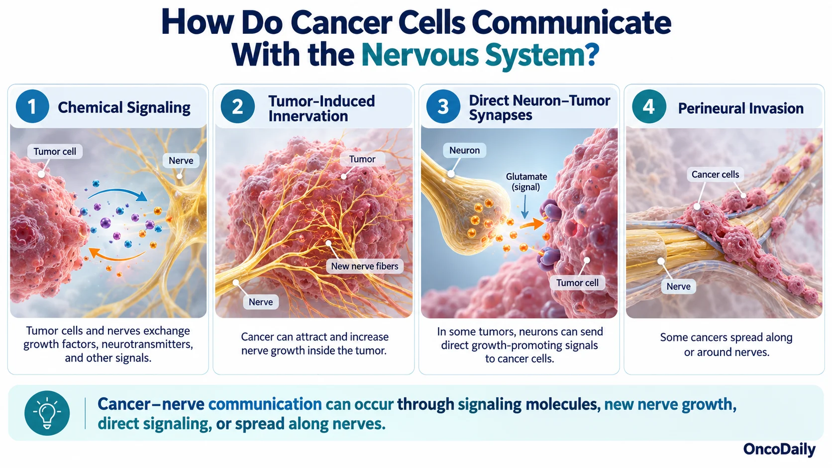
Cancer cells communicate with the nervous system through several biologically distinct mechanisms. Rather than acting as passive structures within or around a tumor, nerves can exchange molecular, electrical, and metabolic signals with malignant cells.
The major forms of cancer–nerve communication include:
- Neurotrophic and paracrine signaling: Tumor cells can release NGF, BDNF, GDNF, axon-guidance molecules, and other factors that alter nearby nerves. Nerves, in turn, release neurotransmitters and neuropeptides that act on cancer, immune, stromal, and vascular cells.
- Tumor-induced innervation: Cancer-derived extracellular vesicles and growth factors can promote axon growth toward and within tumors. In head and neck cancer models, tumor-derived exosomes containing EphrinB1 promoted tumor innervation (Madeo et al., 2018).
- Direct neuron–tumor synapses: In gliomas, neurons form functional glutamatergic synapses with malignant cells, allowing neuronal electrical activity to directly influence tumor biology (Venkataramani et al., 2019; Venkatesh et al., 2019).
- Perineural invasion: Some cancers migrate along or around nerves through coordinated interactions among tumor cells, Schwann cells, extracellular matrix, and neural signals. This is particularly relevant in pancreatic, prostate, and head and neck cancers.
Breast cancer cells that metastasize to the brain can also exploit existing neuronal signaling. Rather than forming the same type of direct synapse seen in glioma, metastatic cells position themselves close to glutamatergic synapses and activate NMDA receptors using neuron-derived glutamate (Zeng et al., 2019).
These mechanisms show that communication between cancer and the nervous system can occur through soluble molecules, direct electrical signaling, nerve remodeling, or physical interaction with neural structures.
How Can Nerves Promote Tumor Growth and Cancer Progression?
Nerves can influence tumor progression both by signaling directly to malignant cells and by changing the surrounding tumor microenvironment. Neurotransmitters released by sympathetic, parasympathetic, and sensory nerves can affect proliferation, invasion, vascular function, metabolism, and immune activity. These effects are highly tissue dependent; a neural pathway that promotes progression in one cancer may behave differently in another.
One of the best-characterized examples is sympathetic adrenergic signaling. Sympathetic nerves release norepinephrine, which can activate β-adrenergic receptors on tumor, stromal, vascular, and immune cells. In prostate cancer models, autonomic innervation was required for efficient tumor development, while β2-adrenergic signaling in endothelial cells promoted metabolic changes that supported angiogenesis and tumor expansion (Magnon et al., 2013; Zahalka et al., 2017).
Sensory neurons also participate in cancer biology. They release neuropeptides such as calcitonin gene-related peptide (CGRP) and substance P, which can modify tumor and immune-cell behavior. In a genetically engineered model of pancreatic ductal adenocarcinoma, ablation of sensory neurons delayed cancer initiation and progression, providing experimental evidence that sensory innervation can contribute functionally to tumor development rather than simply accompany it (Saloman et al., 2016).
Tumors can reinforce these effects by promoting their own innervation. Neurotrophic factors, axon-guidance signals, and extracellular vesicles can stimulate nerve growth within the tumor microenvironment. Increased neural input can then provide additional neurotransmitter and neuropeptide signaling.
Neural pathways can also affect immune cells, stromal cells, and blood vessels. This means that the nervous system may influence cancer without acting directly on the malignant cell itself.
What Are Cancer–Neuron Synapses and How Do They Work?
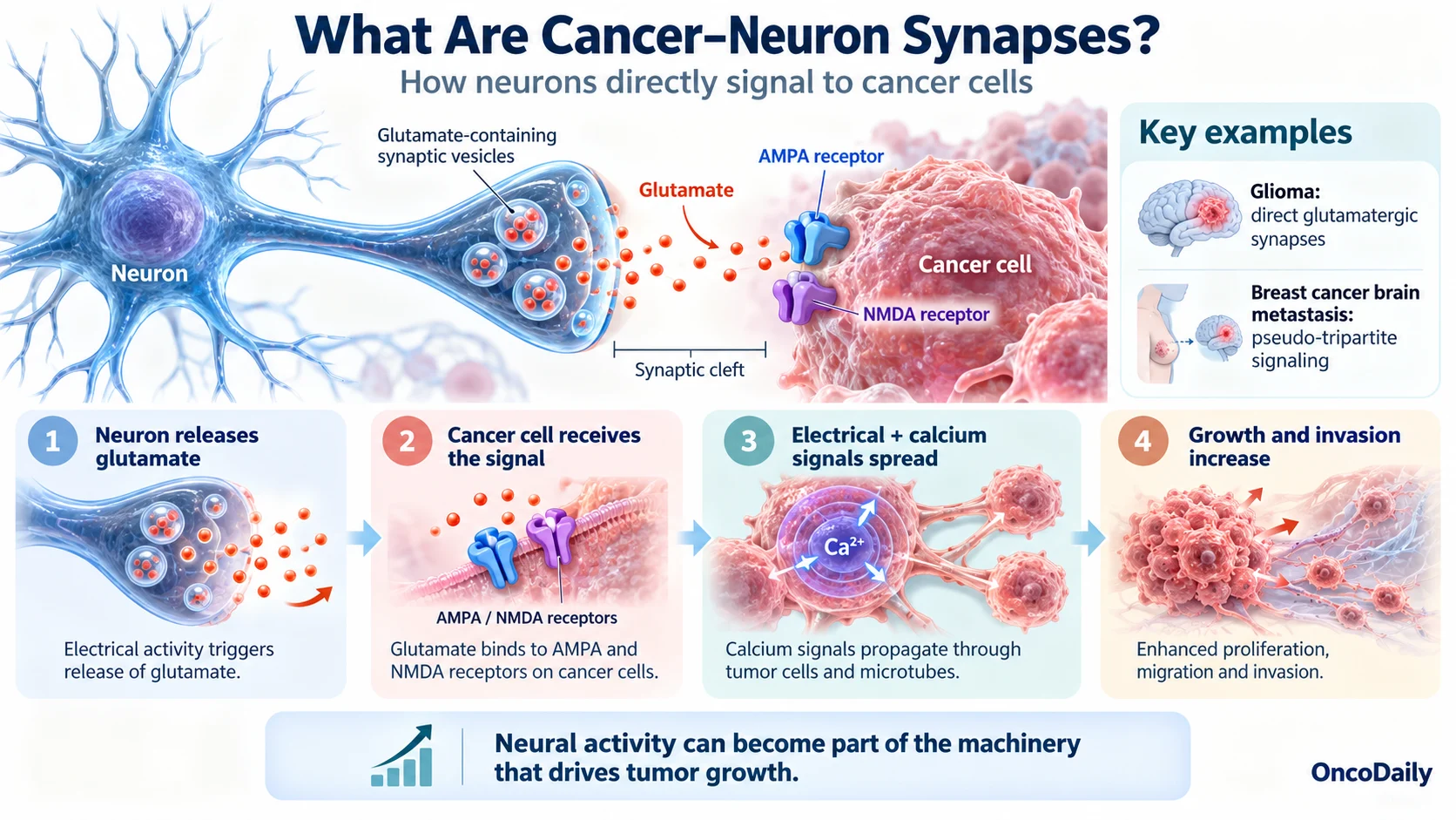
Cancer–neuron synapses are functional connections through which neuronal activity can be transmitted directly to malignant cells. They provide some of the strongest evidence that certain tumors can become integrated into neural circuitry.
In gliomas, presynaptic neurons release glutamate onto tumor cells expressing AMPA-type glutamate receptors. This produces excitatory postsynaptic currents and membrane depolarization within glioma cells. Electrical activity can then influence proliferation and invasion (Venkataramani et al., 2019; Venkatesh et al., 2019).
Importantly, glioma cells do not operate as isolated units. Many are linked through long tumor microtubes and gap junctions, creating multicellular networks through which electrical and calcium signals can propagate. Neuronal input to one part of this network can therefore influence cells beyond those receiving direct synaptic input.
Neural activity also promotes glioma growth through non-synaptic mechanisms. Neuroligin-3 (NLGN3) is released from neural cells in an activity-dependent manner and stimulates growth-promoting pathways in high-grade glioma. Experimental inhibition of NLGN3 signaling markedly suppresses tumor growth, making it one of the most compelling neural-derived therapeutic targets identified to date (Venkatesh et al., 2015; Venkatesh et al., 2017).
A related interaction occurs in breast cancer brain metastases. Metastatic cells can position themselves beside existing neuronal synapses, creating so-called pseudo-tripartite synaptic structures. Neuron-derived glutamate activates NMDA receptors on the cancer cells and supports metastatic colonization and outgrowth in the brain (Zeng et al., 2019).
These findings have changed the traditional view of brain tumors. In some cancers, neural activity is not merely occurring around the tumor; it becomes part of the signaling machinery that supports malignant growth.
Can Tumors Manipulate Neurotransmitters and Neural Signaling?
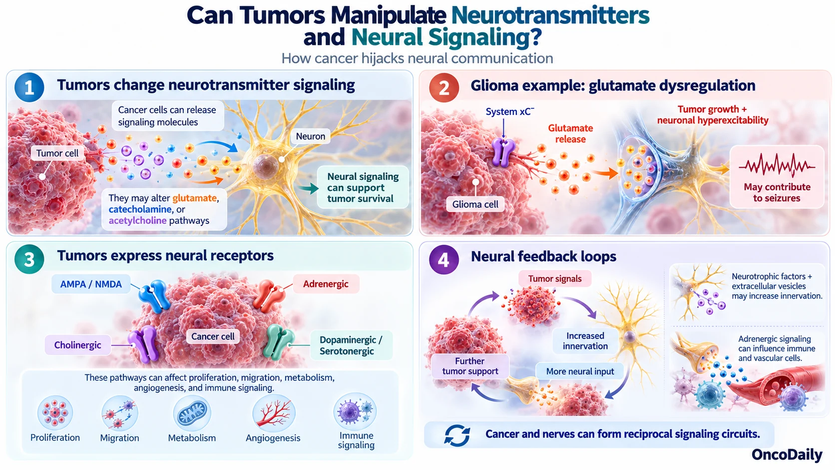
Tumors can actively alter neurotransmitter signaling rather than simply responding to neural input. Cancer cells may change neurotransmitter availability, express receptors normally associated with neural cells, increase local innervation, and alter the electrical behavior of nearby neurons.
One of the clearest examples is glutamate dysregulation in glioma. Glioma cells release glutamate through the cystine–glutamate antiporter system xC−. Increased extracellular glutamate can promote tumor-cell migration and survival while damaging surrounding neurons and increasing neuronal excitability. This altered neuronal environment contributes to the high incidence of seizures seen in patients with glioma (Biegański and Szeliga, 2024).
Cancer cells can also express neurotransmitter receptors that allow them to respond to signals normally used by the nervous system. Gliomas and brain metastases can express AMPA and NMDA receptors, while peripheral cancers may express adrenergic, cholinergic, dopaminergic, serotonergic, or neuropeptide receptors. Activation of these pathways can affect proliferation, migration, metabolism, angiogenesis, and immune function, although the direction and magnitude of the effect vary considerably between cancers (Huang et al., 2025).
How Do Tumors Create Neural Feedback Loops?
Tumors can create self-reinforcing interactions with surrounding nerves. Cancer-derived neurotrophic factors and extracellular vesicles can increase tumor innervation. Those nerves then provide additional neural signals that act on cancer and stromal cells.
Glioma provides an especially clear example. Tumor-associated glutamate signaling contributes to neuronal hyperexcitability, while increased neuronal firing releases glutamate and activity-dependent factors that further stimulate glioma cells. Functional neuron–glioma synapses strengthen this coupling between neural activity and tumor growth.
Neurotransmitters can also modify immune and vascular compartments. Adrenergic signaling, for example, can alter immune-cell trafficking, inflammatory responses, endothelial behavior, and stromal signaling.
The result is not a simple one-way pathway from nerve to tumor. Instead, cancer and the nervous system can form reciprocal signaling circuits, with each altering the biology of the other.
Which Cancers Are Most Strongly Linked to Neural Signaling?
Neural regulation has now been described in many malignancies, but the biological evidence is considerably stronger in some cancers than others. The clearest examples involve different forms of neural interaction rather than one universal mechanism.
- Glioma and glioblastoma: The strongest evidence for direct neural integration. Neurons form glutamatergic synapses with tumor cells, and neuronal activity promotes tumor growth and invasion.
- Prostate cancer: A major extracranial model of autonomic regulation. Sympathetic adrenergic signaling contributes to tumor initiation, vascular remodeling, and disease progression (Magnon et al., 2013; Zahalka et al., 2017).
- Pancreatic ductal adenocarcinoma: Characterized by extensive neural remodeling and perineural invasion. Interactions among nerves, Schwann cells, tumor cells, and stroma contribute to invasion and cancer-related pain.
- Breast cancer brain metastases: Metastatic cells exploit glutamatergic neuronal signaling and NMDA receptors to support growth within the brain (Zeng et al., 2019).
- Head and neck cancers: Perineural invasion is an important pathological feature associated with increased risk of local recurrence and adverse clinical outcomes.
Neural signaling has also been implicated in gastric, colorectal, lung, and several other cancers. However, evidence for the exact mechanisms and their therapeutic relevance remains less mature.
It is therefore more accurate to think of cancer neuroscience as a collection of tumor-specific neural dependencies rather than one pathway shared across all cancers.

Could Targeting Cancer–Nerve Communication Become a New Cancer Treatment?
The discovery that neural signaling can support cancer progression has created interest in targeting the tumor–nerve axistherapeutically. Most strategies remain experimental or in early clinical development, and none should yet be considered a broadly established cancer treatment.
One approach is β-adrenergic blockade. Drugs such as propranolol inhibit sympathetic signaling through β-adrenergic receptors. In a randomized phase II study in patients with operable breast cancer, short-term preoperative propranolol altered tumor gene expression associated with metastatic potential and increased several immune-cell populations within the tumor. The study was not designed to determine whether propranolol reduces recurrence or improves survival, and larger outcome trials would be required for that conclusion (Hiller et al., 2020).
Which Neural Pathways Are Being Targeted in Cancer?
Glutamatergic signaling is one of the most direct targets in brain tumors. Because neuron-to-glioma synapses depend largely on AMPA receptors, investigators are testing whether disrupting this signaling can reduce tumor–neuron connectivity.
The antiepileptic drug perampanel, an AMPA-receptor antagonist, is being evaluated in the randomized phase IIa PerSurge/NOA-30 study in progressive glioblastoma. The trial is specifically examining whether AMPA-receptor inhibition alters tumor-cell network connectivity and tumor growth dynamics (Heuer et al., 2024).
Other promising approaches include:
- NLGN3 targeting: Inhibition of neuronal activity-dependent NLGN3 release strongly suppresses high-grade glioma growth in preclinical models (Venkatesh et al., 2017).
- Neurotrophic signaling inhibition: NGF, BDNF, GDNF, and their receptors are being explored as targets for disrupting tumor-induced innervation and neural support.
- Adrenergic pathway inhibition: β-blockers and other modulators of autonomic signaling are being studied in selected tumor types.
- Neurotransmitter-receptor blockade: AMPA, NMDA, cholinergic, and other neural receptors represent potential targets depending on the cancer.
- Neuroimmune targeting: Neural pathways that suppress antitumor immunity may eventually provide opportunities to combine cancer-neuroscience approaches with immunotherapy.
A major challenge is specificity. Sympathetic, parasympathetic, sensory, and central neural pathways can have different and sometimes opposing effects depending on the cancer, tissue, and stage of disease. Broad neurological suppression would therefore be unlikely to provide a rational therapeutic strategy.
The more plausible future is precision targeting of specific neural dependencies: identifying tumors that rely on a particular neurotransmitter receptor, synaptic pathway, neurotrophic factor, or nerve subtype and disrupting that interaction alongside conventional cancer therapy.
FAQ
Can cancer cells really communicate with neurons?
Yes. Some tumors can respond to neurotransmitters, neurotrophic factors, and other neural signals, while glioma cells can even form functional synaptic connections with neurons.
How does the nervous system help tumors grow?
Neural signals can influence cancer-cell proliferation, invasion, angiogenesis, immune activity, and metabolism. The exact effect depends on the tumor type and the nerve pathway involved.
Which cancers are most strongly linked to neural signaling?
The strongest evidence is seen in gliomas and glioblastoma, prostate cancer, pancreatic cancer, head and neck cancers, and breast cancer metastases to the brain.
What are cancer–neuron synapses?
They are functional connections in which neurons transmit electrical and chemical signals directly to tumor cells. These have been demonstrated most clearly in gliomas.
Can cancer–nerve communication be targeted with treatment?
Potentially. Researchers are studying β-blockers, glutamate-receptor antagonists, NLGN3 inhibition, and other neural-targeted approaches, but most remain experimental or in early clinical development.