
Brain metastases remain one of the most difficult complications of metastatic breast cancer. Approximately 30% of patients with metastatic disease will eventually develop central nervous system involvement, and despite major improvements in systemic therapy, CNS progression continues to influence survival, quality of life, and treatment sequencing.
A 2026 study published in ESMO Open now adds another dimension to this challenge: breast cancer brain metastases may contain a substantial number of clinically actionable genomic alterations that are not necessarily captured by profiling the primary tumor or extracranial disease.
Griguolo, Collesei and colleagues performed comprehensive genomic profiling of surgically resected breast cancer brain metastases and found ESCAT I/II actionable alterations in 76.8% of samples. Alterations involving homologous recombination repair and the PI3K/AKT/PTEN pathway were particularly frequent, suggesting that the biology of CNS disease may contain therapeutic vulnerabilities that deserve specific attention.
The study does not establish that genomic sequencing of brain metastases should immediately dictate treatment. No patient received therapy based on the sequencing results, and prospective evidence demonstrating benefit from CNS genotype–matched treatment remains limited. But the findings raise an increasingly relevant question for precision breast oncology:
Are we adequately characterizing the tumor that is actually growing in the brain?
Brain Metastases May Not Be Genomically Equivalent to the Primary Tumor
The traditional approach to metastatic breast cancer often relies on molecular information obtained from the primary tumor or an extracranial metastatic biopsy. That approach assumes sufficient biological continuity across different disease sites. Brain metastases challenge that assumption.
Previous studies have documented discordance in hormone receptor and HER2 status between primary breast tumors and matched brain metastases, with reported discordance rates of approximately 21% for hormone receptors and 10% for HER2. Molecular evolution during CNS dissemination can extend beyond receptor status, producing changes in gene expression and potentially actionable genomic alterations.
The brain is not simply another metastatic site. Tumor cells reaching the CNS must adapt to a distinct microenvironment, overcome or exploit the blood–brain barrier, and interact with neural and immune components that differ substantially from those present in extracranial organs.
This evolutionary pressure may create or select molecular features that are particularly relevant to CNS disease.
The Study Focused on Clinically Actionable Alterations
The investigators retrospectively identified patients with breast cancer who underwent neurosurgery at three institutions between 2003 and 2019. Among 88 archived brain metastasis samples, 56 had sufficient material for genomic analysis. Whole-exome sequencing was performed using the Agilent SureSelect V6 platform, and genomic alterations were classified according to the updated ESMO Scale for Clinical Actionability of Molecular Targets (ESCAT).
Only alterations meeting ESCAT level I or II criteria for metastatic breast cancer or tumor-agnostic indications were considered clinically actionable. The 56 brain metastases represented several biologically distinct populations: 33.9% were HR-negative/HER2-negative, 25.0% were HR-positive/HER2-negative, and 38% were HER2-positive.
Most patients had a single brain metastasis, reflecting an important characteristic of the cohort: these were patients selected for neurosurgical resection rather than an unselected population with CNS metastatic disease.
More Than Three-Quarters of Brain Metastases Had an ESCAT I/II Alteration
The headline result was the high prevalence of potentially actionable molecular findings. Among the 56 evaluable brain metastases, 43 tumors—76.8%—contained at least one ESCAT I/II actionable alteration.
The frequency remained high across breast cancer subtypes:
- 85.7% in HR-positive/HER2-negative disease, 68.4% in HR-negative/HER2-negative disease, and
- 78.3% in HER2-positive disease.
This does not mean that three-quarters of patients currently have an approved CNS-active targeted treatment available. Actionability according to ESCAT describes the strength of evidence linking an alteration to a therapeutic strategy. In brain metastases, another critical variable must be added: can the relevant drug achieve sufficient intracranial exposure to exploit that target?
The study therefore identifies therapeutic opportunities rather than demonstrating therapeutic benefit.
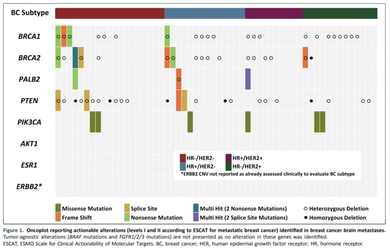
Homologous Recombination Repair Alterations Were Strikingly Common
The most frequent genomic abnormalities involved the homologous recombination repair pathway. At least one alteration in BRCA1, BRCA2, or PALB2 was identified in 53.6% of samples.
Importantly, not every heterozygous alteration necessarily indicates functional homologous recombination deficiency. The investigators therefore separately evaluated biallelic inactivation, which more convincingly reflects loss of normal HRR function and may be more biologically relevant to PARP inhibitor sensitivity.
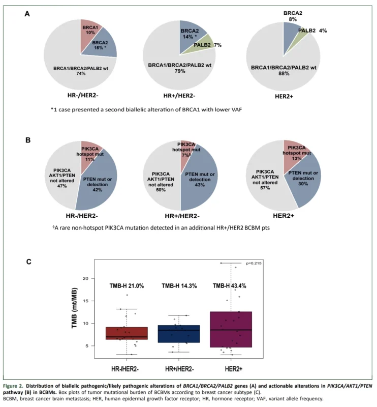
Biallelic BRCA1, BRCA2, or PALB2 alterations were detected in 19.6% of all brain metastases.
The prevalence was higher in HER2-negative disease: 26% in HR-negative/HER2-negative tumors and 21% in HR-positive/HER2-negative tumors, compared with approximately 12% in HER2-positive brain metastases.
The distribution displayed in Figure 2 on page 5 of the paper visually reinforces this subtype difference, showing a greater proportion of biallelic BRCA1/2/PALB2 abnormalities among HER2-negative brain metastases.
Could PARP Inhibition Become More Relevant to CNS Disease?
The HRR findings raise an obvious therapeutic question.
PARP inhibitors are established therapies for selected patients with BRCA-associated HER2-negative metastatic breast cancer, and activity has also been demonstrated in certain somatic BRCA and germline PALB2 contexts. But their specific efficacy against active breast cancer brain metastases selected according to the genomic profile of the CNS lesion itself remains poorly defined.
The authors review the limited clinical evidence. In OlympiAD, patients with germline BRCA-mutated HER2-negative metastatic breast cancer and CNS metastases had a higher overall response rate with olaparib than with physician’s-choice therapy, although the subgroup contained only 26 patients. Evidence for active intracranial disease remains even more limited.
The present study therefore provides biological rationale rather than clinical validation.
A particularly interesting future strategy would be prospective testing of CNS-penetrant PARP inhibitors or combinations in patients selected according to HRR alterations detected directly in brain metastases.
That distinction matters because a mutation detected in the brain lesion may not necessarily be represented in the primary tumor or plasma.
The PI3K/AKT/PTEN Pathway Was Altered in Nearly Half of Cases
A second major genomic signal involved the PI3K/AKT/PTEN pathway. ESCAT I/II alterations involving PIK3CA or PTEN were present in 48.2% of brain metastases. No actionable AKT1 alterations were detected.
The prevalence was remarkably consistent across biological subgroups: pathway alterations were identified in 53% of HR-negative/HER2-negative, 50% of HR-positive/HER2-negative, and 43% of HER2-positive brain metastases.
Most abnormalities involved PTEN. PTEN mutations or deletions were observed in approximately: 42% of HR-negative/HER2-negative disease, 43% of HR-positive/HER2-negative disease, and 30% of HER2-positive brain metastases.
This pattern may be particularly relevant biologically. PTEN loss has previously been implicated in brain metastatic progression, and experimental evidence suggests that PTEN deficiency may influence interactions between breast cancer cells and the CNS microenvironment.
For therapeutic development, the high prevalence of downstream pathway abnormalities may make AKT inhibition particularly interesting, provided that drugs with adequate CNS penetration can be developed.
PIK3CA May Carry Additional Significance in HER2-Positive Brain Metastases
Among 23 HER2-positive brain metastases, three contained hotspot PIK3CA mutations, while seven had PTEN deletion. Most genomic alterations assessed in the study were not significantly associated with survival. However, hotspot PIK3CA mutation in HER2-positive brain metastases was associated with worse overall survival after brain metastasis diagnosis.
This finding is biologically plausible because activation of the PI3K/AKT/mTOR pathway can maintain downstream proliferative signaling independently of HER2 and has been implicated in resistance to HER2-directed treatment.
The authors therefore propose a rationale for studying CNS-penetrant PI3K-pathway inhibitors together with anti-HER2 therapy in HER2-positive breast cancer brain metastases.
The prognostic observation should nevertheless remain exploratory. Only three HER2-positive cases carried hotspot PIK3CA mutations, making the analysis far too small to define a clinically actionable prognostic subgroup.
TMB-High Disease Was More Common Than Expected
Another intriguing finding was the frequency of tumor mutational burden–high disease. Median TMB across the cohort was 8.8 mutations/Mb, and 28.6% of brain metastases met the threshold for TMB-high disease of ≥10 mutations/Mb.
The prevalence was particularly high among HER2-positive brain metastases, where 43.4% were TMB-high, compared with 21.0% of HR-negative/HER2-negative and 14.3% of HR-positive/HER2-negative samples. Figure 2 on page 5 clearly illustrates this distribution across the three biological subtypes.
In principle, TMB-high status can create eligibility for tumor-agnostic immune checkpoint inhibition in relevant regulatory settings. But this finding should not be overinterpreted.
The authors emphasize that it remains uncertain whether TMB-high breast cancer brain metastases derive meaningful intracranial benefit from checkpoint inhibition. The immune microenvironment of brain metastases differs from extracranial disease, and clinical evidence supporting intracranial immunotherapy activity in breast cancer remains limited.
Again, genomic actionability and actual CNS treatment efficacy are not synonymous.
An Unexpected Absence of ESR1 Mutations
One of the more surprising observations was the absence of actionable ESR1 mutations in the HR-positive/HER2-negative brain metastases. No actionable ESR1 alteration was identified among the 14 HR-positive/HER2-negative samples.
At first glance, this appears inconsistent with contemporary metastatic HR-positive breast cancer, where ESR1 mutations are a well-recognized mechanism of acquired resistance to aromatase inhibitors. The authors appropriately caution against broad conclusions.
The HR-positive/HER2-negative subgroup was small, and selection for neurosurgical resection may have enriched for tumors with specific biological characteristics. Because brain metastases in HR-positive disease often develop relatively late, this selected population might also represent cancers less dependent on classical endocrine pathways.
The absence of ESR1 should therefore be considered hypothesis-generating rather than evidence that ESR1 biology is unimportant in HR-positive CNS disease.
Survival Remained Strongly Dependent on Breast Cancer Subtype
The study also illustrates how dramatically prognosis after brain metastasis differs according to breast cancer biology. At a median follow-up of 46.9 months, median overall survival from brain metastasis diagnosis was 28.6 months in the entire cohort.
By subtype, however, median OS was:
- 53.0 months in HER2-positive disease
- 33.4 months in HR-positive/HER2-negative disease
- only 9.4 months in triple-negative breast cancer
These differences likely reflect multiple factors, including underlying tumor biology and the availability of increasingly CNS-active systemic therapies, particularly for HER2-positive disease. They also illustrate why genomic discovery may be particularly important in TNBC brain metastases, where prognosis remains markedly poor despite therapeutic advances elsewhere in breast oncology.
Why Plasma ctDNA May Not Be Enough
The study raises another practical issue: how should CNS-specific genomic information be obtained? Plasma ctDNA has transformed molecular monitoring in metastatic breast cancer, but CNS disease presents a particular biological challenge. The authors note that plasma-derived ctDNA may not reliably capture genomic alterations present in brain tumors.
This may be especially relevant when intracranial disease is progressing while extracranial disease remains controlled. When neurosurgery is clinically indicated, the resected brain metastasis provides an opportunity for direct molecular profiling.
When surgery is not indicated, the problem becomes substantially more difficult. Cerebrospinal fluid ctDNA appears to represent CNS genomic alterations more accurately than plasma ctDNA, but lumbar puncture or other CSF acquisition is invasive and cannot simply replace routine blood-based testing for every patient.
The authors also highlight emerging noninvasive approaches such as radiomics, although these remain investigational for breast cancer brain metastases. The future may therefore require a dedicated CNS molecular-monitoring strategy rather than assuming that peripheral blood provides a complete representation of intracranial cancer biology.
An Actionable Mutation Does Not Automatically Mean an Actionable Patient
This is perhaps the most important clinical caution from the study. Discovering an ESCAT I/II alteration in a brain metastasis does not automatically mean that the corresponding targeted treatment will work in the CNS.
A drug must cross, or sufficiently penetrate, a disrupted blood–brain barrier, achieve therapeutic concentrations within the metastatic lesion, and retain activity within the distinct CNS microenvironment.
The authors specifically emphasize that HRR and PI3K/AKT pathway abnormalities are biologically attractive, but their clinical significance will ultimately depend on the development and prospective evaluation of therapies with adequate intracranial exposure.
This distinction is essential for translating genomic reports into clinical decisions. The study demonstrates target prevalence. It does not demonstrate target-matched treatment efficacy.
Important Limitations Keep the Findings Hypothesis-Generating
The study is retrospective and includes only 56 genomically evaluable brain metastases.
Selection bias is substantial because every tumor came from a patient undergoing neurosurgery. Most patients had favorable characteristics for surgical treatment, including single brain metastases and relatively early CNS involvement. These patients may not represent the broader population with diffuse or rapidly progressive brain metastatic breast cancer.
Gene fusions and microsatellite instability were not assessed because of technical limitations. Archival FFPE material introduced potential sequencing artifacts, although the investigators used a variant allele frequency threshold designed to minimize their influence. Germline and somatic BRCA1/2/PALB2 alterations could not be distinguished.
Most importantly, the brain metastases were not systematically compared with matched primary tumors or extracranial metastases. The study therefore cannot determine which abnormalities were truly acquired during CNS evolution.
And because sequencing was retrospective, none of the patients received treatment based on these genomic findings. Prospective trials will be necessary before brain-metastasis sequencing can be shown to improve outcomes.
The Bottom Line
This ESMO Open study demonstrates that breast cancer brain metastases are not molecularly barren therapeutic endpoints.
Among 56 surgically resected BCBMs, 76.8% contained an ESCAT I/II actionable genomic alteration. HRR abnormalities involving BRCA1, BRCA2, or PALB2 were found in 53.6%, with biologically more compelling biallelic inactivation in 19.6%. PI3K/AKT/PTEN pathway alterations were present in nearly half of tumors, while 28.6% of brain metastases were TMB-high.
The findings do not establish new treatment standards. Rather, they challenge an important assumption in metastatic breast cancer: that molecular information obtained outside the CNS necessarily describes the tumor growing within the brain.
When brain-metastasis tissue is available, genomic profiling may reveal additional therapeutic vulnerabilities, particularly involving HRR and PI3K/AKT/PTEN biology, that would otherwise remain invisible. The next step is not simply more sequencing. It is proving that these genomic findings can be matched to CNS-penetrant therapies that actually improve intracranial outcomes.
That is where precision oncology for breast cancer brain metastases will ultimately be tested.
Reference
- Griguolo G, Collesei A, Lazzarini E, et al. Clinically actionable genomic alterations in breast cancer brain metastases. ESMO Open. 2026;11(10):108551. doi:10.1016/j.esmoop.2026.108551.