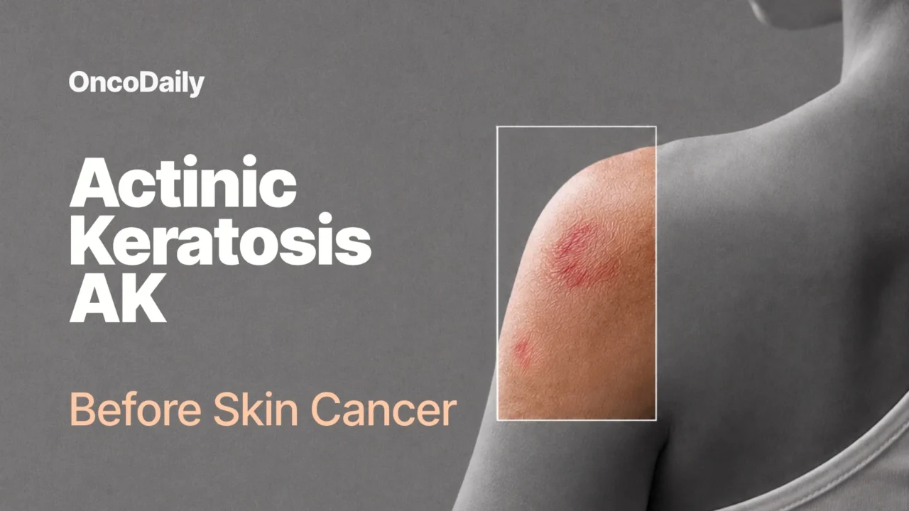
Through “Before Skin Cancer: 5 Precancerous Conditions to Know,” OncoDaily highlights the importance of recognizing persistent or unusual skin changes, seeking timely medical evaluation, and understanding how early diagnosis and treatment may help prevent progression to skin cancer. The first article in this series focused on Bowen disease, a form of squamous cell carcinoma in situ that can resemble common inflammatory skin conditions. The second article focuses on actinic keratosis (AK), a common precancerous skin lesion caused primarily by cumulative ultraviolet (UV) exposure that can progress to squamous cell carcinoma if left untreated.
What Is Actinic Keratosis AK?
Actinic Keratosis (AK), also called solar keratosis, is a common precancerous skin lesion caused by cumulative ultraviolet (UV) radiation exposure. It develops on chronically sun-exposed areas, such as the face, scalp, neck, forearms, and backs of the hands, and appears as a rough, gritty, scaly, or erythematous patch caused by abnormal growth and maturation of epidermal keratinocytes.
Actinic keratosis, also called solar keratosis, was described by Dubreuilh in 1826. Later, the term “keratoma senilis” was proposed by Freudenthal, and in 1958 Pinkus renamed the lesions as actinic keratoses. It literally means a condition (-osis) of excessive horny (kerat-) tissue induced by a ray of light (aktis), presumably ultraviolet (UV) light.
Although classically categorized as preneoplastic lesions, some authors suggest considering them as in situ neoplasms, since they derive from clonal DNA modifications in keratinocytes. [Clarissa Prieto Herman Reinehr, Renato Marchiori Bakos, 2019 ]
Is AK Cancer or Precancer?
AK is a UV-induced atypical or dysplastic keratinocytic proliferation confined to the epidermis. It is generally classified as a premalignant lesion, although some authors regard it as an early form of keratinocyte neoplasia.
The distinction is based on the depth of abnormal cell growth. In AK, atypical keratinocytes may be limited to the basal layer or extend through part of the epidermis, but they have not invaded the underlying dermis. When atypical cells involve the full thickness of the epidermis, the lesion is classified as squamous cell carcinoma in situ, also known as Bowen disease. [D. de Berker et al, 2016] The boundary between severe AK and SCC in situ can be difficult to determine clinically and histologically.
Is AK the Same as Squamous Cell Carcinoma?
Actinic keratosis (AK) is not the same as invasive squamous cell carcinoma (SCC), but these two conditions are closely related. AK is an area of abnormal keratinocyte proliferation and epithelial dysplasia caused by chronic ultraviolet exposure and other risk factors. In AK, atypical cells remain within the different portions of the epidermis and do not invade the basal membrane and dermis. SCC occurs when malignant keratinocytes invade deeper tissue, giving the tumor greater potential for local destruction and, in some cases, metastasis. AK may regress, persist, or progress to invasive SCC.
What Does AK Look Like?
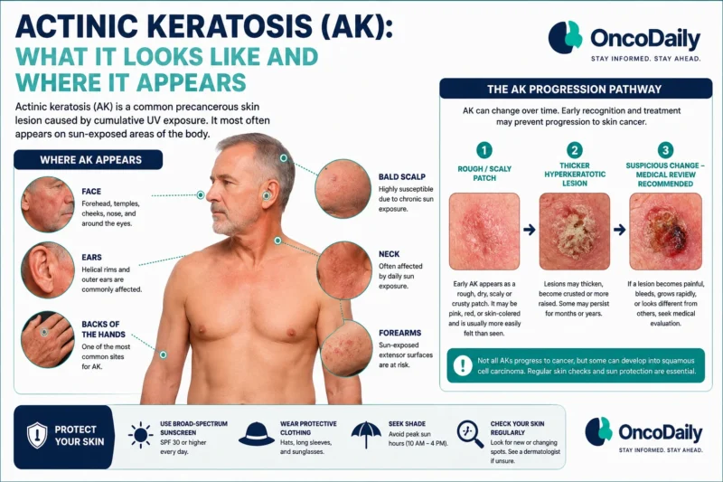
AKs most often are found on chronically sun-exposed sites of the body, such as the bald scalp, face, ears, neck, forearms, and dorsal hands. They are mostly asymptomatic, but may come along with pruritus, burning or stinging pain, bleeding, and crusting. The typical AK lesion, sometimes called the erythematous AK, presents most commonly as a 1-2 cm, erythematous, flat, rough, gritty or scaly papule. It is usually more easily felt than seen.
In some cases, multiple AKs may affect a large area of the sun-exposed skin. In addition, alongside clinically detectable AKs, multiple subclinical lesions may be present. This concept is known as field cancerization and is crucial for the therapeutic approach. Clinically visible AKs may coexist with molecularly altered but apparently normal skin, creating a field at risk for additional AKs and keratinocyte cancers. Some AKs have a pigmented brownish appearance and may be difficult to distinguish from other dermatoses of the elderly, such as seborrheic keratosis. A hypertrophic AK presents as a thicker, scaly, rough papule or plaque that is skin- colored, gray-white, or erythematous. [Fitzpatrick’s Dermatology, page 1857-1867]
Where Does AK Appear?
Actinic keratosis most commonly appears on areas of skin that receive repeated, cumulative ultraviolet exposure. Typical sites include the face, scalp, neck, forearms, and the backs of the hands.These lesions are especially common on chronically sun-exposed skin in fair-skinned adults, although they may also occur in people with darker skin, particularly when there is substantial sun exposure or immunosuppression.
AK vs Other Dermatological Diseases
There are various dermatoses that may be difficult to differentiate from AKs. Spreading pigmented AK may mimic seborrheic keratosis or facial lentigo maligna. In contrast to spreading pigmented AKs, seborrheic keratoses have a velvety to finely verrucous surface that usually feels soft and greasy on palpation. In addition, seborrheic keratoses are also found on the trunk and other body surfaces that are rarely exposed to sunlight.
Dermoscopy can help to distinguish pigmented AK from facial lentigo maligna. Dermoscopic findings of pigmented AK are white and evident follicles, scales, and red color. Intense pigmentation and gray rhomboidal lines are diagnostic clues for lentigo maligna. In rare cases, disseminated superficial actinic porokeratoses may affect the face and be confused with AK. The flat lesions are normally asymptomatic and surrounded by a ridge-like border. Besides other precancerous (eg, arsenical keratosis) or malignant (eg, SCC) lesions, differential diagnoses include melanocytic nevi, senile lentigo, and cutaneous lupus erythematosus. [Fitzpatrick’s Dermatology, page 1857-1867]
What Causes AK?
Actinic keratosis results from the adverse effects of UV radiation on keratinocyte DNA. The changes that occur reduce skin immunity and allow the development of AK. UV-A (320–400 nm) light is the most abundant and penetrates the skin more deeply than UV-B. Here, it causes oxidative damage to nucleic acids, membrane lipids, and cell proteins through the production of reactive oxygen species (ROS). These ROS interrupt normal cellular transduction pathways and cell signaling, causing altered proliferation. UV-B (290–320 nm) irradiation directly causes the formation of cyclobutane pyrimidine dimers and DNA photoproducts, which in turn give rise to the characteristic mutations.
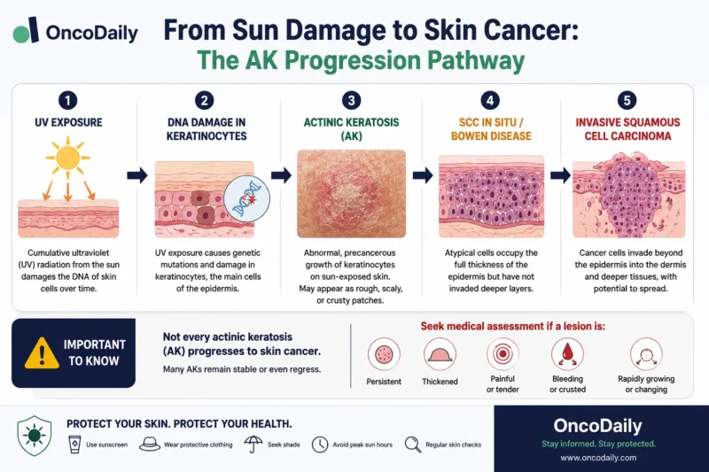
The main mechanisms involved in the formation of actinic keratosis (AK) are inflammation, oxidative stress, immunosuppression, impaired apoptosis, mutagenesis, dysregulation of cell growth and proliferation, and tissue remodeling. Understanding these pathogenic mechanisms forms the basis for the medical management of AK. [Annabel Dodds et al, 2014]
Who Is Most at Risk of AK?
Actinic keratosis is most common in fair-skinned individuals, risk increases with age, especially in older adults, because lesions develop after years of cumulative ultraviolet exposure. People with a previous history of AK or skin cancer are at increased risk of developing additional lesions. Chronic immunosuppression is another important risk factor, especially after organ transplantation or during long-term treatment for inflammatory or rheumatologic diseases. Individuals with photosensitive genetic conditions, such as xeroderma pigmentosum or Bloom syndrome, are also particularly vulnerable. Additional risks include chronic tanning-bed exposure. Regular skin checks and consistent sun protection are especially important for these higher-risk groups. [D. de Berker et al, 2016]
Can AK Become Invasive Skin Cancer?
Actinic Keratosis can progress to invasive cutaneous squamous cell carcinoma (SCC), although most individual lesions do not necessarily undergo malignant transformation. AK may follow several possible courses: it can spontaneously regress, remain stable, or develop into invasive SCC. [Annabel Dodds et al, 2014 ] Although atypical keratinocytes may penetrate the basement membrane and become invasive, the annual progression rate of a single lesion to SCC is very low, but varies substantially according to lesion characteristics, number of lesions, immunosuppression, and patient history.
Many SCCs arise in areas of actinic damage, but not every SCC can be shown to have developed from a clinically recognized AK. The risk for SCC formation increases with the duration of presence, total lesion count (>5), and individual patient characteristics, such as suppressed immune status. [Fitzpatrick’s Dermatology, page 1857-1867]
Warning Signs That The Disease Is Progressing
Changes in an actinic keratosis should be assessed because they may indicate progression toward squamous cell carcinoma (SCC). Warning signs include increasing thickness, development of a hyperkeratotic surface, greater redness, persistent scaling, rapid enlargement, induration, ulceration, bleeding, a cutaneous horn, or a lesion that becomes painful, tender, or itchy. Grade 3 AKs are particularly important because their thicker appearance can make them difficult to distinguish clinically from small, early invasive SCCs. A lesion that enlarges, becomes more prominent, or fails to respond to appropriate treatment also deserves medical review, although these features alone cannot confirm malignant transformation. [D. de Berker et al, 2016]
You Can Also Read Brooke Shields: From Hollywood Icon to Powerful Skin Health Advocate by OncoDaily

How Is AK Diagnosed?
Actinic keratoses are diagnosed clinically in the majority of the cases by examining the lesion’s appearance, texture, location, and degree of thickness. Typical lesions are single or multiple, ill-defined macules, papules, or plaques that appear pink, red-brown, or erythematous and have a dry, adherent scale. They usually occur on chronically sun-exposed skin. Dermoscopy has been shown to be extremely important in increasing the level of confidence and accuracy in equivocal lesions.
Other noninvasive imaging methods, such as confocal microscopy (CM), may also be useful in specific situations when available. Finally, doubtful cases will require histopathological study to confirm the diagnosis. To know the clinical characteristics of the main differential diagnoses of actinic keratoses and how to use the auxiliary methods for diagnosis is crucial in this process.
Dermoscopy
Dermoscopy is a fast-performing, noninvasive method that helps in the diagnosis of actinic keratoses and allows them to be differentiated from their differential diagnoses; moreover, actinic keratoses have well established dermatoscopic criteria.
In facial actinic keratoses four dermoscopic findings are described as essential: (1) erythema forming a pink-reddish vascular pseudonetwork surrounding hair follicles, (2) yellowish-white scales, (3) thin and wavy vessels surrounding the follicles, and (4) follicular openings filled with keratotic plugs.
These structures define the so-called “strawberry” pattern described for most of the facial actinic keratoses. Another finding is the “rosette” sign, seen only with polarized light dermoscopy, a figure resembling a four-leaf clover, formed by four whitish points surrounding the follicular opening. [Clarissa Prieto Herman Reinehr and Renato Marchiori Bakos, 2019]
Does AK Require a Biopsy?
In most cases, actinic keratosis (AK) can be diagnosed based on clinical and dermoscopic features. However, when the diagnosis remains uncertain or the lesion presents with atypical features, a biopsy and subsequent histopathological examination are indicated. The histopathological examination of actinic keratoses is characterized by atypical and pleomorphic keratinocytes in the basal layer of epidermis and by defective maturation of keratinocytes in superficial layers, with abnormal architecture of the epidermis. The number of mitosis is increased and polarity of keratinocytes is lost.
How Is AK Treated?
Treatment for actinic keratosis depends on the number, thickness, location, and clinical appearance of the lesions, as well as the patient’s preferences and overall health. Actinic keratosis is treated with a combination of preventive, lesion-directed, and field-directed approaches. Strict sun protection, including regular sunscreen use, protective clothing, sunglasses, and shade, is essential to prevent new lesions and reduce the risk of malignant transformation. Lesion-directed therapy is generally used for a small number of isolated lesions and may include cryotherapy, which is rapid and effective, or surgical excision when there is a high suspicion of squamous cell carcinoma, particularly in hyperkeratotic lesions.
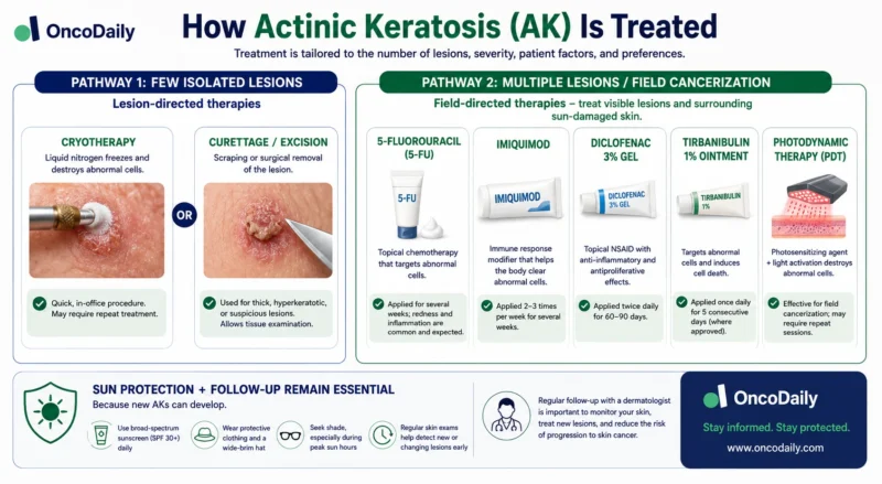
Field-directed treatment is preferred for multiple lesions or areas of widespread sun damage and includes topical 5-fluorouracil, imiquimod, diclofenac, tirbanibulin where locally approved and photodynamic therapy. These treatments can target both clinically visible and subclinical lesions. No single treatment is universally superior. Treatment should be individualized according to lesion number, thickness, location, cosmetic considerations, patient adherence, and suspicion of SCC. [Komal Agarwal et al, 2022]
Which AK Treatment Is Best?
As described above, there are a number of effective lesion-targeted and field treatments available to choose from to decrease the burden of AKs. The individual patient’s needs and expectations, the physician’s skills, the mechanisms of action of the various treatments and their side-effect profiles, and the costs of the agents and procedures should all be considered when choosing a treatment strategy. To help physicians in their choice several national and international guidelines for the management of AKs have been proposed.
Can AK Come Back After Treatment?
Established AKs usually take a chronic course and lesions may persist, spontaneously regress, or progress to invasive SCCs. Spontaneous resolution without treatment may occur. For instance, limiting sun exposure and the use of sunscreen may promote regression. AK is a marker of chronic photodamage, so new lesions and recurrence after treatment are common. However, it is often not entirely clear if these lesions are true local relapses or if they have developed de novo. The presence of AKs indicates long-term sun damage and identifies individuals who are at high risk for developing actinic skin lesions. Thus, AKs are sometimes considered a chronic skin condition where lesions come and go. [Fitzpatrick’s Dermatology, page 1857-1867]
When Should You See a Doctor?
You should see a doctor whenever a new or persistent rough, scaly lesion appears on sun-exposed skin, particularly if the diagnosis is uncertain or the lesion does not improve with standard treatment. Prompt medical assessment is especially important when an actinic keratosis becomes thicker, painful, tender, itchy, bleeds, grows significantly, or develops a more prominent, protuberant appearance, as these features may indicate possible progression to squamous cell carcinoma. [D. de Berker et al, 2016]
Can AK Be Prevented?
Measures of primary prevention are primarily based on the avoidance of exposure to UV, because it is the main risk factor for the development of AKs. Educational and preventive efforts should be directed toward all patients, including children and high-risk populations such as organ transplant recipients or outdoor workers. Minimizing UV radiation is the single most effective means of decreasing the risk of AKs. Because complete avoidance of the sun is impractical, the next best preventive measures are to avoid exposure to intense midday sun; consistently apply and reapply broad-spectrum sunscreens; wear UV-protective cloth- ing, hats, and sunglasses; install UV-protective windows where indicated. Tanning beds should be avoided.
In selected high-risk patients, oral nicotinamide may help reduce the development of new keratinocyte cancers and lower AK counts, but it should be considered only under clinician guidance; nicotinamide is not the same as niacin and should not replace sun protection.
You Can Also Read Bowen Disease: Essential Facts About Skin Precancer Symptoms, Causes, Diagnosis, and Treatment by OncoDaily

Written by Written by Lily Tumanyan MD
FAQ
What is actinic keratosis (AK)?
A common precancerous, rough, scaly patch caused by long-term sun exposure on areas like the face, scalp, and hands.
Is AK cancer or a precancer?
AK is a precancerous lesion; abnormal cells are confined to the epidermis and have not invaded deeper skin layers.
Is AK the same as squamous cell carcinoma (SCC)?
No. AK stays in the epidermis, while SCC invades the dermis and deeper tissues.
What does AK look like?
A small, rough, gritty, or scaly pink/red patch that is often easier to feel than see.
Who is most at risk for AK?
Fair-skinned, older adults, people with high sun exposure, organ transplant recipients, and tanning bed users.
Can AK become invasive skin cancer?
Yes, but most lesions do not; risk rises with many lesions, long duration, and immunosuppression.
How is AK diagnosed, and is biopsy required?
Usually by clinical exam and dermoscopy; biopsy is used only for uncertain or suspicious lesions.
Can AK come back after treatment?
Yes; AK is chronic and linked to sun damage, so recurrence or new lesions are common.
How can AK be prevented?
By limiting UV exposure, using sunscreen, wearing protective clothing, avoiding tanning beds..
How is AK treated?
With spot treatments (e.g., freezing) for few lesions or field therapies (e.g., creams, light therapy) for widespread damage.