






At OncoDaily, we aim to raise awareness about skin cancer prevention, early detection, and the warning signs that should not be overlooked. According to the International Agency for Research on Cancer, more than 1.5 million new cases of skin cancer were estimated worldwide in 2022, including approximately 330,000 cases of melanoma.Many people know to watch for suspicious moles, but fewer are familiar with precancerous skin conditions that may appear before invasive cancer develops.
Through “Before Skin Cancer: 5 Precancerous Conditions to Know,” OncoDaily highlights the importance of recognizing persistent or unusual skin changes, seeking timely medical evaluation, and understanding how early diagnosis and treatment may help prevent progression to skin cancer. The first article in this series focuses on Bowen disease, an early, non-invasive form of squamous cell carcinoma that can resemble common inflammatory skin conditions and may progress if left untreated.
The Skin Patch That Does Not Go Away
For many cases skin lesions start with a small area of change: a small patch of skin that looks dry, red, flaky, or mildly irritated. You may assume it is eczema, simple dryness, or a reaction to soap, weather, or friction. But when a patch persists for weeks or keeps coming back in the same spot, it deserves a closer look. It can be the first sign, ranging from the mildest skin problems to severe precancerous conditions.
These are considered as early cancer or cancer in situ and may clinically mimic eczema, psoriasis, or dermatitis but do not resolve with standard symptomatic treatments. Persistent patches that remain for weeks or months should be carefully assessed, especially if they are scaly, red, or slowly enlarging.
What Is Bowen Disease?
Bowen disease (BD) is a distinct type of squamous cell carcinoma (SCC) in situ and was originally described in 1912 by John T. Bowen, a Boston dermatologist. It may affect both skin and mucous membranes and has the potential to progress to Bowen carcinoma, that is, an invasive squamous cell carcinoma (SCC).
An annual incidence of 15 per 100 000 has been suggested in the U.K. However, this figure was derived from an American study that primarily examined internal neoplasia associated with Bowen Disease, and was the annual incidence rate for the 1980 U.S. white population – this may not be the same as in the less sun-exposed U.K. population. [N.H. Cox et al, Guidelines for management of Bowen’s disease: 2006 update]
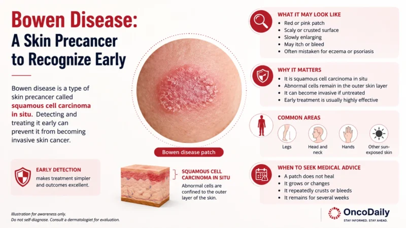
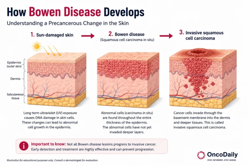
Bowen disease is known as carcinoma in situ which means that atypical cells are located in the outer layer of the skin, not invading the basal membrane, the thin supportive layer that lies between the epidermis and the dermis.
Is Bowen Disease Cancer or Precancer?
It is best understood as an early, non-invasive form of skin cancer, often described as stage 0 squamous cell carcinoma, because the abnormal cells are confined to the epidermis and have not invaded deeper tissue. At the same time, it is frequently discussed alongside precancerous skin conditions because it behaves as a very early lesion with a meaningful risk of progression.
This dual description can be confusing, but both labels point to the same clinical idea: the cells have cancerous appearance and behavior, yet they remain on the surface layer of the skin. That is why some sources call it carcinoma in situ, while others emphasize its precancerous nature. [Thorsten Neubert and Percy Lehmann, 2008]
Is Bowen Disease the Same as Squamous Cell Carcinoma?
The key distinction is invasion. In Bowen Disease, atypical squamous cells stay above the basement membrane and do not enter the dermis; if they cross that barrier, the lesion becomes invasive squamous cell carcinoma. Invasive disease is clinically more serious because it can go through deeper tissue and, in some cases, spread to other parts of the body causing metastatic cancer.
What Does Bowen Disease Look Like?
Clinically, Bowen Disease appears as a well-defined erythematous plaque with a scaly or crusted surface, but variants include verrucous and pigmented Bowen Disease. Histopathology shows full-thickness epidermal dysplasia without dermal invasion. Bowen Disease is usually indolent and slow-growing.
Early Signs of Bowen Disease
Bowen Disease lesions grow slowly and may be accompanied by pruritus, but are usually asymptomatic. Lesions typically present as erythematous plaques with irregular, clearly visible demarcated borders. The surface may be scaling, crusting, or hyperkeratotic. Over time, lesions may grow in size and measure up to several centimeters.
The flat plaque may transform to a nodular or verrucous form. Bowen disease usually appears pink or reddish, whereas crusty or hyperkeratotic lesions may take a gray or brownish color. In very few cases, lesions are pigmented. Lesions of Bowen Disease are usually solitary, but may be multiple in up to 20% of individuals. [Cox et al 2007]
Where Does Bowen Disease Appear?
Sites of predilection include sun-exposed areas such as the head and neck and lower legs, although any site of the body may be affected: less commonly genital or other non-sun-exposed skin. Kossard and Rosen studied 1001 patients with BD and found head and neck (44%) as the most common site followed by lower extremity (29.8%), upper extremity (19.8%), and trunk (6.5%) In intertriginous areas, Bowen disease can present as an oozing, erythematous, or pigmented patch or plaque. Bowen Disease can also damage the mucosal surfaces and appear as verrucous or polypoid papules and plaques, erythroplakia, or a velvety erythematous plaque.
Bowen Disease or ․․․ ?
Bowen disease should be considered in the differential diagnosis of chronic inflammatory skin lesions, particularly eczema, psoriasis, and lichen planus, when a solitary patch persists despite appropriate topical treatment. Clinically, it may closely resemble localized chronic eczema, psoriasis, superficial basal cell carcinoma, actinic keratosis, seborrheic keratosis, or other hyperkeratotic inflammatory lesions, which makes accurate diagnosis truly challenging.
Bowen Disease vs Eczema
From a clinical standpoint, eczema is usually present with additional supportive features, such as pruritus, multifocal involvement, characteristic distribution, or a history of recurrent inflammatory disease, whereas Bowen Disease often remains a slowly enlarging, isolated lesion without the broader pattern typical of inflammatory dermatosis.
Bowen Disease vs Psoriasis
It is necessary to compare clinical patterns of psoriasis and Bowen Disease. Psoriasis commonly affects the extensor surfaces of the body, scalp, and nails and as many inflammatory dermatosis is frequently associated with itch and steroid responsiveness; the lack of these features should prompt reconsideration of the diagnosis.
Other Conditions That Can Look Similar
Hyperkeratotic or verrucous lesions of Bowen Disease can be misinterpreted as viral warts, seborrheic keratosis, and squamous cell carcinoma. For both pigmented and unpigmented Bowen Disease lesions, it is important to consider melanoma and amelanotic melanoma as differential diagnoses, respectively. Superficial basal cell carcinoma may also mimic Bowen disease, but it more often shows a subtle elevated border or translucent edge, which can help distinguish it clinically. Because of their very similar histologic picture, the clinical setting is crucial in differentiating Bowen Disease from sexually transmitted bowenoid papulosis.
What Causes Bowen Disease?
The most important etiologic factors for the development of Bowen Disease include a long history of significant UV exposure and infection with HPV. Significant roles play damaged immune systems of the patient with organ transplants, HIV patients and others with long-term immunosuppression. Other etiologic factors include arsenic exposure and ionizing radiation.
Sun and UV Exposure
Accumulated ultraviolet damage plays a central role in Bowen Disease by producing chronic injury to epidermal keratinocytes over many years. Repeated UV exposure causes DNA damage, oxidative stress, and local immunosuppression, which together reduce the skin’s ability to repair abnormal cells and eliminate emerging atypical clones. Bowen Disease has been described with increased frequency in patients undergoing psoralen plus UVA therapy. In contrast, it is rarely seen in stronger-pigmented individuals.
HPV Infection
Up to 30% of extragenital Bowen Disease lesions have been found to harbor HPV DNA. Several HPV subtypes, such as HPV types 16, 18, 31, 34, 35, 54, 58, 61, 62, and 73 have been detected in BD lesions. Infection with high-risk HPV subtypes such as HPV 16 may be responsible for Bowen Disease lesions on hands or fingers after anodigital infection in patients that simultaneously have anal and genital lesions. However, the prevalence of HPV-associated BD lesions is higher on sun-protected than on sun-exposed body areas.
Who Is Most at Risk of Bowen Disease?
Bowen Disease typically occurs in individuals above 60 years of age. It is rare in individuals below 30 years of age. Immunocompromised individuals are at risk of developing disease at a younger age. Most studies have revealed a slight female preponderance. The incidence is high in Caucasians (1.42/1000). As mentioned earlier, extensive sun exposure is a major risk factor for Bowen disease.
Patients with lighter skin phototypes or a prior skin cancer history are therefore considered at higher risk and should be monitored carefully for persistent scaly plaques or non-healing lesions. [Vijayasankar Palaniappan and Kaliaperumal Karthikeyan, 2022]
Can Bowen Disease Become Invasive Skin Cancer?
Bowen Disease is an intraepidermal neoplastic lesion that initially remains in the outer layer of the skin. Although it does not invade deeper tissue at the beginning, it may progress to invasive squamous cell carcinoma if left untreated. The development of invasive SCC is due to destruction of basement membrane mediated by metalloproteinases. The appearance of a large group of flat epithelial cells without keratin production in the dermis favors the progression into invasive SCC.
How Often Does Bowen Disease Progress?
The development of invasive carcinomas is more common among elderly people and immunocompromised individuals. The HIV infection, by producing Tat protein and decreasing the expression of proteins such as HPV E6 and E7, directly promotes invasive SCC in-situ transformation.
The overall risk that untreated Bowen Disease will progress to invasive carcinoma is estimated at approximately 5%. The invasive SCC of genitals tends to be more aggressive and metastatic. Almost 100% of the invasive SCCs arising from photo-exposed areas are from underlying Bowen Disease. When confined to the epidermis, Bowen Disease does not carry any risk of metastasis. However, one-third of the Bowen Disease which progressed to invasive SCC can metastasize.
Warning Signs of Invasive Squamous Cell Carcinoma
The clinical signs suggestive of malignant transformation are ulceration, bleeding, and nodule formation. Warning signs that Bowen disease may have transformed into invasive squamous cell carcinoma include rapid enlargement, increasing thickness or a raised/nodular component, ulceration, bleeding, crusting, and pain or tenderness. A lesion that develops a more exophytic or papillomatous appearance, or shows a central keratin mass, is also more suspicious for invasion.
How Is Bowen Disease Diagnosed?
The clinical picture together with the typical history of a slowly growing, mostly asymptomatic lesion that remains resistant to topical corticosteroid treatment is highly suspicious for BD. However, final diagnosis requires biopsy and histologic examination.
Skin Examination and Dermoscopy
The dermatologist begins by taking a detailed history of the lesion, including its duration, rate of growth, associated symptoms such as itching, pain, bleeding, or crusting, and any history of sun exposure or previous skin cancer. The lesion is then carefully examined for size, border, scale, thickness, and distribution, since Bowen disease often presents as a slowly enlarging, well-demarcated erythematous scaly plaque on a sun-exposed site. If the diagnosis remains uncertain, dermoscopy and biopsy are performed to distinguish Bowen disease from inflammatory dermatoses and to exclude invasive squamous cell carcinoma.
There are no standard dermoscopic criteria mentioned in the literature for the diagnosis of BD. First described dermoscopic features are glomerular vessels and scaly plaques. The commonly observed features are scaly surface, small brown globules with patchy distribution, homogenous grey brownish and reticular pigmentation. However, dermoscopy is necessary to make a differential diagnosis from actinic keratosis, seborrheic keratosis, basal cell carcinoma, lichen planus-like keratosis, mammary Paget’s disease, and lentigo malignant melanoma. [I. Zalaudek et al, Journal of the European Academy of Dermatology and Venereology, 2006]
Does Bowen Disease Require a Biopsy?
Histopathology is the gold standard diagnostic modality to confirm Bowen disease [Vijayasankar Palaniappan and Kaliaperumal Karthikeyan, 2022]. A biopsy is often required when the clinical diagnosis is uncertain, when the lesion resembles eczema or psoriasis, or when there are features suspicious for invasion such as ulceration, bleeding, or progressive thickening. By demonstrating whether atypical squamous cells remain confined to the epidermis, histology also helps exclude invasive squamous cell carcinoma and guides appropriate management.
How Is Bowen Disease Treated?
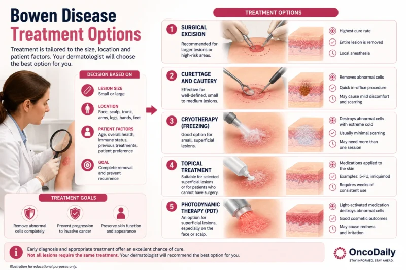
The treatment modality is individual and depends on factors such as the tumor size, location, thickness, number of lesions, patient’s age, immune status, comorbidities, concomitant medication intake, compliance, esthetic outcome, equipment availability, and preference of the patient along with clinician’s expertise. The available therapeutic options are surgical modality, light-based procedures, destructive modality, topical chemotherapy.
Surgical Excision
Simple wide excision of the lesion and primary closure is an acceptable method of treating Bowen Disease of small size, single lesion, and perianal Bowen Disease. It is helpful by histopathologically ruling out the invasive disease. The BD is excised with a minimum of 4 mm margin in well-defined tumors of <2 cm in diameter and at least 6 mm margin for larger lesion or less-differentiated tumors or lesions in high-risk locations (e.g., scalp, eyelids, ears, nose, and lips).
Curettage and Cautery
Curettage and cautery is a minor skin procedure in which the lesion is first scraped away with a curette and then treated with heat to stop bleeding and destroy any remaining abnormal cells. It is a simple, cheap, safe, and one of the most effective treatment modalities suitable in single, small Bowen Disease. It is preferred in patients who have large hyperkeratotic lesions, intolerant to cryotherapy, and those who cannot apply topical agents for prolonged periods of time. The cure rate ranges from 81% for curettage and up to 93% to 98% in case of curettage and cautery.
Cryotherapy
Cryotherapy is a treatment that uses extreme cold, usually liquid nitrogen, to freeze and destroy abnormal skin tissue. It is a quick, simple, inexpensive, outpatient procedure, and the treated area usually forms a blister or scab before healing over the next days to weeks. It is used as a treatment modality in case of single, small Bowen Disease located at well healing sites. The efficacy of cryotherapy differs depending on technique and regimens used. It is avoided in poorly vascularized areas and legs where wound healing is prolonged.
Medicated Creams
Imiquimod and 5-fluorouracil are topical treatments used for Bowen Disease, especially when lesions are in difficult anatomical areas or when surgery is less desirable. Imiquimod works as an immune response modifier by stimulating local cytokine production and has shown clinical efficacy in roughly 57% to 86% of cases after 6 weeks. It can be useful for lesions on the lower leg, penile shaft, glans penis, and large facial lesions. Common side effects of imiquimod include inflammation, erythema, and pigmentation changes.
5-fluorouracil is a topical cytotoxic agent typically applied once or twice daily for 3 to 4 weeks, with repeat treatment if needed. Reported complete clinical response rates range from 48% to 83% with this regimen. Its effectiveness may be improved by occlusion or other adjunctive methods such as laser pretreatment or iontophoresis. The main adverse effects of 5-fluorouracil are pain, erythema, burning, and sometimes ulceration at the application site.
Photodynamic Therapy(PDT)
Photodynamic therapy for Bowen Disease involves topical application of Methyl Amino-levulinate (MAL) under occlusion for three hours followed by illumination using red light from a narrowband light-emitting diode source. The use of fluorescence helps in lesion delineation, recurrence detection with 100% sensitivity and 85% specificity. It is of immense benefit in large lesions (>3 cm2), leg BD, and in difficult-to-treat sites. Topical PDT is noninvasive, tissue sparing, highly efficacious, and good esthetic therapeutic modality.
Radiotherapy options (external beam, radioactive skin patch, Grenz rays) are effective at both high and low doses and suit hard-to-treat areas such as scalp, penile, and perianal BD, but carry drawbacks of cost, inconvenience, and poor wound healing on the legs. Lasers are mainly used for genital and nail BD.
CO2 laser achieved 86% clearance in a large retrospective series after one treatment but may spare deep follicular epithelium and risk recurrence. Combining diode laser as a final pass after multiple CO2 passes has been suggested to reduce recurrence and treatment failure. [Vijayasankar Palaniappan and Kaliaperumal Karthikeyan, 2022]
Monitoring Without Immediate Treatment
In practice, monitoring is most appropriate for older or frail patients, those with lesions located in areas where treatment risks (functional or cosmetic) are high, or when patient preference favors conservative management—provided they are not in a high-risk group. Surveillance should include regular clinical exams focusing on lesion size, morphology, and any new symptoms, with a low threshold for biopsy if changes suggest progression or lateral spread.
Which Bowen Disease Treatment Is Best?
The treatment varies from topical agents to surgical methods. The first choice is often decided by the physician based on the site, size, immunity, and available facilities. In Bowen Disease of less than 2 cm diameter with proper wound healing, curettage is the best option available followed by cryotherapy. In case of poor wound healing, PDT is preferred followed by 5-FU and imiquimod. Cryotherapy is the first choice of treatment modality in case of multiple Bowen Disease. In facial Bowen Disease, curettage is the initial choice followed by topical therapy and PDT. Combination therapies using PDT, CO2-laser, surgery, imiquimod 5% cream, and radiation have been described.
Can Bowen Disease Come Back After Treatment?
Bowen Disease can recur after treatment, so follow-up visits are important even when the treated area looks healed. Recurrence may happen months or years later, which is why the area should be checked regularly for any new change.
In general, recurrence of Bowen Disease is rare and estimated to be approximately 6% within 5 years after sufficient treatment. The theory that persistent Bowen Disease within the deep portion of the follicle is mainly responsible for recurrence could not be confirmed. In fact, recurrent Bowen Disease may be more likely caused by subclinical lateral spread, as excision with narrower lateral margin is associated with a higher risk of recurrence. Once Bowen Disease occurs, the risk of developing subsequent nonmelanoma skin cancer is particularly higher.
There is no evidence, however, that Bowen Disease is associated with a higher risk for internal malignancies. Also, Bowen Disease involving the vulvar region in females and the perianal region in males may be associated with an increased risk of uterine, cervical, vaginal, and anal cancer, most likely as a consequence of HPV infection.
What Does Recurrent Bowen Disease Look Like?
At follow-up, clinicians look for renewed scaling, redness, crusting, thickening, or a new raised growth in the same spot. These changes can mean recurrent Bowen Disease or that there is still atopic appearance of the cells. Patients should also self-check the treated area between appointments and report any persistent irritation, bleeding, or enlargement. Early review matters because recurrent disease is often easier to manage when found promptly.
When Should You See a Doctor?
It is recommended to see a doctor if a skin patch is persistent, growing, bleeding, painful, crusting, or not healing. These changes can signal that the lesion needs assessment and possible treatment.
Can Bowen Disease Be Prevented?
Bowen Disease can be helped with prevention steps that reduce UV damage and improve early detection. It is about protecting skin from the sun by avoiding direct midday exposure, wearing protective clothing and a broad-brimmed hat, using a high-SPF, high-UVA sunscreen on exposed areas. Regular skin checks are important because people who have had Bowen Disease are at higher risk of developing another patch or other skin cancers. Any persistent, growing, bleeding, crusting, painful, or non-healing change should be assessed early. Although these steps do not guarantee prevention, they can lower risk of the disease and help find suspicious changes in early stages.
You Can Also Read Total Body Photography and Automated Lesion Mapping: The Next Stage in Skin Cancer Surveillance by OncoDaily

Written by Lily Tumanyan MD
FAQ
Is Bowen Disease Dangerous?
Bowen disease can be serious because it is a squamous cell carcinoma in situ with a small risk of becoming invasive if untreated. Most cases are treatable, especially when found early.
Does Bowen Disease Spread?
Bowen disease does not spread to deeper tissue at the start, but it can extend sideways within the skin and, in some cases, progress into invasive squamous cell carcinoma. If that happens, the cancer becomes more serious and can potentially spread further.
Does Bowen Disease Hurt or Itch?
Many people have no symptoms, but some lesions can itch, feel irritated, or become tender as they grow. Pain, bleeding, crusting, or increasing thickness are warning signs that need assessment.
How Quickly Does Bowen Disease Grow?
Bowen disease usually grows slowly, which is why it is sometimes mistaken for eczema, psoriasis, or a chronic rash. A patch that keeps enlarging or does not heal over time deserves medical review.
Does Bowen Disease Always Need Treatment?
Not always immediately; in some cases, doctors may monitor it if it is not troublesome and treatment risks are higher. But because there is a chance of progression, suspicious or changing lesions are usually treated rather than ignored.
Can Bowen Disease Return After Removal?
Yes, it can come back after treatment, so follow-up matters. People who have had Bowen disease also have a higher chance of getting another patch in the future.
Is Bowen Disease Contagious?
No, Bowen disease is not infectious and cannot be spread from person to person.
What Happens If Bowen Disease Is Left Untreated?
If untreated, Bowen disease may slowly enlarge and, in a minority of cases, progress into invasive squamous cell carcinoma. That is why persistent, growing, bleeding, crusting, painful, or non-healing patches should be assessed early.