
Sendurai Mani, Associate Director of Translational Oncology at the Legorreta Cancer Center, is presenting a series exploring some of the most influential scientific papers that have shaped the field of cancer research.
Paper 1: The Origins of Proto-Oncogenes
“I recently shared an AI-generated (Claude) list of the top 200 papers that have shaped cancer research.
Starting today, I’ll highlight one paper each day, beginning with N1.
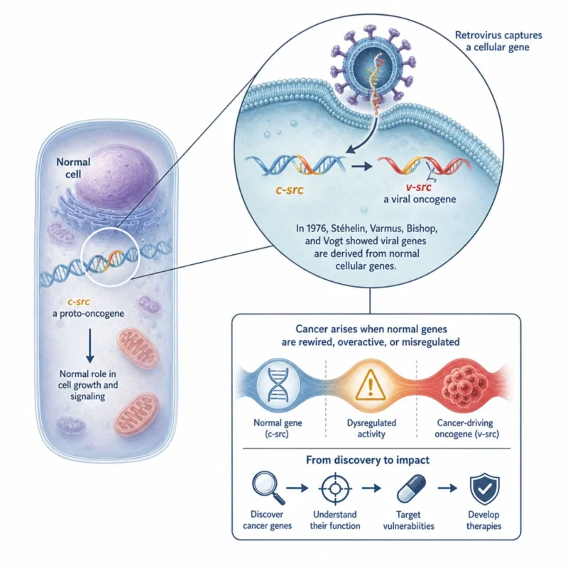
What if some of cancer’s most dangerous genes are not foreign invaders, but normal genes operating in the wrong context?
In 1976, Dominique Stéhelin, Harold Varmus, J. Michael Bishop, and Peter Vogt showed that DNA sequences related to the transforming gene of avian sarcoma viruses were also present in normal bird cells.
This finding was a foundational shift in cancer biology. It helped establish the proto-oncogene concept: viruses can capture and alter normal cellular genes, turning them into oncogenes that drive cancer. The viral gene later became known as v-src; its normal cellular counterpart, c-src, is a proto-oncogene.
The lesson remains strikingly relevant today: cancer often arises not from wholly foreign biology, but from normal growth-control machinery that has been rewired, deregulated, or activated at the wrong time.
This insight opened the path to identifying cancer genes – and ultimately to developing therapies that target their vulnerabilities.
Title: DNA related to the transforming gene(s) of avian sarcoma viruses is present in normal avian DNA
Authors: Dominique Stéhelin, Harold E. Varmus, J. Michael Bishop, and Peter K. Vogt
Paper 2: When Cancer Needs Two Hits: Knudson’s Tumor Suppressor Breakthrough
What if cancer requires not one failure – but two?
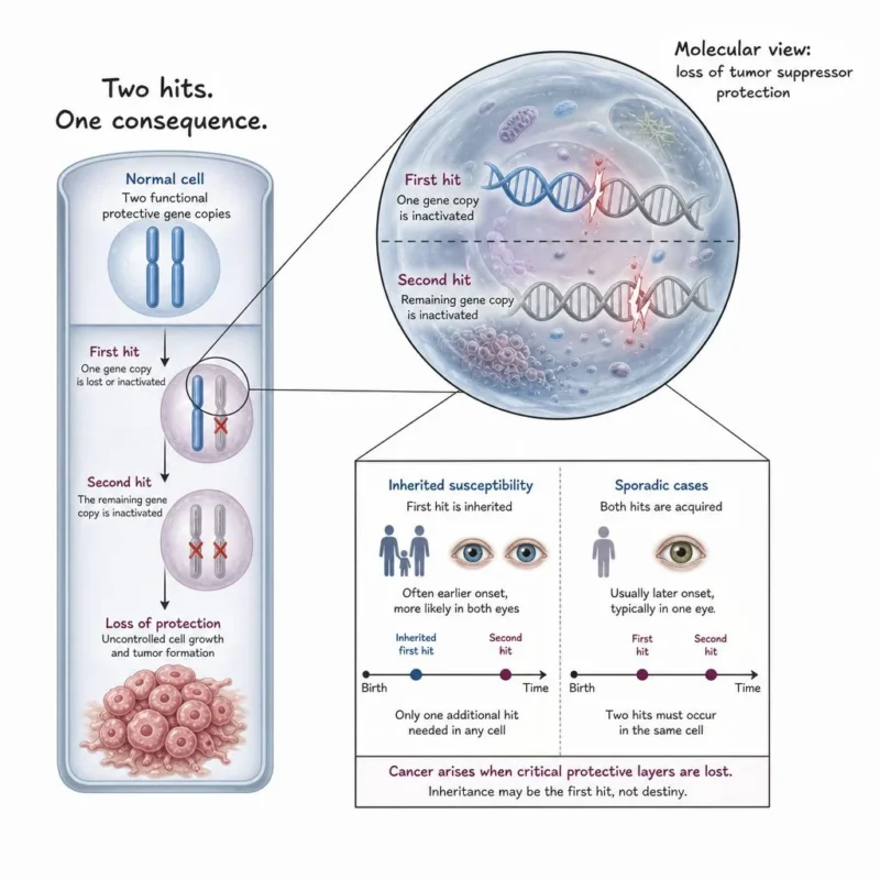
In 1971, Alfred Knudson proposed a deceptively simple explanation for a rare childhood eye cancer, retinoblastoma: tumor formation occurs after both copies of a protective gene are lost or inactivated.
This became known as the two-hit hypothesis.
Knudson noticed that inherited and non-inherited retinoblastoma followed different patterns. Children with inherited susceptibility often develop tumors earlier and in both eyes, consistent with inheriting one damaged gene copy and needing only one additional “hit.” Sporadic cases generally required two acquired hits in the same retinal cell.
The idea fundamentally changed how we understand cancer.
Until then, cancer research had largely focused on genes that become abnormally active and drive growth. Knudson revealed the equally important opposite: genes whose normal role is to restrain proliferation, repair DNA damage, or protect cellular order. Cancer can emerge when those safeguards are removed.
This framework became the foundation of tumor suppressor biology, helping guide discoveries such as RB1, TP53, BRCA1, and BRCA2.
The enduring lesson remains central to precision oncology: inherited risk is often not destiny. It can be the first hit – while cancer develops when critical layers of cellular protection are lost.
Title: Mutation and Cancer: Statistical Study of Retinoblastoma
Author: Alfred G. Knudson, Jr
Paper 3: The Experiment That Helped Launch Immune Checkpoint Blockade
What if the immune system already knew how to attack cancer, but cancer survived because the immune response had its brakes on?
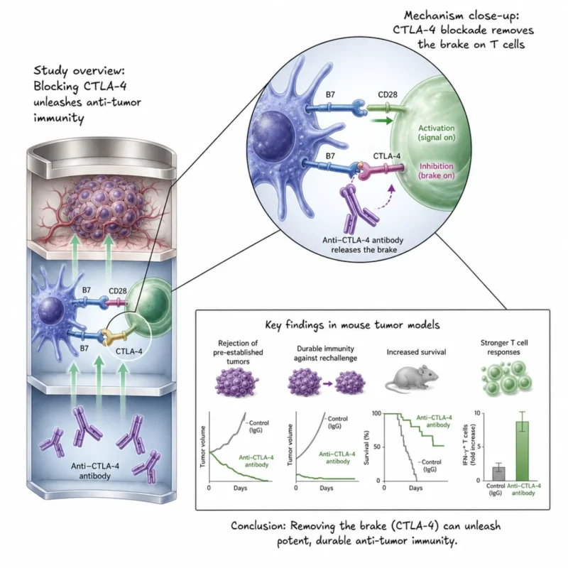
In 1996, Dana Leach, Matthew Krummel, and James Allison published a landmark Science paper testing a bold idea: instead of trying only to stimulate the immune system, could they fight cancer by removing an inhibitory signal?
That signal was CTLA-4, a negative regulator of T-cell activation.
In mouse tumor models, the researchers showed that blocking CTLA-4 with antibodies could lead to tumor rejection, including pre-established tumors. Even more remarkably, animals that rejected their tumors developed immunity against subsequent exposure to the tumor cells.
The conceptual shift was profound. Cancer immunotherapy did not necessarily require teaching the immune system an entirely new response. Sometimes, the immune response was already there; it simply needed to be unleashed.
This experiment became a crucial foundation for immune checkpoint blockade, ultimately contributing to the development of CTLA-4-targeting therapies such as ipilimumab and helping establish immunotherapy as a major pillar of modern cancer treatment.
Title: Enhancement of Antitumor Immunity by CTLA-4 Blockade
Authors: Dana R. Leach, Matthew F. Krummel, and James P. Allison
Paper 4: The Discovery of PD-1, the Second Pillar of the Checkpoint Inhibitor Revolution
What if one of the most important targets in modern cancer therapy was discovered while scientists were studying cell death rather than cancer?
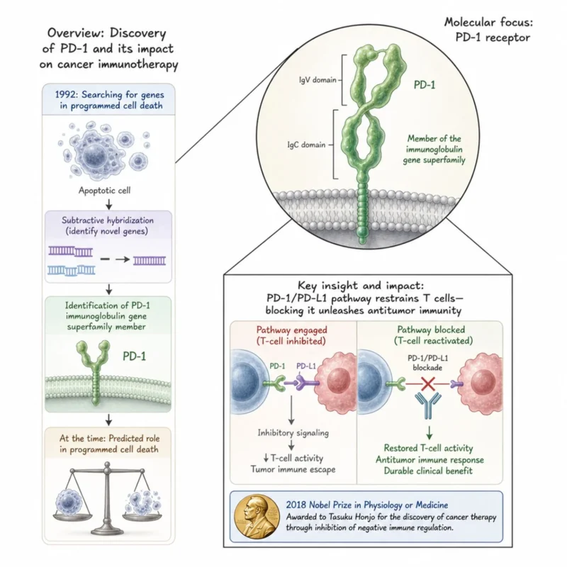
In 1992, Yasumasa Ishida, Yasutoshi Agata, Keiichi Shibahara, and Tasuku Honjo reported the discovery of a previously unknown gene that they called PD-1 (Programmed Death-1).
Using subtractive hybridization, the researchers sought genes associated with programmed cell death. They identified PD-1 in two different mouse cell lines undergoing classical programmed cell death and found that the predicted protein belonged to the immunoglobulin gene superfamily.
At the time, its future importance in cancer was impossible to foresee.
The original paper suggested that PD-1 might participate in programmed cell death. Subsequent work, however, revealed something even more consequential: PD-1 functions as an inhibitory receptor that helps restrain immune responses.
That discovery eventually transformed cancer immunology.
Tumors can exploit the PD-1/PD-L1 pathway to suppress T-cell activity and escape immune attack. Blocking this checkpoint can restore antitumor immune responses, a principle that ultimately led to the development of PD-1/PD-L1 checkpoint inhibitors and durable responses in patients with multiple cancer types.
Together with CTLA-4, PD-1 became one of the central pillars of the immune checkpoint revolution. Tasuku Honjo later shared the 2018 Nobel Prize in Physiology or Medicine for the discovery of cancer therapy through inhibition of negative immune regulation.
The enduring lesson is one of my favorites in science: a discovery need not begin with cancer to transform cancer treatment. Fundamental biology, followed far enough, can open therapeutic doors that were never visible at the start.
Authors: Y. Ishida, Y. Agata, K. Shibahara and T. Honjo
Paper 5 of 200: Imatinib for CML – The Paper That Helped Launch the Targeted Therapy Era
What if cancer treatment could target the specific molecular abnormality driving a tumor rather than broadly killing rapidly dividing cells?
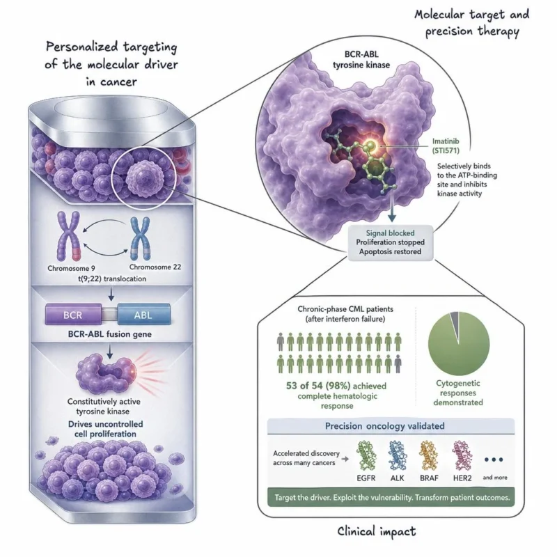
In 2001, Brian Druker and colleagues published a landmark New England Journal of Medicine study showing the remarkable clinical activity of imatinib (STI571) in chronic myeloid leukemia (CML).
CML had already provided one of the clearest molecular stories in cancer biology.
The Philadelphia chromosome, created by the t(9;22) translocation, encodes the BCR-ABL fusion protein, a constitutively active tyrosine kinase that drives disease.
Imatinib was designed to inhibit this abnormal kinase.
In patients with chronic-phase CML in whom interferon therapy had failed, the results were striking: 53 of 54 patients achieved a complete hematologic response, and cytogenetic responses were also observed.
This was more than a successful drug trial.
It provided powerful clinical proof of a new therapeutic philosophy: Identify the molecular driver – understand its biology – design a drug that selectively inhibits it – treat the cancer based on that vulnerability.
Imatinib helped transform CML from a life-threatening leukemia into a disease that, for many patients, can be controlled for years with targeted therapy.
Its impact also extended far beyond CML.
The success of imatinib strengthened the concept of precision oncology and accelerated the search for actionable molecular alterations across cancer, including EGFR, ALK, BRAF, HER2, and many others.
The enduring lesson: understanding what drives a cancer can reveal its Achilles’ heel. Modern targeted oncology owes an enormous debt to this principle.
Authors: Brian J. Druker, Moshe Talpaz, Debra J. Resta, Bin Peng, Elisabeth Buchdunger, John M. Ford, Nicholas B. Lydon, Hagop Kantarjian, Renaud Capdeville, Sayuri Ohno-Jones, and Charles L. Sawyers
Paper 6: HER2 Amplification – Connecting a Cancer Gene to Patient Outcomes
What if a genetic alteration inside a tumor could tell us not only how the cancer works but also how aggressively it might behave?
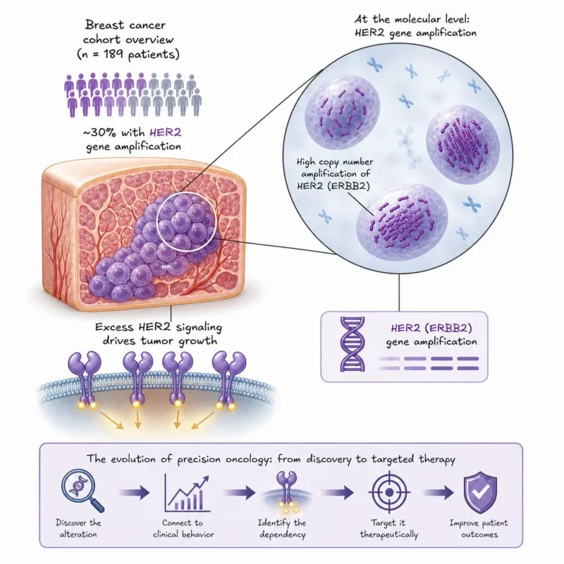
In 1987, Dennis Slamon and colleagues published a landmark paper in Science examining the HER2/neu (ERBB2) gene in human breast cancer.
They analyzed tumors from 189 patients with primary breast cancer and found HER2 gene amplification in roughly 30% of cases.
But the most important observation was the clinical connection.
HER2 amplification was significantly associated with shorter time to disease relapse and reduced overall survival. In patients with lymph-node-positive breast cancer, HER2 amplification emerged as an important predictor of outcome.
This helped establish a powerful concept in oncology:
A molecular alteration could define a biologically and clinically distinct subgroup of cancer.
At the time, HER2 was primarily a clue about tumor biology and prognosis.
But that clue eventually became a therapeutic opportunity.
Recognition that some breast cancers were driven by excessive HER2 signaling provided the biological rationale for targeting HER2. This ultimately contributed to the development of trastuzumab (Herceptin) and, later, an entire generation of HER2-directed therapies.
The story of HER2 captures the evolution of precision oncology beautifully:
Discover the alteration – connect it to clinical behavior – identify the dependency – target it therapeutically.
Today, HER2 is not simply a prognostic marker. It is one of the clearest examples of how molecular classification can fundamentally change cancer treatment.
The enduring lesson: when we divide cancers by their molecular drivers rather than only by where they arise, entirely new therapeutic possibilities emerge.
Authors: Dennis J. Slamon, Gary M. Clark, Steven G. Wong, Wendy J. Levin, Axel Ullrich, and William L. McGuire
Paper 7: Trastuzumab + Chemotherapy – When HER2 Went From a Bad Prognostic Marker to a Therapeutic Target
What if one of the features that made a cancer more aggressive could also become its greatest therapeutic vulnerability?
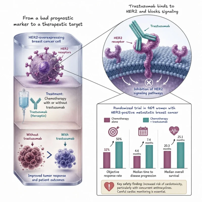
In 2001, Dennis Slamon and colleagues published a pivotal New England Journal of Medicine trial evaluating trastuzumab (Herceptin), a monoclonal antibody targeting HER2, in women with HER2-overexpressing metastatic breast cancer.
The biological foundation had been laid years earlier.
HER2 amplification has been linked to aggressive breast cancer and poorer outcomes. The next question was transformative:
Could we directly target HER2 and change the course of the disease?
In this randomized trial, 469 women received either standard chemotherapy alone or chemotherapy plus trastuzumab.
The results provided a striking answer.
Adding trastuzumab increased the objective response rate from 32% to 50%, extended the median time to disease progression from 4.6 to 7.4 months, and improved the median overall survival from 20.3 to 25.1 months, despite patients in the chemotherapy-only group being allowed to receive trastuzumab after progression.
This was more than a positive breast cancer trial.
It was powerful clinical proof of a principle that now sits at the heart of precision oncology:
Find the molecular alteration – identify the dependency – build a therapy against it – select the patients whose tumors carry the target.
The HER2 story is especially remarkable when viewed alongside Paper 6.
1987: HER2 amplification helps identify a subgroup of breast cancers with worse outcomes.
2001: That same molecular feature helps identify patients who can benefit from a therapy specifically directed against it.
That transition, from biomarker to drug target, captures the promise of translational cancer research.
The enduring lesson: the molecular abnormality that makes a cancer dangerous can sometimes also reveal how to defeat it.
Authors: Dennis J. Slamon, Brian Leyland-Jones, Steven Shak, Hank Fuchs, Virginia Paton, Alex Bajamonde, Thomas Fleming, Wolfgang Eiermann, Janet Wolter, Mark Pegram, Jose Baselga, and Larry Norton
Paper 8: The Philadelphia Chromosome: The First Consistent Chromosomal Abnormality Linked to Human Cancer
What if cancer could be traced to a visible abnormality in a chromosome?
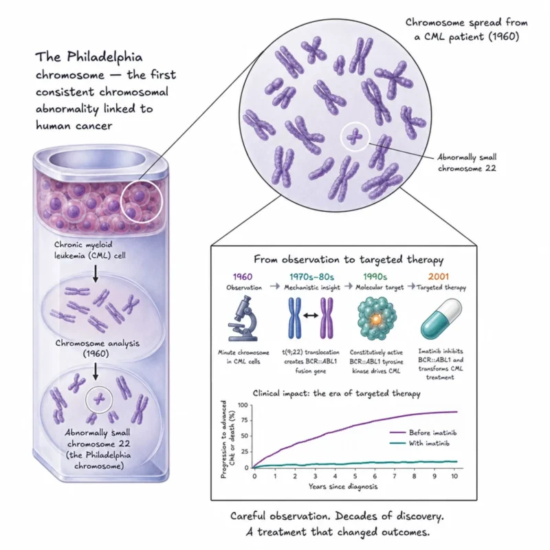
In 1960, Peter Nowell and David Hungerford reported a remarkable observation while studying cells from patients with chronic myeloid leukemia (CML). They repeatedly found an unusually small chromosome in leukemic cells.
At the time, the tools of chromosome analysis were primitive compared with today’s sequencing technologies. Yet the consistency of this abnormality suggested something profound.
Cancer might be associated with specific genetic changes rather than simply representing uncontrolled cell growth. The abnormal chromosome became known as the Philadelphia chromosome, named after the city where Nowell and Hungerford made their discovery.
The original observation could not explain how the chromosome caused leukemia.
That story unfolded over the following decades.
Researchers later demonstrated that the Philadelphia chromosome results from a reciprocal translocation between chromosomes 9 and 22 — t(9;22). This rearrangement creates the BCR::ABL1 fusion gene, producing a constitutively active tyrosine kinase that drives CML.
And that molecular understanding eventually led to something extraordinary: a drug designed to inhibit the driver itself.
The progression is a beautiful example of how cancer research builds across generations:
- 1960: See the abnormal chromosome.
- 1970s–80s: Understand the translocation and genes involved.
- 1990s: Develop an inhibitor against the molecular driver.
- 2001: Demonstrate remarkable clinical activity with imatinib.
What began as an unusual chromosome under a microscope ultimately became a blueprint for precision oncology.
The enduring lesson: careful observation can precede understanding by decades, but once the biology is understood, an observation can become a treatment.
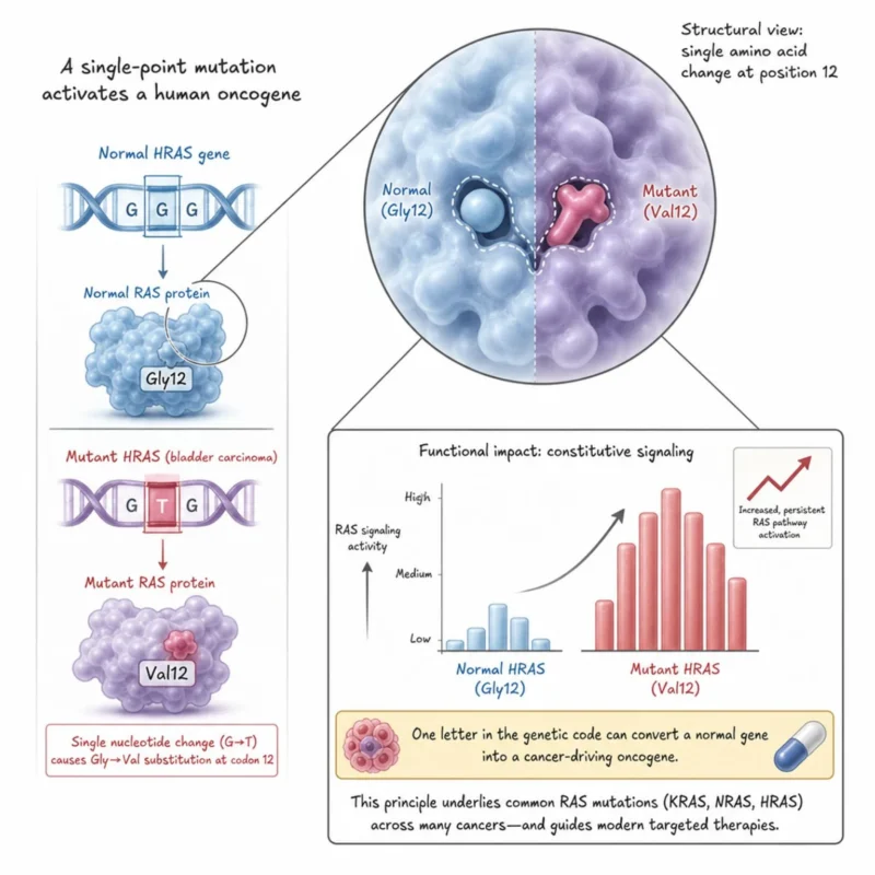
Paper 9: A Single-Point Mutation That Helped Explain How a Human Oncogene Is Activated
Can changing a single nucleotide turn a normal human gene into a cancer-driving oncogene?
In 1982, Clifford Tabin, Robert Weinberg (my postdoctoral mentor), and colleagues published a landmark paper in Nature that helped answer this question.
Researchers already knew that DNA from certain human cancer cells could transform normal cells in laboratory experiments. But a fundamental mystery remained:
What was actually different about the cancer-causing gene?
The Weinberg and team compared the transforming HRAS gene from a human bladder carcinoma with its normal counterpart.
The difference was remarkably small.
A single nucleotide substitution changed codon 12 of HRAS, resulting in the replacement of glycine with valine in the RAS protein.
One tiny change in DNA was enough to dramatically alter the gene’s biological behavior. This was a major conceptual advance.
Cancer did not necessarily require the acquisition of completely foreign genes or massive genetic alterations. A normal cellular gene could become oncogenic through something as subtle as a single-point mutation.
The discovery helped establish a principle that now sits at the center of cancer genomics: Specific mutations can activate proto-oncogenes and drive malignant transformation. We now know that RAS pathway mutations are among the most common oncogenic alterations in human cancer, with KRAS, NRAS, and HRAS playing important roles across many tumor types.
And four decades later, the story has come full circle.
RAS was once considered extraordinarily difficult to target therapeutically. Today, drugs targeting specific mutant forms of RAS, particularly KRAS G12C and, more recently, other RAS alterations, have translated this foundational biology into new treatment strategies.
The enduring lesson: sometimes the difference between normal growth and cancer is not an entirely new gene, it can be a single letter in the genetic code.
Title: Mechanism of activation of a human oncogene
Authors: Clifford J. Tabin, Scott M. Bradley, Cornelia I. Bargmann, Robert A. Weinberg, Alex G. Papageorge, Edward M. Scolnick, Ravi Dhar, Douglas R. Lowy, Esther H. Chang
Paper 10: The Discovery of Telomerase – The Enzyme Cancers Exploit for Immortality
What if one of the fundamental limits on how long a cell can divide could be overcome by a single enzyme?
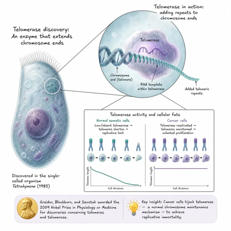
In 1985, Carol Greider and Elizabeth Blackburn published a landmark paper in Cell describing an enzyme capable of adding DNA sequences to the ends of chromosomes.
That enzyme would become known as telomerase. The discovery emerged from studies of the single-celled organism Tetrahymena, not from cancer research.
Chromosome ends, or telomeres, create a fundamental biological problem. Because conventional DNA replication cannot fully copy the ends of linear chromosomes, telomeres progressively shorten as cells divide.
Greider and Blackburn identified an enzymatic activity that could extend these chromosome ends by adding telomeric repeats.
This solved a fundamental puzzle in chromosome biology:
How can cells maintain the ends of their chromosomes despite repeated rounds of DNA replication?
The implications for cancer became profound.
Most normal human somatic cells have limited telomerase activity. As telomeres progressively shorten, cells eventually encounter barriers to continued proliferation.
Cancer cells must somehow escape those limits.
- And many do so by reactivating telomerase, allowing them to maintain their telomeres and continue dividing.
- This ultimately helped establish replicative immortality as one of the defining capabilities of cancer.
- The discovery is also a beautiful example of why fundamental science matters.
An enzyme discovered while studying chromosome biology in a unicellular organism ultimately revealed one of the mechanisms that enables human cancer cells to achieve seemingly unlimited proliferation.
Greider and Blackburn, together with Jack Szostak, were awarded the 2009 Nobel Prize in Physiology or Medicine for discoveries concerning telomeres and the enzyme telomerase.
The enduring lesson: cancer cells do not invent immortality from scratch; they hijack normal mechanisms of chromosome maintenance to escape the limits placed on cellular lifespan
Title: Identification of a Specific Telomere Terminal Transferase Activity in Tetrahymena Extracts
Authors: Carol W. Greider and Elizabeth H. Blackburn
Paper 11: HPV-16 in Cervical Cancer – Connecting a Virus to One of the World’s Major Cancers
What if a common viral infection could initiate the molecular events that eventually lead to cancer, and preventing that infection could therefore prevent cancer itself?
In 1983, Matthias Dürst, Lutz Gissmann, Harald Ikenberg, and Harald zur Hausen published a landmark PNAS paper providing compelling evidence linking human papillomavirus type 16 (HPV-16) to cervical cancer.
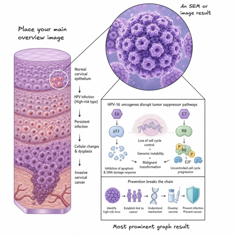
At the time, the idea that specific human papillomaviruses might cause cervical cancer was far from established. The researchers examined cervical cancer biopsies for HPV DNA. Their finding was striking.
They detected DNA from the newly characterized papillomavirus HPV-16 in approximately half of the cervical cancer samples examined. Importantly, HPV-16 was distinct from the papillomavirus types commonly associated with benign genital warts.
This helped establish a crucial idea: Not all HPV types carry the same cancer risk. Certain ‘high-risk’ HPV types are specifically associated with malignant transformation.
Subsequent research revealed how these viruses drive cancer.
The viral proteins E6 and E7 interfere with two of the cell’s most important tumor-suppressor pathways, p53 and RB, disrupting normal control of cell proliferation and survival.
The implications extended far beyond understanding cervical cancer.
Once a causal link between high-risk HPV infection and cervical cancer became firmly established, an extraordinary possibility emerged: If infection could be prevented, perhaps the cancer could be prevented too.
That biological insight ultimately contributed to the development of HPV vaccines, giving us something remarkably powerful in oncology: a vaccine capable of preventing infections responsible for multiple cancers.
Harald zur Hausen received half of the 2008 Nobel Prize in Physiology or Medicine for discovering that human papillomaviruses cause cervical cancer.
The story is a powerful progression:
Identify the virus – establish the cancer association – understand the molecular mechanism – develop a vaccine – prevent cancer before it begins.
The enduring lesson: some of the greatest advances in cancer treatment are not treatments at all; they are ways of preventing cancer from developing in the first place.
Authors: M Dürst, L Gissmann, H Ikenberg, and H zur Hausen
You don’t want to miss this: Sendurai Mani: Join Us for the 2026 Translational Oncology Research Symposium at Brown University