





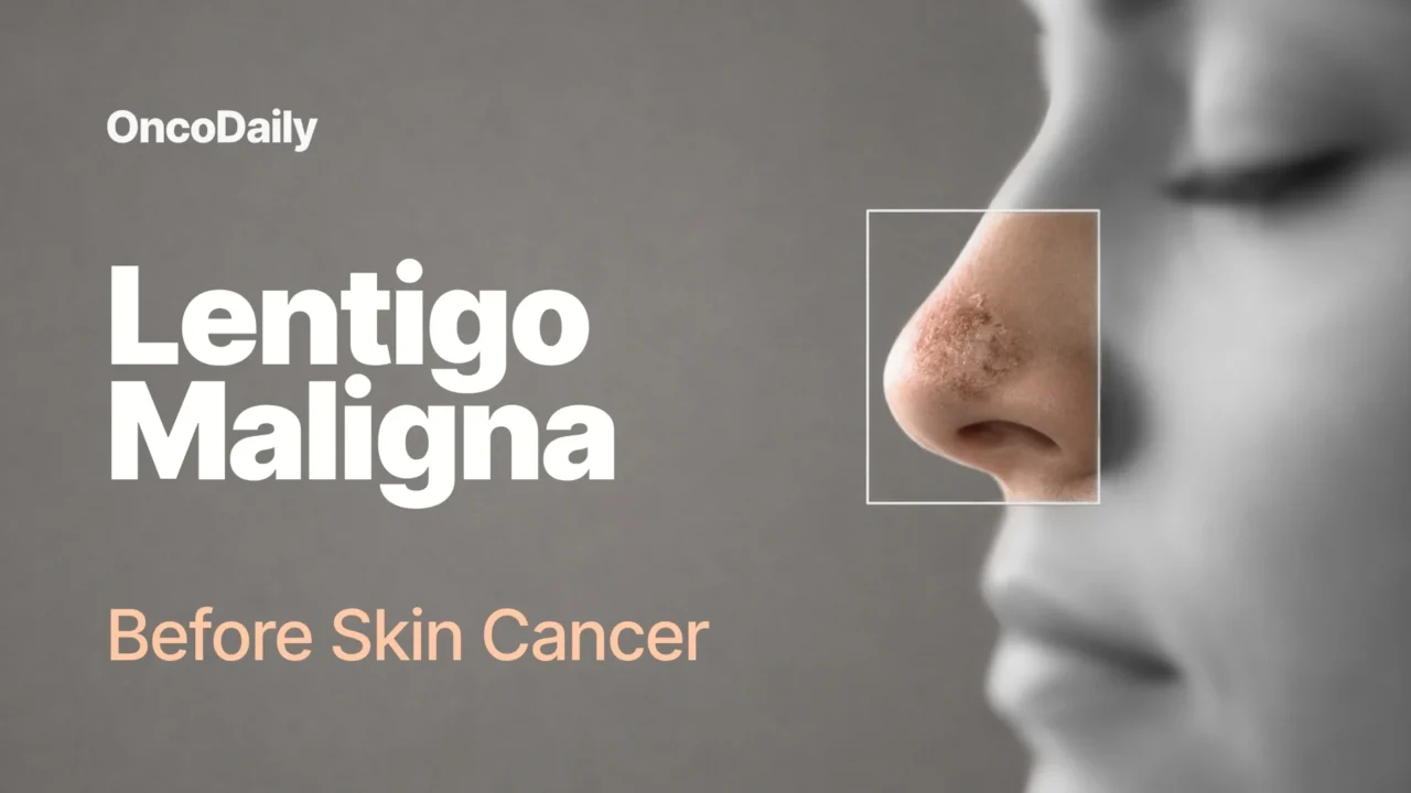
Lentigo maligna (LM) is a subtype of melanoma in situ, meaning it is an early, non-invasive form of melanoma that remains confined to the epidermis. It arises on chronically sun-damaged skin, most often on the head and neck of older individuals, and typically appears as a slowly enlarging, irregularly pigmented macule or patch. Hutchinson first described this lesion in 1890, referring to it as “Hutchinson’s melanotic freckle.” LM usually follows an indolent course, remaining within the epidermis for years or even decades. The incidence of LM has been increasing worldwide, and it is now recognized as one of the more common forms of melanoma in situ, particularly among older, fair-skinned populations. [Ana-Maria Forsea et al., 2026]
Is LM Cancer or Precancer?
LM is best classified as an early (stage 0) melanoma melanoma in situ rather than a “precancer” in the same sense as actinic keratosis. This means that it is already a malignant melanoma confined to the epidermis, before it has invaded the deeper layers of the skin. Although it grows slowly and has a good prognosis at this stage, it carries the potential to progress into an invasive tumor with potentially aggressive behavior, known as lentigo maligna melanoma (LMM).
Although some patient materials and academic publications describe melanoma in situ as “pre-cancer,” current classifications treat LM as stage 0 melanoma—already malignant but non-invasive. In that sense, it behaves like an early-stage malignancy rather than a benign precursor: the main clinical concern is detecting it and treating it before invasion occurs.
Is LM the Same as Melanoma?
Lentigo maligna is a subtype of melanoma, but it is not the same as invasive melanoma. Lentigo maligna is best classified as an early, non-invasive form of skin cancer, specifically melanoma in situ, rather than a precancerous condition. By definition, it remains confined to the epidermis, and the diagnosis changes to lentigo maligna melanoma only once the lesion becomes invasive. In other words, lentigo maligna is an early, non-invasive stage of melanoma rather than a separate disease.
Once dermal invasion occurs (lentigo maligna melanoma), the lesion may develop papules, nodules, or thickened areas that can be palpable. Because it can progress to invasive lentigo maligna melanoma, which carries a significantly worse prognosis, accurate diagnosis and complete treatment are essential to prevent progression to invasive disease.
What Does LM Look Like?
Lentigo maligna typically appears as a slowly enlarging, irregularly pigmented macule or patch on chronically sun-exposed skin, most often on the face, neck, and scalp. Its clinical features are well captured by the ABCDE rule. The lesion is usually asymmetrical in shape and color, and it becomes more asymmetrical as it enlarges. The border is irregular, often jagged or notched. The color is frequently variegated, with different shades of brown and black, and occasionally pink or white areas.
The lesion is usually greater than 6 mm in diameter and tends to evolve over time. Because it is an in situ lesion, it is typically smooth and non-palpable, and it may be misdiagnosed for years as a benign lesion such as a solar lentigo.
What Causes LM?
The major risk factor for developing lentigo maligna is ultraviolet radiation, particularly cumulative lifetime exposure to UVR. In contrast to nodular melanoma and superficial spreading melanoma, which are associated with intense intermittent sun exposure, lentigo maligna is strongly linked to chronic, long-term UVR exposure. This explains why it most often arises on the face and other chronically sun-damaged sites, and why it tends to occur in older individuals, who have accumulated greater lifetime sun exposure.
Studies have shown that the risk increases with the number of years spent in sunny regions, total hours of sunlight, the amount of actinic damage, and a prior history of nonmelanoma skin cancer. Other suggested risk factors include X-ray irradiation, estrogen and progesterone, and non-permanent hair dyes. Lentigo maligna is also more likely in people with genetic conditions that predispose them to sun sensitivity, such as oculocutaneous albinism, xeroderma pigmentosum, Werner syndrome, and porphyria cutanea tarda. No association with smoking or alcohol has been demonstrated. [Yana Puckett et al., NIH, 2025]
Who Is Most at Risk of LM?
Risk factors associated with LM include increased age, chronic sun exposure and sun-damaged skin, a greater number of lentigines, a greater number of actinic keratoses, and a history of previous keratinocyte carcinomas. LM occurs predominantly in older individuals, particularly those over 60 years of age, and shows a male predominance. [Yana Puckett et al., NIH, 2025]
Genetic conditions such as xeroderma pigmentosum, oculocutaneous albinism, Werner syndrome, and porphyria cutanea tarda are also associated with LM. Larger LM lesions have been proposed as a risk factor for transformation into lentigo maligna melanoma. Compared with other melanoma subtypes, LM has a distinct epidemiologic profile, with a stronger association with chronic and occupational sun exposure and a weaker association with a high nevus count. [Helena Iznardo et al., 2020]
Can LM Become Invasive Skin Cancer?
Lentigo maligna can progress to invasive skin cancer. By definition, lentigo maligna is melanoma in situ, meaning the atypical melanocytes remain confined to the epidermis. However, if left untreated, it can invade the dermis, and at this point the diagnosis changes to lentigo maligna melanoma, an invasive form of melanoma that carries a significantly worse prognosis.
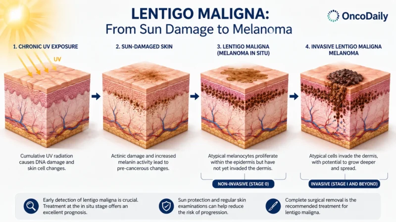
The timeframe for progression varies widely. Features that may suggest transformation into invasive disease include the development of a nodule, an increasing number of colours, especially blue or black, and ulceration or bleeding. Because lentigo maligna may remain in situ for years or even decades and can be misdiagnosed as a benign lesion, timely recognition and complete treatment are important to prevent progression to invasive melanoma.
How Is LM Diagnosed?
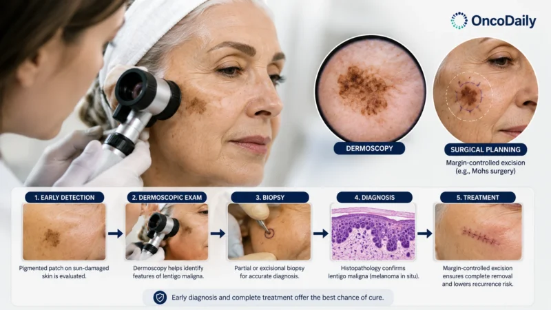
Lentigo maligna is diagnosed through clinical examination, dermoscopy, and, when available, reflectance confocal microscopy. Dermoscopy is used to distinguish lentigo maligna from other pigmented facial lesions, using established criteria such as the pseudonetwork and the inverse approach.
Reflectance confocal microscopy can improve diagnostic accuracy, particularly for clinically and dermoscopically equivocal lesions, and it may also help identify areas of invasion and delineate surgical margins. However, histopathological evaluation remains the diagnostic gold standard. Excisional or partial biopsies are used for pathological diagnosis, and dermoscopy, or reflectance confocal microscopy when available, should guide site selection for partial biopsies to maximize the likelihood of sampling the most diagnostically representative area.
How Is LM Treated?
In 2026, International Dermoscopy Society published consensus recommendations for the management of lentigo maligna. The consensus states that treatment of lentigo maligna (LM) is primarily surgical. Complete surgical excision is the recommended first-line treatment, with the goal of achieving histopathologically negative margins.
Many guidelines describe 5–10 mm clinical margins as commonly used for melanoma in situ, but the 2026 International Dermoscopy Society consensus notes that no single margin width is definitively proven for LM.Because LM often extends subclinically beyond its visible borders on chronically sun-damaged skin, accurate margin estimation is difficult and incomplete excision is relatively common. Margin-controlled techniques are therefore preferable for large or ill-defined lesions, particularly those on the head and neck. Lesions on the head and neck with ill-defined borders or a diameter of ≥2 cm frequently require larger margins, staged excision, or Mohs micrographic surgery.
When surgery is not feasible or desirable, radiotherapy and topical imiquimod are recommended as alternative treatments. Imiquimod cream has shown high clinical clearance rates and can be used as primary treatment, as adjuvant therapy after excision with narrow or positive margins, or as neoadjuvant therapy to reduce lesion size before surgery. Radiotherapy is likewise an effective option for patients who cannot undergo surgery. In contrast, cryotherapy and laser therapy are not recommended for LM.
Because LM carries a substantial risk of local recurrence, which varies with treatment modality and margin status, regular clinical and dermoscopic follow-up is essential to detect recurrence, disease progression, and new primary tumors early. [Ana-Maria Forsea et al., JEADV, 2026]
Can LM Be Prevented?
Prevention of lentigo maligna centers on limiting chronic sun exposure, since LM develops on chronically sun-damaged skin and chronic solar exposure is considered an important pathway in the development of its invasive form, lentigo maligna melanoma. Because LM arises predominantly on the head and neck and its incidence is higher in sunnier latitudes, consistent sun protection of chronically exposed areas is the main primary preventive measure.
Secondary prevention relies on early detection: LM progresses slowly and has the potential to transform into invasive melanoma, so regular skin examinations and skin cancer awareness are important, particularly for individuals at high risk.
You Can Also Read Actinic Keratosis (AK) : The Precancerous Skin Lesion You Shouldn’t Ignore by OncoDaily

Written by Written by Lily Tumanyan MD
FAQ
Is lentigo maligna dangerous?
Lentigo maligna (LM) is an early, non‑invasive melanoma (melanoma in situ). It has an excellent prognosis when treated, but can progress to invasive melanoma if left untreated.
How fast does lentigo maligna grow?
LM grows very slowly, often over years or decades, which is why it is frequently misdiagnosed as a benign sun spot.
Can lentigo maligna turn into invasive melanoma?
Yes. Untreated LM can invade the dermis and become lentigo maligna melanoma (LMM). Recent data suggest a relatively low short‑term risk (around 1% over 10 years), higher for head/neck lesions and older age.
What are the warning signs that LM is becoming invasive?
Watch for a new raised nodule or thickening, new colors (especially blue/black), ulceration, bleeding, or rapid change in size or shape. These suggest dermal invasion and need urgent evaluation.
How is lentigo maligna different from a sun spot?
Solar lentigines are benign, uniformly colored, and stable. LM is typically asymmetrical, has irregular borders and multiple colors, and slowly enlarges over time.
Do I need a biopsy if LM is suspected?
Yes. Histopathology is the gold standard. Because LM can be patchy, clinicians often take multiple partial biopsies from the most suspicious areas (guided by dermoscopy).
What is the best treatment for lentigo maligna?
Complete surgical removal is the first‑line. Wide local excision with 5–10 mm margins is common; margin‑controlled techniques (staged excision or Mohs) are preferred for large or ill‑defined facial lesions.
Are there non‑surgical options?
Yes. For patients who cannot have surgery, topical imiquimod and/or radiotherapy are effective alternatives. Cryotherapy or laser alone are not recommended.
Will lentigo maligna come back after treatment?
Recurrence is possible, especially if margins are not fully clear. Regular clinical and dermoscopic follow‑up is essential to detect recurrences or new tumors early.
How can I reduce my risk of developing LM?
Limit cumulative sun exposure: avoid tanning, limit midday sun, wear protective clothing and a hat, and use broad‑spectrum SPF 30+ sunscreen daily. High‑risk patients should have regular skin checks.