
Muna Al-Khaifi, GP Oncologist at Mount Sinai Hospital (Toronto), Sinai Health. This article explores whether body fat percentage may better predict vertebral fracture risk than BMI in women with early breast cancer receiving aromatase inhibitor therapy. The findings highlight that excess adiposity was independently associated with a higher risk of vertebral fracture progression, while higher bone mineral density and muscle mass were associated with lower risk. The article also emphasizes the potential importance of assessing body composition alongside traditional measures such as BMI and BMD to better identify patients at increased fracture risk during aromatase inhibitor therapy.
Body Fat Percentage May Better Predict Vertebral Fracture Risk During Aromatase Inhibitor Therapy
Body fat percentage may better identify women with early breast cancer at increased risk of vertebral fractures than body mass index (BMI) during aromatase inhibitor (AI) therapy, according to a new study by Schivardi et al. (2026) published in JAMA Network Open.
The findings suggest that excess adiposity, defined as a fat mass percentage (FM%) greater than 40.8%, is independently associated with vertebral fracture progression, challenging the traditional assumption that higher body weight is protective for bone health.
What is the objective of the study?
(AIs greatly reduce estrogen levels and can speed up bone loss in postmenopausal women with early breast cancer (EBC). Although obesity based on BMI has traditionally been considered protective for bone health, growing evidence suggests that higher body fat may actually increase fracture risk.
The study aimed to determine whether a FM% greater than 40.8%, measured using dual-energy x-ray absorptiometry (DXA), is associated with new or worsening vertebral fractures (VFs) in patients with EBC receiving AI therapy.
Who participated in the study?
The study included 769 women with early breast cancer (EBC), with a median age of 63 years and a median body mass index (BMI) of 24.6. Participants were followed for a median of approximately 46 months.
How common was vertebral fracture progression?
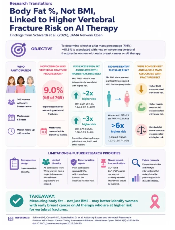
During follow-up, vertebral fracture (VF) progression occurred in 69 of 769 patients (9.0%). This included patients who developed a new vertebral fracture or experienced worsening of an existing fracture. Most events occurred within the first 60 months of follow-up.
Was excess body fat associated with vertebral fracture progression?
Patients with a FM% greater than 40.8% had approximately double the risk of vertebral fracture progression compared with patients without excess adiposity, even after accounting for factors such as age, previous fractures, and bone mineral density (BMD) (HR, 2.00; 95% CI, 1.44-2.82; P<.001).
A separate analysis supported this finding, showing that excess adiposity was associated with nearly 3 times the risk of vertebral fracture progression (HR, 2.77; 95% CI, 1.30-5.93; P=.01).
Did BMI identify the same fracture risk as body FM%?
No. BMI alone was not significantly associated with vertebral fracture progression. Importantly, some women with a BMI considered to be within the normal range still had excess body fat.
Among women with a BMI below 25 who had an FM% greater than 40.8%, the risk of vertebral fracture progression was more than 6 times higher (HR, 6.63; 95% CI, 1.93-21.85; P<.001). These findings suggest that measuring body composition may identify at-risk patients who could be missed when BMI is used alone.
Were bone density and muscle mass associated with fracture risk?
Yes. Higher BMD was associated with a lower risk of vertebral fracture progression (HR, 0.79; 95% CI, 0.66-0.94; P=.01).
Higher appendicular lean mass index (ALMI), a measure of muscle mass in the arms and legs relative to height, was also associated with a substantially lower risk of fracture progression (HR, 0.38; 95% CI, 0.22-0.65; P<.001). In contrast, having more body fat relative to muscle mass was associated with increased fracture risk.
Why might excess body fat increase fracture risk during AI therapy?
The findings challenge the long held assumption that obesity protects against fractures. Adipose tissue can normally support bone health partly through peripheral estrogen production. However, aromatase inhibitors strongly suppress estrogen production, which may reduce this protective effect.
In this setting, excess adiposity may instead contribute to chronic inflammation and poorer bone quality. The findings suggest that body composition, not simply body weight or BMI, may be important when assessing fracture risk in women receiving AI therapy.
What are the limitations and future research priorities?
- Retrospective design: The study cannot establish a direct causal relationship between excess adiposity and vertebral fractures.
- Bone-targeting agents: Most participants received BTAs, which may have lowered the observed fracture rate.
- Limited diversity: The study included White women from a single Italian center. Future studies should include more diverse racial, ethnic, and geographic populations to improve generalizability.
- Newer weight-loss medications: GLP-1 and GLP-1/GIP receptor agonist use was not routinely recorded. Their effects on body composition and fracture risk should be explored.
- Future research: Larger, prospective studies should confirm whether measuring body fat and muscle mass improves fracture risk assessment beyond BMI and BMD and whether lifestyle interventions that reduce fat while preserving muscle can lower fracture risk.
Reference
Schivardi G, Cosentini D, Scartabellati G, et al. Adiposity Excess and Vertebral Fractures in Patients With Breast Cancer Taking Aromatase Inhibitors. JAMA Netw Open. 2026;9(7):e2626450. doi:10.1001/jamanetworkopen.2026.26450
You may also be interested in: Muna Al-Khaifi: Menopausal Symptoms and Quality of Life After Cancer