Key takeaways
- The placenta is selectively permeable, so many chemotherapy drugs can reach the fetal circulation to some extent.
- The amount of placental transfer varies considerably between chemotherapy classes and individual drugs, making agent selection important.
- Gestational timing is critical: chemotherapy exposure during the first trimester carries the greatest risk of miscarriage and major congenital malformations.
- During the second and third trimesters, several established chemotherapy regimens can be used when clinically necessary, although fetal and neonatal risks remain.
- Cancer treatment during pregnancy requires individualized multidisciplinary care, with close fetal monitoring, treatment planning, and coordination of delivery.
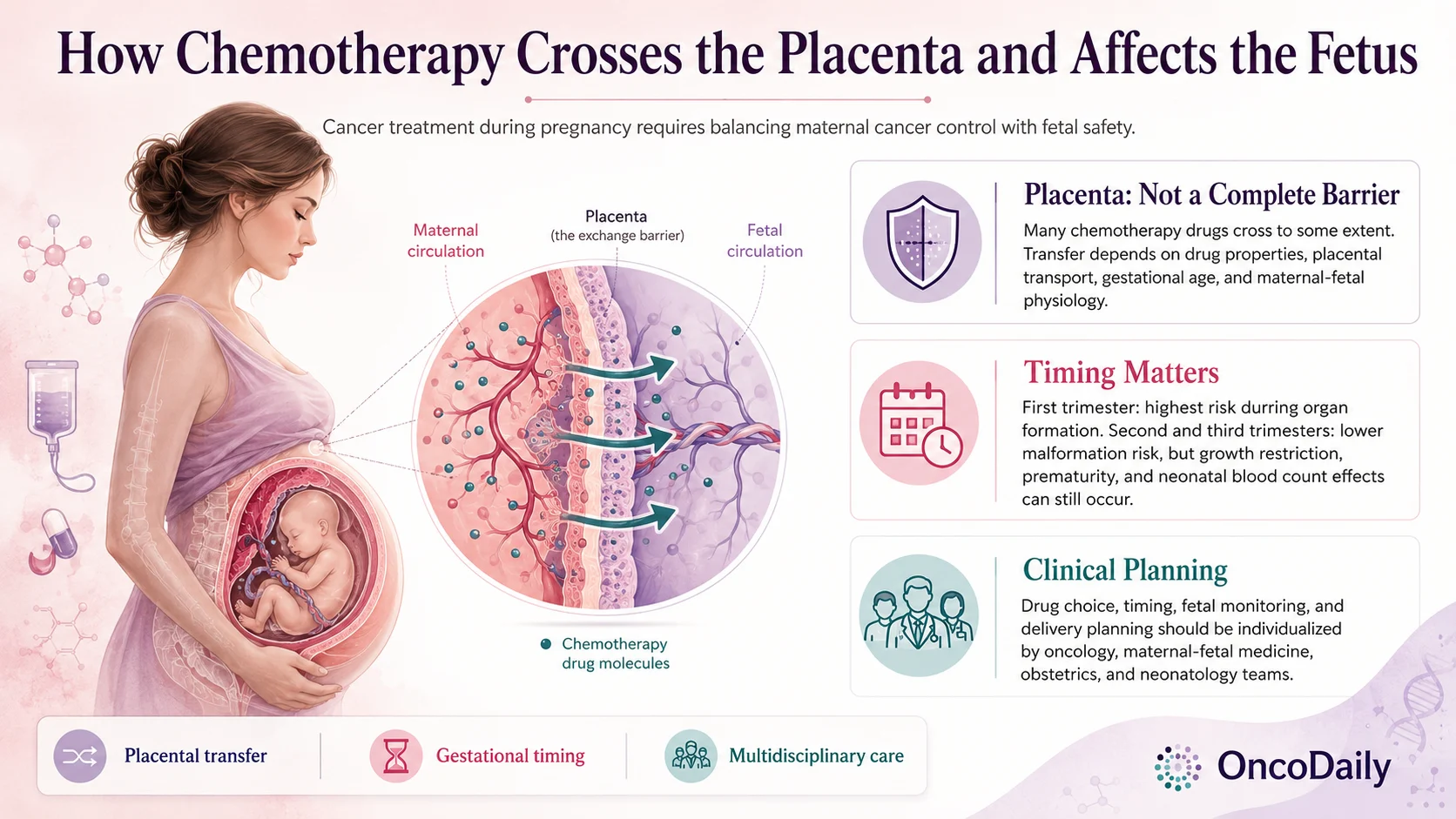
The placenta plays a central role in protecting the developing fetus, but it is not an absolute barrier. It regulates exchange between the maternal and fetal circulations while still allowing some medications, including chemotherapy agents, to cross. The extent of this transfer varies according to the drug’s physicochemical properties, placental transport mechanisms, gestational age, and maternal–fetal physiology.
This becomes especially important when cancer is diagnosed during pregnancy. The effects of chemotherapy depend not only on which drug is used, but also on when exposure occurs. Treatment during the first trimester carries the greatest risk because organogenesis is taking place, whereas chemotherapy administered during the second and third trimesters is associated with a substantially lower risk of major congenital malformations. Later exposure can still influence fetal growth, pregnancy duration, and neonatal blood counts.
Understanding how chemotherapy interacts with the placental barrier is therefore essential for treatment planning. Drug selection, treatment timing, fetal surveillance, and delivery planning require an individualized multidisciplinary approach involving oncology, maternal–fetal medicine, obstetrics, neonatology, and other relevant specialists.
Overview of Placental Drug Transfer
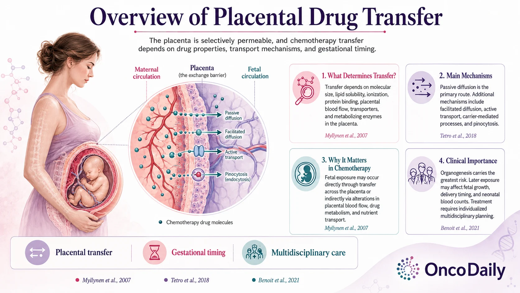
The placenta is a selectively permeable organ that regulates exchange between the maternal and fetal circulations. Placental drug transfer depends on factors including molecular size, lipid solubility, ionization, protein binding, placental blood flow, and the activity of placental transporters and metabolizing enzymes (Myllynen et al., 2007; Tetro et al., 2018).
Passive diffusion along a maternal–fetal concentration gradient is the principal mechanism by which many drugs cross the placenta. However, facilitated diffusion, active transport, carrier-mediated uptake and efflux, transcytosis, and other transport processes can also contribute. Some drugs may additionally accumulate within placental tissue, meaning that fetal exposure cannot be predicted solely from maternal drug concentrations (Tetro et al., 2018).
Chemotherapy can affect the fetus both directly and indirectly. Direct exposure occurs when a drug or its metabolites cross the placenta, while indirect effects can result from changes in placental development, vascularization, metabolism, or nutrient transport. The degree and clinical significance of these effects differ considerably among individual anticancer agents (Benoit et al., 2021; Depoix et al., 2020).
These differences explain why placental transfer cannot be considered a uniform property of chemotherapy as a whole. Treatment decisions must take into account the specific agent, gestational age, treatment schedule, and available maternal and fetal safety data.
Chemotherapy Agents That Cross the Placenta
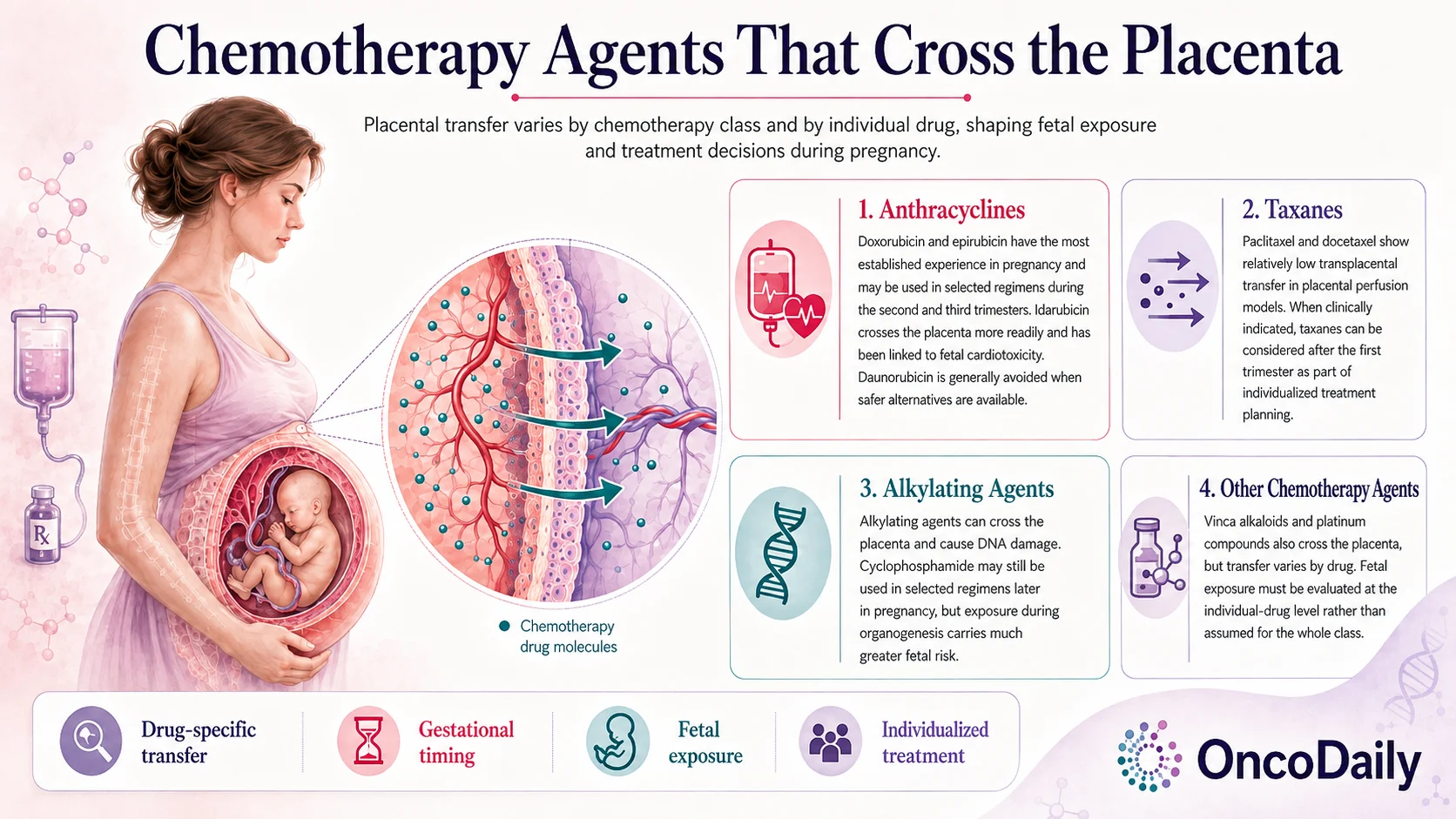
Placental transfer varies considerably between chemotherapy classes and even between drugs within the same class. Anthracyclines, taxanes, vinca alkaloids, platinum compounds, and alkylating agents have all demonstrated some degree of transplacental passage, but fetal exposure differs substantially between individual agents (Berveiller et al., 2016; Benoit et al., 2021).
Anthracyclines
Doxorubicin and epirubicin have some of the most established clinical experience among anthracyclines used during pregnancy and can be incorporated into selected chemotherapy regimens during the second and third trimesters. Their fetal exposure appears relatively limited compared with some other anthracyclines (Benoit et al., 2021; Nanda et al., 2025).
Idarubicin is more lipophilic and has greater placental permeability than daunorubicin. Fetal and neonatal cardiotoxicity has been reported following idarubicin exposure, making it less suitable during pregnancy.
Daunorubicin should be considered separately. It remains an established treatment option for selected hematologic malignancies during the second and third trimesters and is generally preferred over idarubicin when an anthracycline is required in these settings (Benoit et al., 2021; Nanda et al., 2025).
Taxanes
Paclitaxel and docetaxel show relatively low transplacental transfer in ex vivo perfusion studies using term human placentas. Their high protein binding and interactions with placental transport mechanisms may contribute to relatively limited fetal exposure (Berveiller et al., 2012).
Clinical experience with taxanes during pregnancy has also increased, particularly in breast cancer. When taxane therapy is clinically indicated, it can be considered after the first trimester as part of an individualized treatment strategy (Benoit et al., 2021; Nanda et al., 2025).
Alkylating Agents
Alkylating agents can cross the placenta and produce DNA damage. Cyclophosphamide has nevertheless been incorporated into established chemotherapy regimens during the second and third trimesters when treatment is necessary (Berveiller et al., 2016; Benoit et al., 2021).
The timing of exposure remains critical. Alkylating-agent exposure during organogenesis carries substantially greater concern for miscarriage and congenital abnormalities than exposure later in pregnancy (Berveiller et al., 2016; Benoit et al., 2021).
You can also read Chemotherapy During Pregnancy: What Can Be Used in Each Trimester and What Should Be Avoided on OncoDaily.
Other Chemotherapy Agents
Vinca alkaloids and platinum compounds can also cross the placenta, although transfer varies by drug. Available preclinical and clinical studies demonstrate that fetal exposure cannot be assumed to be equivalent across agents within these classes. Treatment therefore needs to be assessed at the individual-drug level rather than according to chemotherapy class alone (Berveiller et al., 2016; Benoit et al., 2021).
Fetal Risks Associated With In Utero Exposure
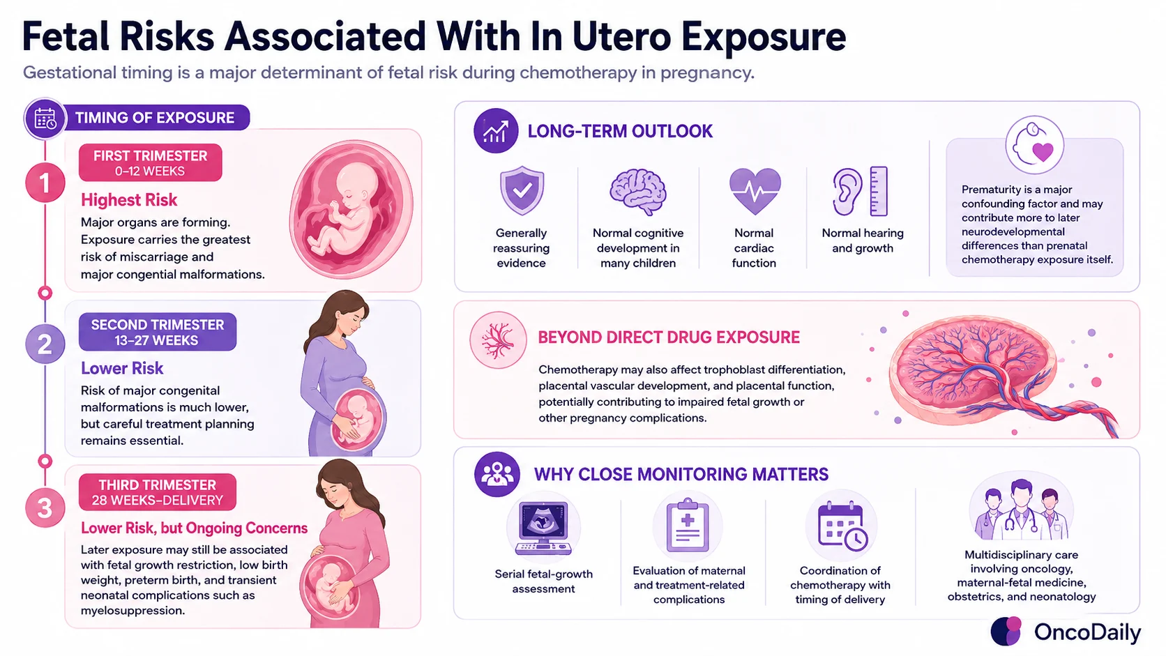
The fetal risks associated with chemotherapy depend strongly on gestational timing. Exposure during the first trimester, particularly while major organs are forming, carries the greatest risk of miscarriage and major congenital malformations. In a multicenter cohort, chemotherapy initiated before 12 weeks of gestation was associated with a substantially higher rate of major congenital malformations than chemotherapy started later in pregnancy (van Gerwen et al., 2021).
When chemotherapy is administered during the second or third trimester, the risk of major congenital malformations is considerably lower. However, treatment later in pregnancy can still be associated with fetal growth restriction, low birth weight, preterm birth, and transient neonatal complications such as myelosuppression (Nanda et al., 2025; Gulersen et al., 2026).
Available long-term evidence is generally reassuring. Studies following children exposed to maternal cancer treatment during pregnancy have reported broadly reassuring cognitive, cardiac, growth, and general developmental outcomes. Prematurity is an important confounding factor and appears to contribute more strongly to adverse neurodevelopmental outcomes than prenatal chemotherapy exposure itself (Amant et al., 2015).
Fetal risk is also not limited to direct drug exposure. Experimental evidence suggests that chemotherapy can affect trophoblast differentiation, placental vascular development, and pathways involved in placental function, potentially contributing to impaired fetal growth or other pregnancy complications (Depoix et al., 2020).
For this reason, treatment during pregnancy requires close multidisciplinary monitoring, including serial assessment of fetal growth, evaluation of maternal and treatment-related complications, and careful coordination of chemotherapy with the timing of delivery (Gulersen et al., 2026).
Clinical Implications for Cancer Treatment in Pregnancy
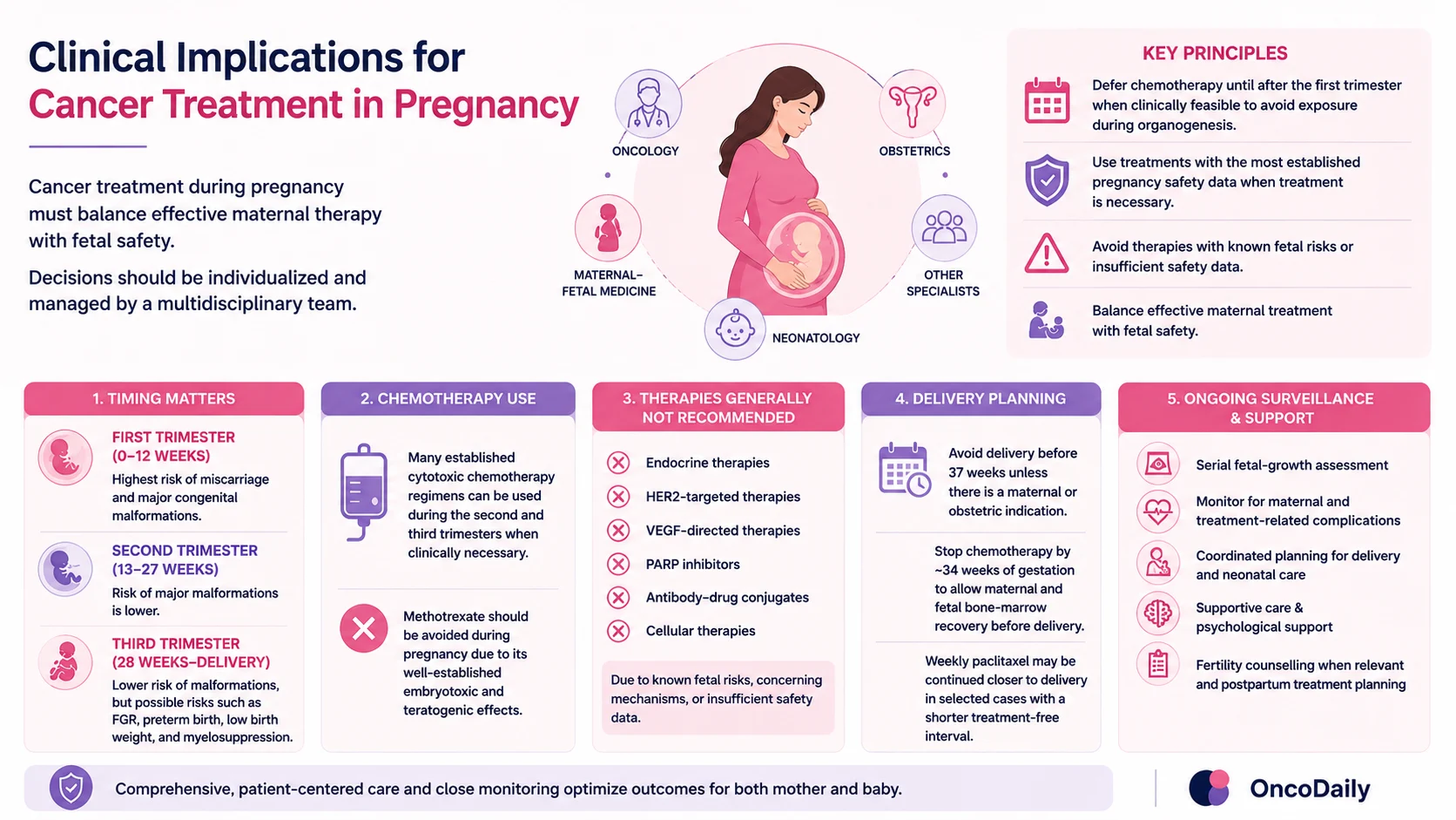
Cancer treatment during pregnancy must be individualized according to cancer type and stage, maternal prognosis, gestational age, available treatment alternatives, and the patient’s preferences. Multidisciplinary management involving oncology, maternal–fetal medicine, obstetrics, neonatology, and other relevant specialists is central to care.
Current guidance emphasizes that effective maternal cancer treatment should not be withheld solely because of pregnancy. When clinically feasible, chemotherapy is deferred until after the first trimester to avoid exposure during organogenesis (Loren et al., 2026; Gulersen et al., 2026).
Many established cytotoxic chemotherapy regimens can be administered during the second and third trimesters when treatment is necessary. However, methotrexate is contraindicated during pregnancy because of its embryotoxic and teratogenic effects.
Endocrine therapies, HER2-targeted therapies, VEGF inhibitors, PARP inhibitors, antibody–drug conjugates, and cellular therapies are also contraindicated during pregnancy because of known fetal risks, concerning mechanisms of action, or insufficient pregnancy safety data (Loren et al., 2026).
Delivery before 37 weeks should be avoided unless there is a maternal or obstetric indication. Most chemotherapy is stopped by approximately 34 weeks of gestation to allow several weeks for maternal and fetal bone-marrow recovery before delivery. Weekly paclitaxel can, in selected circumstances, be administered up to approximately 35–36 weeks because a shorter treatment-free interval can be used (Gulersen et al., 2026).
Ongoing obstetric surveillance is essential throughout treatment. This includes serial fetal-growth assessment, monitoring for maternal and treatment-related complications, and coordinated planning for delivery and neonatal care. Supportive care, psychological support, fertility counselling when relevant, and postpartum treatment planning are also important components of comprehensive management (Loren et al., 2026; Nanda et al., 2025; Gulersen et al., 2026).
Written by Marine Marachlian, MD
FAQ
Can chemotherapy cross the placenta?
Yes. Many chemotherapy drugs can cross the placenta to some extent, although the amount transferred varies between individual agents and drug classes.
When is chemotherapy most dangerous during pregnancy?
The greatest fetal risk occurs during the first trimester, when major organs are forming and the risk of miscarriage and congenital malformations is highest.
Can chemotherapy be given during the second trimester?
Yes. Several established chemotherapy regimens can be used during the second trimester when treatment is clinically necessary.
Is chemotherapy safe during the third trimester?
Some chemotherapy regimens can be used during the third trimester, but treatment must be carefully timed because of risks such as fetal growth restriction, preterm birth, and neonatal myelosuppression.
Which chemotherapy drugs have lower placental transfer?
Paclitaxel and docetaxel have shown relatively low transplacental transfer in human placental perfusion studies. Transfer, however, remains drug-specific.
Which anthracyclines are most commonly used during pregnancy?
Doxorubicin and epirubicin have some of the most established pregnancy experience and can be used in selected regimens after the first trimester.
Which cancer drugs should be avoided during pregnancy?
Methotrexate should be avoided. Endocrine therapies, HER2-targeted therapies, VEGF-directed therapies, PARP inhibitors, antibody–drug conjugates, and cellular therapies are also generally not recommended during pregnancy.
Can chemotherapy affect the placenta itself?
Yes. Chemotherapy may affect placental development, vascularization, trophoblast function, and nutrient transport in addition to directly exposing the fetus to a drug.
How are babies monitored during maternal chemotherapy?
Monitoring commonly includes serial fetal-growth assessments, surveillance for pregnancy complications, and coordinated planning for delivery and neonatal care.
What are the long-term outcomes for children exposed to chemotherapy in utero?
Available evidence is generally reassuring, with many studies reporting normal cognitive development, cardiac function, hearing, and growth. However, long-term data remain limited, and prematurity can influence developmental outcomes.