
The Tumor microenvironment is judged as much by the company it keeps as by its own mutations. Long dismissed as inert scaffolding, the tissue surrounding a tumor is now known to be an active participant in the disease — a shifting ecosystem of stromal cells, fibroblasts, immune cells, extracellular matrix, and blood vessels that can just as easily protect a tumor as attack it. This tumor microenvironment (TME) helps decide whether a cancer is eliminated outright, held in check for years, or given free rein to invade and spread (Guo et al., 2026). Nowhere has that become more consequential than in the immunotherapy era: as checkpoint inhibitors have moved into routine practice, it has become clear that the TME itself often decides who responds and who doesn’t, and understanding how tumors exploit it to dodge immune attack has become one of the most active pursuits in cancer research (Zhu et al., 2025).
Immune evasion is now considered a defining hallmark of cancer in its own right, arising not from any single trick but from a shifting alliance between tumor cells, the surrounding stroma, and the immune infiltrate meant to destroy them (Tufail et al., 2025). This review unpacks that alliance: the principal mechanisms tumors use to escape immune surveillance within the TME, how those mechanisms play out differently across cancer types, and the therapeutic strategies now being built to break them apart.
The Immunoediting Framework
Tumor–immune interactions are often described through three phases: elimination, equilibrium, and escape. In the elimination phase, innate and adaptive immunity detect and destroy transformed cells before a clinically apparent tumor forms. Cells that survive this initial surveillance enter equilibrium, a prolonged period in which the immune system restrains but does not eradicate the tumor. Over time, immunosuppressive mechanisms within the TME accumulate in magnitude, allowing the tumor to enter the escape phase and grow despite ongoing immune pressure (Labani-Motlagh et al., 2020). This framework underlies most contemporary discussion of why some tumors are immunologically “hot” — heavily infiltrated and responsive to checkpoint blockade — while others remain “cold” and resistant (Labani-Motlagh et al., 2020). This distinction now sits at the center of clinical decision-making: hot tumors, dense with cytotoxic T cells, are the ones most likely to respond to PD-1/PD-L1 or CTLA-4 blockade, whereas cold tumors, which lack meaningful T-cell infiltration, and immune-excluded tumors, where T cells are present but physically barred from the tumor core by stroma and aberrant vasculature, largely fail to respond regardless of how potent the checkpoint inhibitor is. Converting cold and excluded tumors into hot ones has therefore become one of the central therapeutic goals in contemporary immuno-oncology, motivating combination strategies that pair checkpoint blockade with radiation, oncolytic viruses, cancer vaccines, or agents that directly remodel the TME.
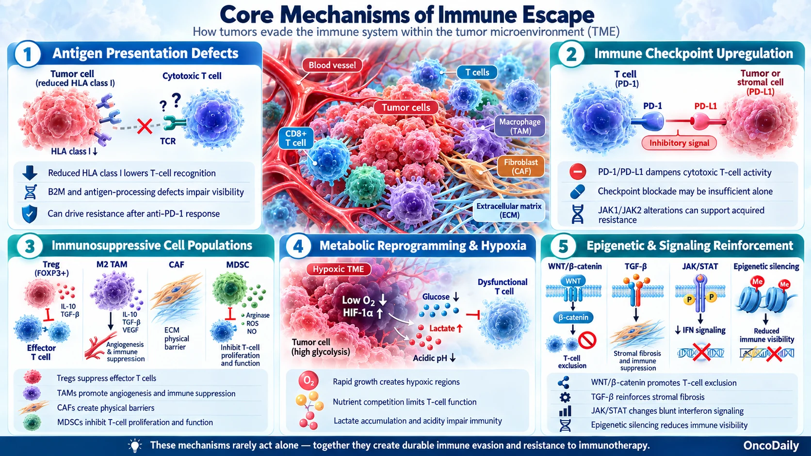
Core Mechanisms of Immune Escape
Antigen Presentation Defects
Malignant transformation is frequently accompanied by altered expression of histocompatibility antigens and other immunologically relevant surface molecules. Reduced or lost expression of HLA class I antigens impairs the ability of cytotoxic T lymphocytes to recognize tumor cells, and altered antigen presentation is identified as one of the core tactics tumors use to evade immune detection (Tufail et al., 2025; Ferrone and Whiteside, 2007). This has direct clinical consequences for checkpoint inhibitor therapy: loss of beta-2 microglobulin, a component required to transport HLA class I molecules to the cell surface, along with HLA allele loss and defects in antigen-processing machinery genes, has been documented in tumors that relapse after initially responding to anti-PD-1 therapy. Once a tumor stops presenting antigen altogether, there is nothing left for a reinvigorated T cell to recognize, which makes these defects a distinct and clinically important route to immunotherapy resistance (Ferrone and Whiteside, 2007; Sade-Feldman et al., 2017).
Immune Checkpoint Upregulation
The PD-1/PD-L1 axis is central to immune escape within the TME. When PD-L1 expressed on tumor or stromal cells engages PD-1 on activated T cells, cytotoxic T lymphocyte function is dampened, allowing malignant cells to resist immune attack (Zhu et al., 2025). This pathway, along with CTLA-4 and other checkpoint molecules, has become the dominant therapeutic target in modern immuno-oncology, though its blockade alone frequently proves insufficient because of the broader immunosuppressive context established by the TME (Tufail et al., 2025). A well-documented clinical illustration is acquired resistance to anti-PD-1 therapy in melanoma: tumors that respond initially can later relapse through loss-of-function mutations in JAK1 or JAK2, genes required for interferon-γ signaling, which blunts the very immune pressure that checkpoint blockade is meant to unleash (Zaretsky et al., 2016). Escape routes like this are a major reason single-agent checkpoint blockade is increasingly combined with other TME-targeted strategies rather than used alone.
Immunosuppressive Cell Populations
The immunosuppressive TME is not the work of a single cell type but of several distinct populations, each disabling antitumor immunity through a different mechanism. Regulatory T cells (Tregs) suppress effector T-cell activity directly, through inhibitory cytokines such as IL-10 and TGF-β and through metabolic competition for IL-2, and their accumulation in tumors is consistently associated with poorer outcomes and blunted responses to checkpoint blockade. M2-polarized tumor-associated macrophages (TAMs) reinforce this suppression from a different angle, secreting anti-inflammatory cytokines, expressing checkpoint ligands such as PD-L1, and promoting the angiogenesis and tissue remodeling that further entrench the tumor. Cancer-associated fibroblasts (CAFs) act more mechanically: they deposit dense extracellular matrix and build physical and chemokine-mediated barriers that keep cytotoxic T cells from ever reaching the tumor core, a process central to the immune-excluded phenotype (Zhang and Wu, 2025). Myeloid-derived suppressor cells (MDSCs) add a further layer, directly inhibiting T-cell proliferation and function; their expansion has been linked to systemic pro-inflammatory states associated with aging and obesity, which predate tumor formation and can prime a more permissive environment for immune evasion before a tumor even forms (Binnewies et al., 2018). Because each of these populations drives resistance through a distinct mechanism, therapies that target only one of them — checkpoint blockade alone, for instance — often leave the others free to sustain an immunosuppressive TME.
Metabolic Reprogramming and Hypoxia
Rapid tumor growth outstrips the local oxygen supply, producing hypoxic regions that are associated with poor clinical outcomes, greater tumor heterogeneity, and the emergence of treatment-resistant clones (Vito et al., 2020). Hypoxia-driven signaling has traditionally been considered tolerogenic, but some hypoxic conditions can also trigger immunogenic cell death, a nuance that complicates simple models of hypoxia as purely immunosuppressive (Vito et al., 2020). Beyond oxygen, the TME’s broader metabolic landscape further constrains the metabolic activity that effector T cells need to function, providing another route through which tumors dampen the host immune response (Tufail et al., 2025). More specifically, nutrient competition, lactate accumulation, and altered transporter expression on tumor and immune cells have been identified as concrete mediators of this metabolic suppression (Yu et al., 2025; Chen et al., 2023).
Epigenetic and Signaling Reinforcement
Beyond these cellular and metabolic mechanisms, several intracellular signaling pathways reinforce immune escape at the level of the tumor cell itself. Aberrant activation of WNT/β-catenin signaling has been linked to T-cell exclusion, in part by suppressing the chemokines tumors need to recruit dendritic cells and T cells into the tumor bed (Spranger and Gajewski, 2015). TGF-β signaling, beyond its direct suppressive effects on T cells described above, also promotes an immune-excluded phenotype by reinforcing stromal fibrosis. JAK/STAT pathway alterations, including loss-of-function mutations that blunt interferon-γ responsiveness, are recurrently implicated in acquired resistance to checkpoint inhibitors. Layered onto these signaling changes, epigenetic silencing of immune-related genes — including antigen-processing components and T-cell-recruiting chemokines — can further mute a tumor’s visibility to the immune system, and these mechanisms frequently co-occur in settings of acquired immunotherapy resistance (Zhang and Wu, 2025). Advances in single-cell and spatial transcriptomic technologies have substantially improved the field’s ability to resolve these overlapping processes at high resolution (Zhang and Wu, 2025).
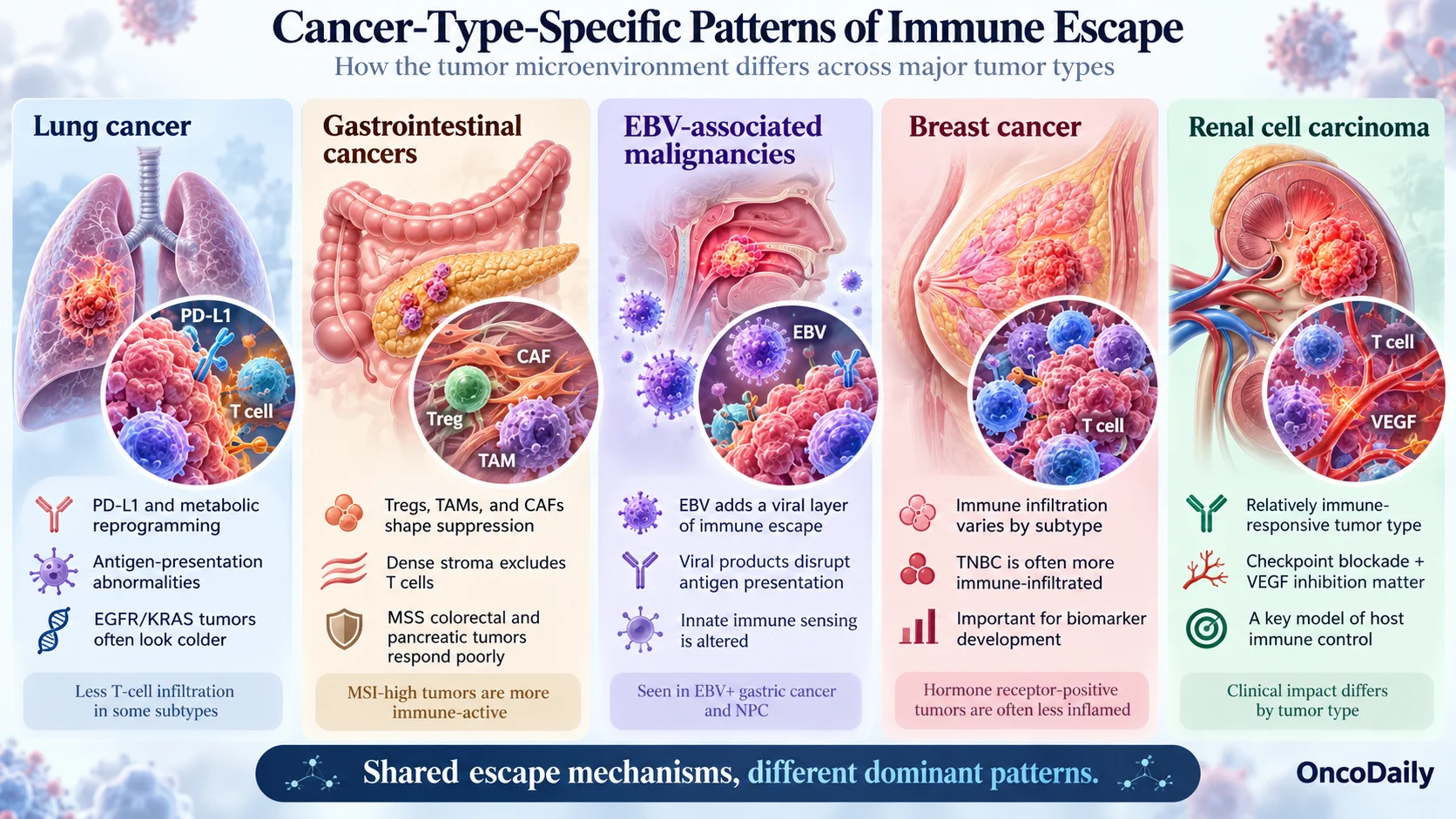
Cancer-Type-Specific Patterns
The general mechanisms above manifest differently across tumor types:
Lung cancer: Immune escape in the lung cancer TME centers on metabolic reprogramming, overexpression of checkpoint molecules such as PD-L1, and abnormalities in antigen presentation. EGFR- and KRAS-driven tumors in particular tend to present a colder, less T-cell-infiltrated phenotype than tumors with a high tumor mutational burden, and this heterogeneity continues to explain why a substantial fraction of patients treated with PD-1/PD-L1 inhibitors either fail to respond or develop resistance (Wang et al., 2025).
Gastrointestinal cancers: GI tumors show a particularly complex immunosuppressive architecture, with Tregs, TAMs, and CAFs cooperating with mechanical stromal stress to exclude T cells. This exclusion is most pronounced in microsatellite-stable colorectal and pancreatic tumors, a pattern that helps explain why these subtypes respond far more poorly to checkpoint inhibition than their microsatellite-instable, T-cell-rich counterparts (Zhang and Wu, 2025).
EBV-associated malignancies: Epstein–Barr virus-driven cancers, including EBV-positive gastric carcinoma and nasopharyngeal carcinoma, add a virological layer to immune escape, in which EBV-encoded gene products and microRNAs actively interfere with antigen presentation and innate immune sensing while reshaping the TME (Bauer et al., 2021).
Breast cancer: The interplay between tumor cells and tumor-infiltrating lymphocytes and TAMs is central to breast cancer progression and metastasis. Triple-negative breast cancer characteristically shows higher levels of immune infiltration than hormone receptor-positive subtypes, and this heterogeneity has become a focus for biomarker development and precision immunotherapy (Kotsifaki et al., 2023).
Renal cell carcinoma: RCC’s relatively strong response to immune-activating therapies, including combination checkpoint blockade and VEGF-pathway inhibition, has made its immune microenvironment a particular focus, underscoring how host immune control can vary sharply by tumor type even when general escape mechanisms are conserved (Shapiro et al., 2023).
Therapeutic Implications
Recognition that immune escape is TME-driven rather than purely tumor-intrinsic has reshaped therapeutic strategy. Current approaches can be grouped by what, specifically, they are trying to change within the TME.
Restoring antigen visibility. Where tumors have downregulated antigen presentation, strategies aim either to restore it directly, for instance through epigenetic modulators that re-express silenced antigen-processing genes, or to bypass classical antigen presentation altogether, as engineered cell therapies such as CAR-T do.
Reversing checkpoint-driven suppression. Beyond single-agent PD-1/PD-L1 or CTLA-4 blockade, combination checkpoint strategies and next-generation targets such as LAG-3 aim to overcome the redundancy of inhibitory pathways that lets tumors escape single-target blockade.
Remodeling the suppressive cell compartment. Agents that deplete or reprogram Tregs, repolarize TAMs from an M2 toward an M1 phenotype, or block MDSC recruitment are designed to dismantle the cellular apparatus of suppression directly, rather than simply overpowering it with more T-cell activation.
Breaking down physical exclusion. Because CAFs and dense stroma physically block T-cell infiltration, strategies targeting stromal signaling, including TGF-β inhibition, and combinations with radiation or oncolytic viruses aim to convert immune-excluded and cold tumors into hot ones.
Correcting the metabolic environment. Approaches targeting hypoxia, lactate metabolism, and nutrient competition seek to restore the metabolic conditions effector T cells need to function within the tumor itself.
Systemic and adjunctive approaches. mRNA cancer vaccines and interventions targeting the gut microbiota are being investigated as ways to broaden and sustain antitumor immune responses alongside these more targeted strategies, all aimed at reversing resistance and remodeling an immunosuppressive TME into one more conducive to effective antitumor immunity (Zhang and Wu, 2025).
Together, these approaches reflect a shift from targeting the tumor cell alone toward remodeling the TME as an integrated system. Artificial intelligence-assisted analysis is also being integrated into TME characterization, supporting automated pathology review, spatial profiling of immune cell distribution, biomarker discovery, and prediction of which patients are likely to respond to a given immunotherapy, and is increasingly used to help stratify patients for more personalized treatment selection (Racacho et al., 2025).
Conclusion
The tumor microenvironment is now central to understanding both why cancers escape immune control and why immunotherapies succeed in some patients and fail in others. Antigen presentation defects, checkpoint upregulation, immunosuppressive cell populations, metabolic and hypoxic stress, and epigenetic reprogramming all converge within the TME to produce durable immune evasion — and, critically, they rarely act alone, which is why single-mechanism therapies so often meet resistance. The clinical trajectory of the field is now toward combination strategies purpose-built to dismantle several of these mechanisms at once: pairing checkpoint blockade with agents that restore antigen presentation, deplete or reprogram suppressive myeloid and stromal populations, or correct the metabolic conditions T cells need to function. As single-cell, spatial, and AI-assisted profiling tools mature, they are moving from research instruments toward practical tools for choosing which combination is likely to work in a given patient’s tumor — the shift from treating cancer as a single disease to treating each tumor microenvironment as its own therapeutic target is likely to define the next phase of cancer immunotherapy.
You can also read Immuno 2026 Day 7 Highlights: Global Access to Immunotherapy and Education by OncoDaily.
Written By Manushak Ghukasyan
FAQ
What is the tumor microenvironment?
The tumor microenvironment (TME) is the ecosystem surrounding cancer cells, including immune cells, fibroblasts, blood vessels, extracellular matrix, and signaling molecules. These components can influence tumor growth, spread, and response to treatment.
How do tumors escape the immune system?
Tumors can evade immune attack through several mechanisms, including loss of antigen presentation, activation of immune checkpoints such as PD-1/PD-L1, recruitment of immunosuppressive cells, metabolic changes, hypoxia, and signaling pathways that prevent immune cells from entering the tumor.
What is the difference between “hot” and “cold” tumors?
Hot tumors contain substantial immune-cell infiltration, particularly cytotoxic T cells, and are generally more likely to respond to checkpoint inhibitors. Cold tumors have little immune infiltration, while immune-excluded tumors contain immune cells that are unable to penetrate the tumor core.
Why do some cancers become resistant to immunotherapy?
Resistance can develop when tumors alter or lose antigen presentation, activate alternative immune-suppressive pathways, develop mutations affecting interferon signaling, or create a microenvironment that physically or metabolically prevents T cells from functioning effectively.
Can targeting the tumor microenvironment improve cancer treatment?
Potentially, yes. Current research is exploring combinations that target immune checkpoints, suppressive immune cells, cancer-associated fibroblasts, hypoxia, metabolism, and other components of the TME to make tumors more responsive to immunotherapy.