
Small-cell lung cancer (SCLC) has remained one of the most difficult settings for durable immunotherapy. First-line platinum–etoposide combined with PD-L1 blockade can produce rapid tumor regression, but disease control is frequently short-lived, and median overall survival in extensive-stage SCLC (ES-SCLC) has remained approximately 12–13 months. Many patients also deteriorate before receiving second-line treatment, making earlier and more durable immune control an important therapeutic objective.
Tarlatamab approaches this problem differently from conventional immune checkpoint inhibition. It is a bispecific T-cell engager that simultaneously binds delta-like ligand 3 (DLL3) on SCLC cells and CD3 on T cells, physically redirecting T-cell cytotoxicity toward DLL3-expressing cancer cells. Instead of relying primarily on an endogenous antitumor response that can be reinvigorated by checkpoint blockade, tarlatamab provides T cells with a pharmacologic connection to the tumor.
The clinical importance of this strategy is becoming increasingly clear. Tarlatamab has already demonstrated superiority over chemotherapy after platinum-based treatment, and the phase III DeLLphi-305 trial now shows that introducing DLL3-directed T-cell engagement during first-line maintenance can substantially extend disease control and survival. At the same time, a new real-world report describing tumor inflammation-associated neurotoxicity (TIAN) in tarlatamab-treated patients with brain metastases raises an intriguing mechanistic question: could the same redirected immune activity responsible for attacking SCLC also produce localized inflammation around intracranial tumor deposits?
Together, these findings provide a useful framework for understanding tarlatamab not simply as another active drug in SCLC, but as a therapy that changes how T cells encounter the tumor.
Tarlatamab (Imdelltra): Uses in Cancer, Side Effects, Dosages, Expectations, and More
From DLL3 Recognition to T-Cell Killing
The central mechanism of tarlatamab is conceptually straightforward: one molecule connects the tumor cell to the T cell.
One binding arm recognizes DLL3 on the cancer cell, while the other engages CD3 on the T cell. This creates close physical contact between the two cells and redirects T-cell activity toward the DLL3-expressing target. The result is activation of T-cell cytotoxicity against the tumor cell rather than dependence solely on naturally occurring T-cell recognition of tumor antigens.
This distinction is important when considering why tarlatamab may be particularly relevant in SCLC. Checkpoint inhibitors such as durvalumab remove inhibitory signaling through the PD-1/PD-L1 axis, but they do not themselves specify which tumor cell a T cell should recognize. Tarlatamab adds that missing physical connection by using DLL3 as the tumor-facing target and CD3 as the T-cell-facing target.
The therapeutic concept can therefore be viewed as a sequence:
DLL3-positive SCLC cell → tarlatamab-mediated DLL3/CD3 bridging → T-cell engagement → redirected cytotoxicity → tumor-cell killing and inflammatory immune activation.
This same sequence also helps explain why the biology of tarlatamab cannot be separated entirely from its characteristic toxicities. Activating T cells against cancer can generate substantial inflammatory signaling, and where that activation occurs may influence how the toxicity presents clinically.
Why Add Tarlatamab After Chemoimmunotherapy?
DeLLphi-305 examined tarlatamab at a particularly interesting point in the SCLC treatment course. Patients had already completed four cycles of platinum–etoposide, with at least three administered concurrently with durvalumab, and were eligible only if their disease had not progressed. Approximately 89% had achieved a complete or partial response to induction chemoimmunotherapy before maintenance randomization.
The trial therefore did not ask whether tarlatamab could rescue immediately refractory disease. Instead, it tested whether DLL3-directed T-cell engagement could improve control of residual SCLC after initial cytoreduction.
This setting is mechanistically attractive because durvalumab and tarlatamab intervene at different points in the immune response. Durvalumab blocks PD-L1-mediated inhibition, whereas tarlatamab directly redirects T cells toward DLL3-expressing cancer cells. Early activity observed with tarlatamab plus a PD-L1 inhibitor in DeLLphi-303 provided the clinical rationale for evaluating this approach in phase III.
However, DeLLphi-305 should not be interpreted as proof of biological synergy between the two agents. Because the randomized comparison was tarlatamab plus durvalumab versus durvalumab alone, the trial establishes the benefit of adding tarlatamab to maintenance durvalumab but cannot determine whether PD-L1 blockade specifically potentiates DLL3/CD3 engagement.
DeLLphi-305: A Large Clinical Effect From Adding T-Cell Engagement
DeLLphi-305 randomized 563 patients with ES-SCLC to tarlatamab plus durvalumab or durvalumab alone after completion of initial durvalumab-based chemoimmunotherapy. Importantly, patients with treated or untreated asymptomatic brain metastases were eligible, allowing the study to include a clinically relevant population often associated with poor outcomes.
At the prespecified interim analysis, the addition of tarlatamab produced a substantial survival advantage. Median OS from maintenance randomization was 21.1 months with tarlatamab–durvalumab versus 12.7 months with durvalumab, corresponding to a hazard ratio for death of 0.42. Twelve-month survival was 79.4% versus 53.3%, respectively.
The effect on disease control was similarly pronounced. Median PFS increased from 1.9 months with durvalumab alone to 7.4 months with tarlatamab–durvalumab, with a hazard ratio for progression or death of 0.34. At 12 months, 38.4% of patients receiving the combination remained progression-free compared with 9.5% receiving durvalumab alone.
Among patients with measurable disease at maintenance randomization, confirmed objective responses occurred in 26.4% versus 4.1%, and median response duration was 12.7 versus 5.6 months. The ability to generate additional tumor regression after induction therapy is particularly notable because these patients had already been exposed to chemotherapy and PD-L1 blockade.
These results establish the clinical value of adding tarlatamab during first-line maintenance. What they do not establish is precisely why the effect is so large. The study did not directly determine whether preceding chemotherapy altered DLL3-directed immunity, whether continued PD-L1 blockade enhanced tarlatamab activity, or whether the benefit predominantly reflected direct DLL3/CD3-mediated T-cell redirection.
DeLLphi-305: Tarlatamab–Durvalumab Redefines First-Line Maintenance in Extensive-Stage SCLC
CRS Shows the Systemic Consequence of T-Cell Engagement
The mechanism responsible for tarlatamab’s antitumor activity also creates a characteristic inflammatory toxicity. When large numbers of T cells are activated through a bispecific engager, inflammatory mediators can be released rapidly, producing cytokine release syndrome (CRS).
In DeLLphi-305, CRS occurred in 49.6% of patients receiving tarlatamab–durvalumab. Importantly, it was predominantly low grade: 41.8% of patients experienced grade 1 CRS and 7.4% grade 2, while only one grade 3 event occurred and there were no grade 4 or fatal events. CRS developed primarily after the first two tarlatamab doses, with a median onset of 22.5 hours after the most recent administration.
Most episodes resolved with supportive treatment, although glucocorticoids were used in 21.6% of patients and tocilizumab in 3.9%. This concentration of CRS around the initial exposures helps explain the step-up dosing strategy used with tarlatamab, beginning with 1 mg before escalation to 10 mg.
CRS can therefore be understood as one systemic manifestation of the inflammatory biology created by redirected T-cell activation. The emerging neurological observations raise the possibility that inflammation may behave differently when immune engagement occurs around tumor deposits within the CNS.
What Happens When the Target Is Inside the Brain?
Brain metastases are common and clinically important in SCLC. DeLLphi-305 included patients with asymptomatic treated or untreated brain metastases, and approximately 20% of the trial population had previous or current brain metastases when maintenance treatment began. The survival and PFS benefit appeared to extend across prespecified subgroups, including patients with brain metastases, although these subgroup analyses were not independently powered.
This activity is consistent with previously reported intracranial activity of tarlatamab in treated SCLC. But intracranial T-cell engagement raises a second question beyond efficacy: could rapid immune attack against a brain metastasis itself generate neurological symptoms through local tumor inflammation?
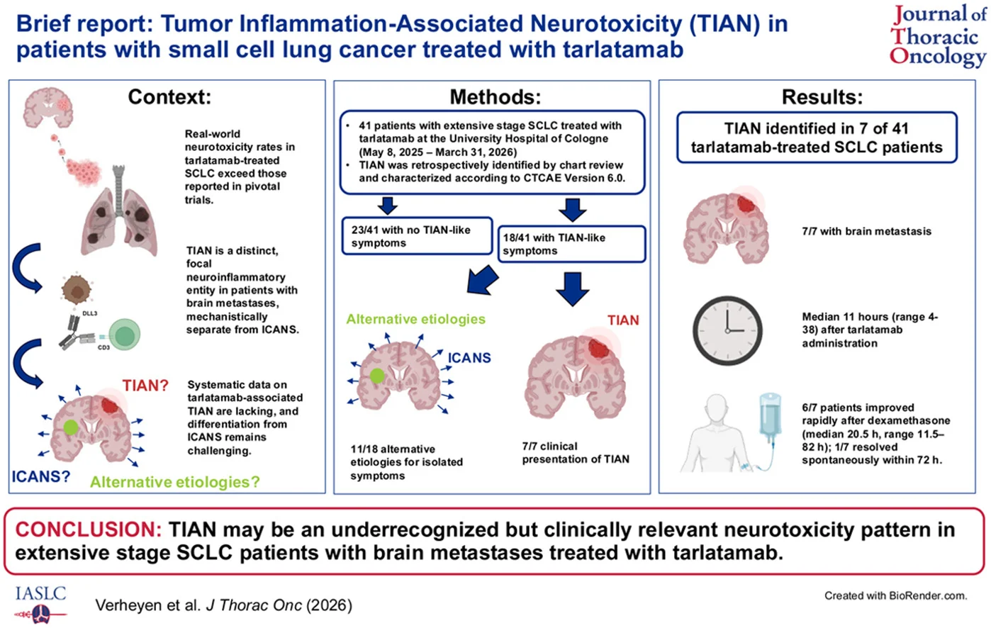
Verheyen and colleagues explored this possibility in a 2026 real-world case series describing tumor inflammation-associated neurotoxicity, or TIAN, among patients receiving tarlatamab. TIAN had previously been described in CAR T-cell therapy and is proposed to represent a localized inflammatory process around CNS tumor sites rather than the predominantly diffuse encephalopathy associated with classical ICANS.
TIAN: A Localized Consequence of Redirected Immunity?
The investigators retrospectively evaluated 41 patients with ES-SCLC treated with tarlatamab at the University Hospital of Cologne. Patients underwent inpatient step-up dosing with structured monitoring, including repeated vital signs, ICE scoring and neurological assessment when clinically indicated.
Seven patients—17% of the cohort—were retrospectively classified as having neurological deterioration consistent with TIAN. A striking common feature was that all seven had brain metastases before tarlatamab initiation. Symptoms emerged rapidly, at a median of 11 hours after administration, with presentations including severe headache, dizziness, confusion, somnolence, paresthesia, psychomotor slowing and worsening of pre-existing focal neurological deficits.
Two patients had imaging findings consistent with pseudoprogression or increased edema around known brain metastases. Six of seven improved following dexamethasone, with a median time to improvement of 20.5 hours, while the remaining patient recovered spontaneously within 72 hours.
The authors interpret this pattern as potentially consistent with rapid inflammation around intracranial tumor deposits following T-cell engagement. In this model, tarlatamab-mediated recognition of DLL3-positive tumor cells could trigger local immune activation, producing edema and focal neurological dysfunction within an anatomically vulnerable compartment.
This interpretation is biologically compelling but remains hypothesis-generating. The study was retrospective, included only 41 patients, lacked systematic neuroimaging at symptom onset in most cases, and could not definitively exclude tumor progression in every patient. The reported 17% frequency should therefore not be interpreted as an established incidence of tarlatamab-associated TIAN.
Why TIAN May Not Be the Same as ICANS
Distinguishing TIAN from immune effector cell-associated neurotoxicity syndrome (ICANS) may have important clinical implications.
In DeLLphi-305, formally reported ICANS occurred in only 1.1% of patients receiving tarlatamab–durvalumab. One event was grade 2, and there were no grade ≥3, serious or fatal ICANS events. A broader analysis capturing potentially associated neurological events identified events in 9.6% of patients receiving tarlatamab–durvalumab and 6.8% receiving durvalumab alone.
The phenotype described in the Cologne series differed from classical diffuse encephalopathy. Symptoms were frequently focal or anatomically related to pre-existing CNS lesions, and two patients demonstrated imaging changes compatible with local inflammation. The investigators also observed very rapid onset, usually within approximately 12 hours of treatment, and relatively rapid improvement after dexamethasone.
Another important difference involved the ICE score. Patients with clinically relevant focal symptoms could retain normal or only mildly abnormal ICE scores because the instrument is primarily designed to detect diffuse encephalopathy rather than localized neurological deficits. The authors therefore propose that patients with pre-existing CNS disease may require careful baseline neurological assessment and repeated examination in addition to conventional ICE monitoring.
These observations do not yet establish TIAN as a formally validated tarlatamab toxicity syndrome. TIAN and ICANS can overlap clinically, and one patient in the series had features potentially consistent with an overlap syndrome. Prospective characterization will be necessary before diagnostic or management algorithms can be clearly separated.
Could TIAN Tell Us Something About Tarlatamab’s Antitumor Activity?
The relationship between tumor inflammation and neurological toxicity creates an intriguing translational question. If TIAN reflects inflammation generated around DLL3-expressing brain metastases, it could provide indirect evidence that redirected T cells are reaching and interacting with intracranial tumor deposits.
Some observations from the case series are compatible with that possibility. Several patients subsequently demonstrated intracranial disease control, including partial responses and stable disease. However, follow-up was incomplete, some patients could not be evaluated, and the cohort was far too small to determine whether neurological inflammation correlated with intracranial efficacy.
It would therefore be premature to interpret TIAN as evidence of therapeutic response or as a biomarker of effective T-cell engagement. A patient does not need to develop TIAN, CRS or another inflammatory toxicity for tarlatamab to work, and neither of the two studies establishes a relationship between toxicity intensity and clinical benefit.
The more interesting hypothesis is broader: the anatomical location of tumor-directed immune activation may influence how the pharmacology of a T-cell engager becomes clinically visible. Systemic activation can manifest as CRS, whereas inflammation occurring around a CNS tumor could theoretically produce focal neurological symptoms because even modest edema can have significant consequences within the brain. This concept now requires prospective biological and radiographic validation.
From Second-Line Therapy to Earlier Immune Intervention
The evolution of tarlatamab in SCLC has been rapid. Its clinical activity was first established after platinum-based therapy, and DeLLphi-305 now demonstrates that DLL3-directed T-cell engagement can produce substantial benefit when introduced during first-line maintenance. The next step is to determine whether this mechanism should be deployed even earlier.
The ongoing phase III DeLLphi-312 trial is evaluating tarlatamab with durvalumab, carboplatin and etoposide in untreated ES-SCLC. Additional studies, including DeLLphi-306 and PrE0512, are evaluating tarlatamab after chemoradiotherapy in limited-stage disease.
These studies will address an important mechanistic and therapeutic question: should T-cell redirection be introduced after chemotherapy has reduced tumor burden, or can it be integrated from the beginning of treatment to generate deeper and more durable disease control?
Earlier treatment may also change the toxicity landscape. Larger tumor burden, different patterns of CNS involvement and simultaneous chemotherapy or checkpoint blockade could influence the magnitude and location of T-cell activation. As tarlatamab moves earlier, understanding not only whether it works but where and how the redirected immune response occurs will become increasingly important.