
Combination immune checkpoint blockade has transformed the treatment of advanced melanoma, but its clinical success comes with a persistent challenge: the immune activation responsible for tumor regression can also cause serious, sometimes life-threatening complications. Combining PD-1 and CTLA-4 inhibition produces broader immune activation than either approach alone, increasing the potential for durable antitumor responses while also raising the risk of immune-related adverse events (irAEs).
For years, the management of these toxicities has largely depended on recognizing inflammation after it develops and controlling it with corticosteroids or other immunosuppressive treatments. Although this approach remains essential, it does not address an increasingly important question in immuno-oncology: could the immune system be modified before treatment to prevent severe toxicity while preserving the mechanisms responsible for cancer control?
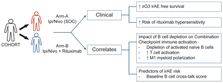
A randomized study published in the Journal of Clinical Investigation in August 2026 by Kavita M. Dhodapkar and colleagues offers an intriguing perspective. By adding rituximab, a B-cell-depleting antibody, to nivolumab and ipilimumab in patients with advanced melanoma, investigators observed fewer severe immune-related adverse events without an apparent reduction in tumor response. Detailed immune profiling further revealed that B-cell depletion was associated with changes in T-cell and myeloid-cell activation, suggesting that immunotherapy toxicity may be influenced by immune interactions extending beyond T cells.
However, the study also revealed an important complication: severe rituximab-associated hypersensitivity reactions occurred frequently enough to require premature trial closure. Its findings therefore represent a promising biological proof of concept rather than an immediately applicable preventive treatment.
Why Are B Cells Important in Checkpoint Inhibitor Toxicity?
PD-1 and CTLA-4 are inhibitory receptors that regulate T-cell activation through different mechanisms. CTLA-4 competes with CD28 for binding to CD80 and CD86, limiting costimulatory signals involved in T-cell activation, while PD-1 suppresses signaling in activated T cells, particularly during persistent antigen exposure. Blocking both pathways can enhance antitumor immunity, but it may also disturb the mechanisms that maintain immune tolerance.
Although T cells are central to checkpoint inhibitor activity, immune-related toxicity involves a wider network of immune cells, including B cells, myeloid cells, and tissue-resident immune populations.
B cells are best known for producing antibodies, but their immunological functions extend considerably further. They present antigens to T cells, express costimulatory molecules, release cytokines, and coordinate interactions between different immune populations. These activities can support effective antitumor responses, particularly within organized immune structures such as tertiary lymphoid structures, but they may also contribute to autoimmune inflammation.
Previous research has identified changes in circulating B-cell populations during combination checkpoint blockade that can precede the development of clinical irAEs. This observation suggests that B cells may participate in the early stages of immune dysregulation, before tissue inflammation becomes clinically apparent.
The biological rationale for the study was therefore not simply to suppress B cells, but to investigate whether removing particular B-cell populations could disrupt the immune interactions associated with severe toxicity.
Rituximab targets CD20, a surface protein expressed on most mature B cells, and induces B-cell depletion through immune-mediated mechanisms. Because B-cell depletion can produce sustained clinical benefits in several autoimmune diseases, the investigators hypothesized that a short course of rituximab might reduce the development of checkpoint inhibitor-associated autoimmune complications.
Yet this strategy involved an important biological uncertainty. B cells can also contribute positively to antitumor immunity, meaning that their depletion could theoretically interfere with the effectiveness of checkpoint blockade. Determining whether toxicity could be reduced without compromising cancer control was therefore central to the investigation.
A Randomized Trial Testing Preventive B-Cell Depletion
The prospective randomized trial (NCT03719131) enrolled patients with unresectable advanced melanoma who were eligible for combination checkpoint blockade. Patients were assigned to receive standard nivolumab plus ipilimumab or the same regimen with an additional course of rituximab.
Checkpoint blockade consisted of nivolumab 1 mg/kg and ipilimumab 3 mg/kg every three weeks for four doses, followed by nivolumab maintenance. In the experimental arm, rituximab was administered as a four-week course around treatment initiation, with the schedule subsequently modified in response to hypersensitivity reactions.
The primary endpoint was the incidence of grade 3 or higher irAEs. Investigators also evaluated tumor responses, survival outcomes, and treatment-associated changes in circulating immune populations.
The trial was initially designed to enroll 22 patients per arm, but recruitment was stopped early because of unexpectedly frequent rituximab hypersensitivity reactions. The reported clinical comparison included seven patients in each treatment group, making the results preliminary despite the randomized design.
Fewer Severe irAEs, but a New Safety Concern
The principal clinical finding was a substantial difference in severe immune-related toxicity between the treatment groups. Grade 3 or higher immune-related adverse events occurred in 57% of patients (4/7) receiving nivolumab plus ipilimumab alone, compared with 14% (1/7) among those receiving additional rituximab.
At two years, estimated grade 3 or higher irAE-free survival was 86% in the rituximab group versus 29% in the control group (p=0.01). The reduction was not restricted to a particular organ system, suggesting that B-cell depletion may influence broader mechanisms of immune dysregulation.
Tumor response was not obviously compromised. Objective response rates were 43% in the rituximab group and 29% in the control group, while overall survival appeared similar. However, the study was not powered to establish equivalent antitumor efficacy, and these small numerical differences cannot demonstrate improved cancer outcomes.
The safety findings also require careful interpretation. Three of seven patients receiving rituximab experienced grade 3 hypersensitivity reactions, corresponding to 43% of the experimental group. Although these reactions were reported as reversible, their frequency led to premature trial closure.
This distinction is crucial: reducing severe irAEs does not necessarily improve overall treatment safety if the preventive intervention introduces serious complications of its own.
The study therefore suggests that B-cell depletion may reduce checkpoint inhibitor-associated toxicity, but it does not establish rituximab as a safe or clinically validated preventive strategy.
Activated Naive B Cells: A Possible Link Between Immune Activation and Autoimmunity
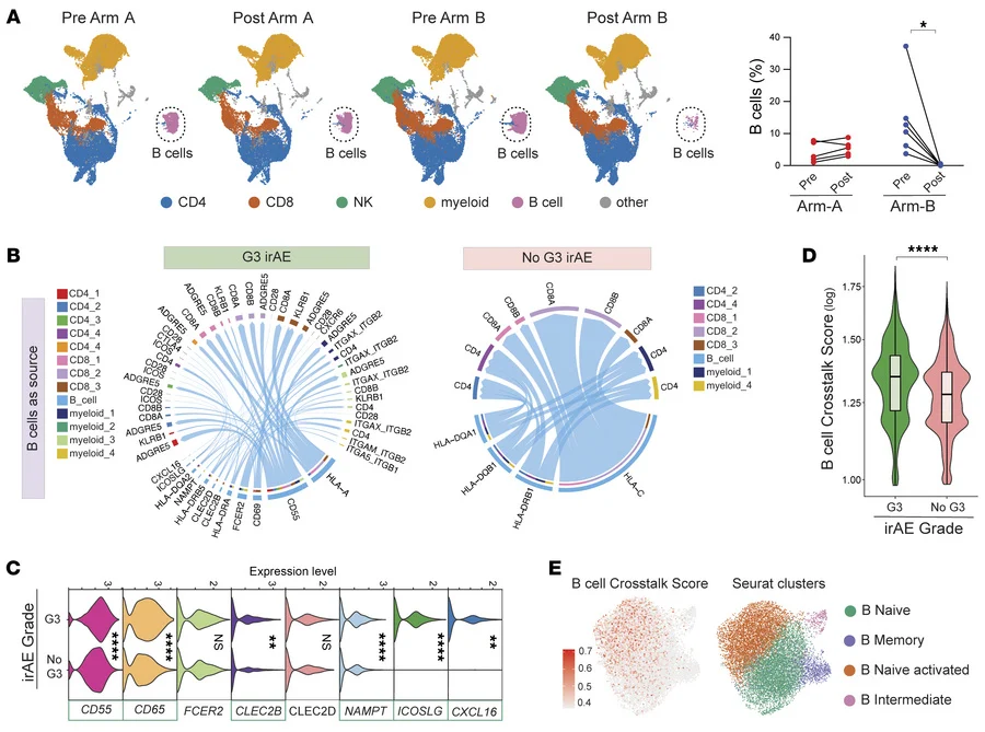
To understand why B-cell depletion might reduce irAEs, the investigators examined blood samples collected before treatment and approximately four weeks after checkpoint blockade initiation. Using mass cytometry and single-cell transcriptomics, they investigated how rituximab affected circulating B cells and their interactions with other immune populations.
As expected, rituximab substantially depleted circulating B cells, particularly naive B cells. However, the most informative observation came from examining the immune characteristics of patients who developed severe toxicity.
Computational analysis of cell-to-cell communication identified a distinct pattern of B-cell signaling associated with grade 3 or higher irAEs. Six genes—CXCL16, ICOSLG, NAMPT, CLEC2B, CD69, and CD55—were incorporated into a B-cell crosstalk signature that correlated with severe toxicity risk before treatment.
This signature was particularly enriched in activated naive B cells, a population with similarities to B cells previously implicated in autoimmune diseases.
The finding is biologically important because it suggests that susceptibility to immunotherapy toxicity may be partly determined by immune interactions already present before the first dose of checkpoint blockade. Rather than developing entirely as a consequence of treatment, severe irAEs may emerge from pre-existing immune networks that become amplified when inhibitory checkpoints are removed.
The investigators also observed an association between the B-cell crosstalk signature and irAE risk in an independent melanoma dataset, providing additional support for the biological relevance of these interactions.
However, these analyses do not establish that the six-gene signature can predict toxicity reliably in individual patients. They also cannot determine whether activated naive B cells directly cause irAEs or serve as markers of a broader immune state associated with autoimmunity.
Nevertheless, the findings introduce a potentially valuable direction for future research: identifying patients at increased risk of severe toxicity through pretreatment immune profiling.
Why Did B-Cell Depletion Not Suppress T-Cell Activation?
One of the most unexpected findings was that rituximab-mediated B-cell depletion did not simply suppress the immune activation induced by nivolumab and ipilimumab.
Instead, the investigators observed increased representation of several activated T-cell populations in the rituximab group.
Mass cytometry identified expansion of proliferating memory CD4+ T cells expressing HLA-DR, CD38, and ICOS, particularly following treatment with rituximab. Single-cell transcriptomics also demonstrated increased CXCL13+ICOS+ CD4+ T cells and cytolytic effector CD8+ T-cell populations.
These observations are especially interesting because CXCL13-expressing T cells have been associated with antitumor immune responses in several cancer settings. CXCL13 is a chemokine involved in immune-cell recruitment and organization, while ICOS provides costimulatory signals that support activated T-cell responses.
The enrichment of these populations suggests that B-cell depletion may alter the organization of checkpoint blockade-induced immunity without eliminating important T-cell activation programs.
This finding challenges the assumption that reducing autoimmune toxicity necessarily requires broad immunosuppression. Instead, it raises the possibility that specific immune-cell interactions contribute disproportionately to toxicity, while other pathways remain capable of supporting antitumor responses.
However, increased circulating T-cell activation does not prove enhanced tumor-specific immunity. The study did not establish whether these T-cell populations directly mediated tumor regression or whether similar changes occurred within the tumor microenvironment.
The Myeloid Compartment Revealed Another Unexpected Effect
B-cell depletion also influenced circulating myeloid cells. Compared with patients receiving nivolumab and ipilimumab alone, those receiving rituximab demonstrated increased expression of inflammatory signaling programs in monocytes, including genes associated with NF-κB activation.
The investigators also observed enrichment of M1-like macrophage-associated transcriptional signatures, suggesting a shift toward inflammatory myeloid activation.
At first glance, this appears paradoxical. If rituximab reduced severe immune-related toxicity, why did some inflammatory pathways become more active?
The findings suggest that the relationship between immune activation and autoimmune injury may depend on more than the overall intensity of inflammation. Different immune-cell populations, signaling pathways, and cellular interactions may contribute differently to tumor control and tissue damage.
B-cell depletion may therefore reshape the immune response rather than uniformly suppress it.
However, the study cannot establish whether increased myeloid inflammation contributed to preserved antitumor activity or reduced toxicity. The immune profiling was exploratory, and the observed transcriptional changes should not be interpreted as proof of a protective mechanism.
Could Temporary B-Cell Depletion Produce Lasting Immune Changes?
Another intriguing observation was that the reduction in severe irAE risk appeared to persist even though rituximab-mediated B-cell depletion was temporary. Circulating B-cell populations returned toward baseline by approximately 12 months, yet the difference in severe irAE-free survival remained evident during follow-up.
The investigators proposed that early B-cell depletion might interrupt immune interactions involved in the initiation of autoimmune toxicity, potentially producing effects that extend beyond the period of active B-cell depletion.
This concept resembles the immune-reset phenomenon described in certain autoimmune diseases, where B-cell-targeted treatment can lead to prolonged clinical improvement despite eventual immune-cell recovery.
In the context of checkpoint blockade, the timing of this intervention may be particularly important. Early changes in immune-cell activation can precede the clinical appearance of irAEs, suggesting that preventive immune modulation might be most effective before pathogenic inflammatory responses become established.
Nevertheless, the study did not directly demonstrate an immune reset, and the durability of the observed effect requires confirmation. The findings support a hypothesis that should be investigated rather than an established mechanism of rituximab action.
What Does This Mean for Clinical Practice?
The clinical implications of this study are promising but limited.
The randomized design provides an important foundation for investigating preventive B-cell depletion, while the accompanying immune analyses offer biological explanations for the observed reduction in severe irAEs. However, the trial was stopped early, involved only seven patients per treatment arm, and identified a substantial risk of rituximab-associated hypersensitivity.
Consequently, the results do not support routine administration of rituximab alongside nivolumab and ipilimumab to prevent immune-related toxicity.
The study also raises questions that future trials will need to address. It remains unclear whether B-cell depletion can reduce severe toxicity in larger and more diverse populations, whether alternative agents or treatment schedules could avoid hypersensitivity reactions, and whether particular B-cell populations can be targeted without disrupting beneficial antitumor immunity.
Another important consideration is the role of B cells within tumors. In several cancer types, intratumoral B cells and tertiary lymphoid structures have been associated with favorable responses to immunotherapy. Therefore, circulating B-cell depletion cannot automatically be assumed to have the same consequences across different tissues or tumor microenvironments.
Future studies will need to evaluate not only the frequency of irAEs but also infection risk, tumor response, survival, and the long-term consequences of modifying B-cell populations.