
Immune checkpoint inhibitors have transformed the treatment of mismatch repair-deficient/microsatellite instability-high (dMMR/MSI) colorectal cancer, producing durable responses in many patients. However, not all patients with dMMR/MSI tumors benefit from immunotherapy, while the majority of patients with mismatch repair-proficient/microsatellite-stable (pMMR/MSS) colorectal cancer remain resistant to conventional checkpoint blockade.
This difference has traditionally been explained by tumor-intrinsic characteristics, including mutation burden, neoantigen availability, and DNA mismatch repair status. Yet these factors do not fully explain why some immunogenic tumors fail to respond or why a subset of MSS tumors demonstrates evidence of immune activation.
Increasing attention is therefore being directed toward the tumor microenvironment, particularly the stromal cells that surround cancer cells and influence immune-cell infiltration, tumor progression, and treatment response.
A study published in Gut, titled Stromal Biomarker-Based Framework for Identifying pMMR/MSS and dMMR/MSI Colorectal Cancers With Poor Outcomes and Limited Benefit From Immunotherapy, by Mar Iglesias Coma, Jordi Badia-Ramentol, Carolina Martinez-Ciarpaglini, and colleagues, investigated whether specific cancer-associated fibroblast populations could improve prognostic assessment and identify tumors less likely to benefit from immune checkpoint inhibitors.
Through integrated molecular profiling, histopathological analyses, clinical cohorts, and experimental models, the investigators identified CTHRC1-positive cancer-associated fibroblasts (CTHRC1+ CAFs) as a stromal population associated with transforming growth factor-beta (TGF-β) signaling, reduced T-cell infiltration into cancer-cell regions, increased relapse risk, and limited response to immunotherapy.
Importantly, these associations were observed in both dMMR/MSI and pMMR/MSS colorectal cancers, suggesting that the stromal environment may provide clinically relevant information beyond mismatch repair status alone.

Pseudoprogression During Immune Checkpoint Inhibitor Therapy
Why Does Immunotherapy Resistance Remain a Challenge in Colorectal Cancer?
DNA mismatch repair status is one of the most important biomarkers guiding immunotherapy in colorectal cancer. Deficient mismatch repair leads to the accumulation of genetic alterations and potentially immunogenic neoantigens, increasing the likelihood that the immune system will recognize tumor cells.
Approximately 10–15% of localized colorectal cancers and 5% of metastatic cases exhibit dMMR/MSI characteristics. These tumors are generally more immunogenic and more likely to respond to PD-1-based immune checkpoint inhibition than pMMR/MSS tumors.
Nevertheless, a substantial proportion of patients with dMMR/MSI metastatic colorectal cancer do not achieve meaningful benefit from checkpoint inhibitors. Primary resistance can occur despite the presence of molecular features typically associated with immune responsiveness, while acquired resistance may develop after an initial response.
Conversely, pMMR/MSS tumors, which account for approximately 85–95% of colorectal cancers, generally demonstrate limited sensitivity to immune checkpoint blockade. Their lower immunogenicity and frequently immunosuppressive microenvironment contribute to the limited efficacy observed in unselected populations.
However, not all MSS tumors have identical immune characteristics. Some demonstrate increased T-cell infiltration and other features of an immune-active microenvironment, suggesting that mismatch repair status alone cannot fully describe the biological diversity of colorectal cancer.
These observations highlight the need for additional biomarkers capable of identifying tumors with effective antitumor immunity and distinguishing them from tumors in which immune responses are restricted by the surrounding microenvironment.
Cancer-Associated Fibroblasts: More Than Structural Support
Cancer-associated fibroblasts are stromal cells that contribute to the organization and remodeling of the tumor microenvironment. Beyond producing extracellular matrix components, CAFs participate in signaling interactions that influence cancer-cell behavior, immune-cell recruitment, and treatment response.
However, CAFs are not a uniform population. Different fibroblast subsets can express distinct molecular programs and may have different effects on tumor progression and immunity.
One pathway closely associated with stromal activation is transforming growth factor-beta (TGF-β), a signaling molecule involved in tissue remodeling, fibrosis, and immune regulation.
In colorectal cancer, increased TGF-β activity has been associated with an immunosuppressive tumor microenvironment in which cytotoxic T cells may accumulate around tumor regions without effectively entering cancer-cell compartments.
This phenomenon, known as immune exclusion, can limit the effectiveness of checkpoint inhibitors even when immune cells are present within the tumor tissue.
The investigators therefore examined whether a specific CAF population could serve as a measurable indicator of stromal TGF-β activity and help identify tumors with poor prognosis or limited immunotherapy responsiveness.
How Was the Study Conducted?
The researchers used a multiomic strategy integrating transcriptomic datasets, single-cell RNA sequencing, computational cell deconvolution, immunohistochemistry, and experimental models to characterize clinically relevant fibroblast populations in colorectal cancer.
Their analysis included more than 3,000 patient samples across multiple retrospective cohorts, covering localized and advanced disease.
An initial transcriptomic metacohort included 2,138 patients from 17 colorectal cancer datasets, providing a large population in which to investigate the relationship between stromal gene expression and clinical outcomes.
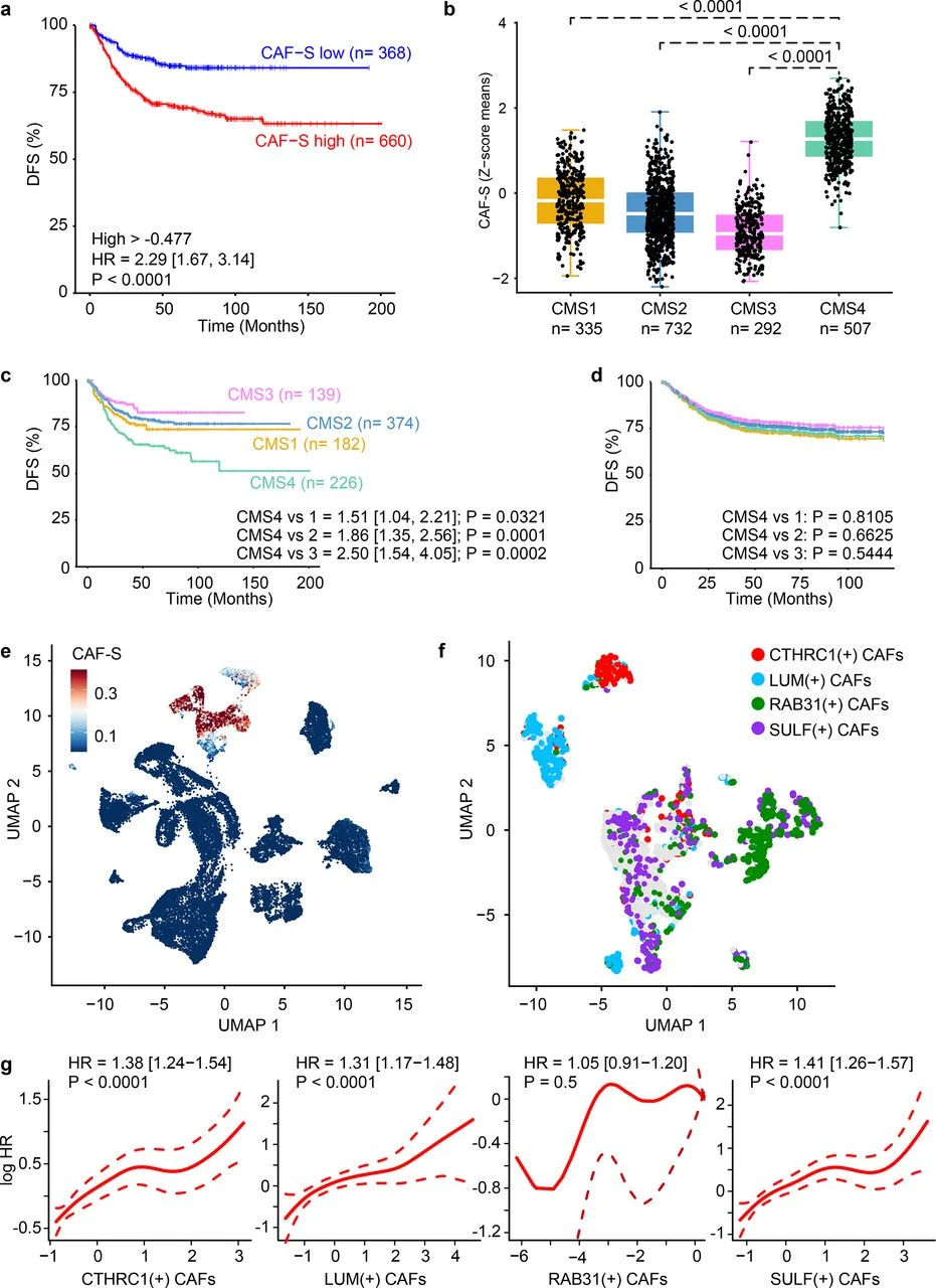
The investigators first developed a cancer-associated fibroblast-enriched gene signature, termed CAF-S, containing 525 genes preferentially expressed in fibroblasts compared with other tumor-associated cell populations.
Among 1,028 patients with localized colorectal cancer and available follow-up, increased CAF-S expression was associated with shorter disease-free survival, independently of established clinical factors including tumor stage, location, age, sex, and adjuvant chemotherapy use.
The researchers subsequently examined individual CAF populations using single-cell RNA sequencing and identified several candidate markers, including CTHRC1, LUM, RAB31, and SULF1.
These markers were further evaluated at the protein level through immunohistochemistry in independent colorectal cancer cohorts. Among them, CTHRC1 emerged as the most consistently informative candidate for identifying fibroblast populations associated with disease relapse.
Additional analyses explored the biological relationship between CTHRC1 expression and TGF-β signaling, the distribution of CD8+ T cells within tumors, and responses to immune checkpoint inhibitors in previously treated clinical cohorts.
CTHRC1-Positive Fibroblasts Identify Colorectal Cancers With Poorer Outcomes
CTHRC1, or collagen triple helix repeat containing 1, is a protein associated with extracellular matrix remodeling and tissue responses. In this study, its expression in cancer-associated fibroblasts emerged as a potentially important indicator of adverse tumor biology.
In an initial immunohistochemical analysis of 266 patients with localized colorectal cancer, the investigators examined the expression of CTHRC1 and other candidate fibroblast markers within tumor tissue.
Although several markers were expressed by CAF populations, increased CTHRC1-positive fibroblast abundance showed the clearest association with disease relapse.
This relationship was subsequently supported by analyses across independent transcriptomic and protein-level cohorts, including patients with stage II and stage III colorectal cancer.
Importantly, CTHRC1-positive CAFs remained associated with shorter disease-free survival after adjustment for conventional clinical factors. The association was observed in patients who received adjuvant chemotherapy and in those who did not, suggesting that the marker reflects an underlying adverse tumor phenotype rather than simply differences in treatment exposure.
However, these findings do not establish that CTHRC1 predicts benefit from adjuvant chemotherapy. Demonstrating treatment-specific predictive value would require prospective studies designed to evaluate whether chemotherapy outcomes differ according to biomarker status.
The researchers also examined CTHRC1 within the established consensus molecular subtype (CMS) classification of colorectal cancer.
CTHRC1-positive fibroblasts were particularly enriched in CMS4 tumors, a molecular subtype characterized by prominent stromal activation and generally unfavorable outcomes. Nevertheless, higher CTHRC1-positive CAF abundance was associated with worse prognosis across multiple CMS categories, suggesting that the marker captures additional stromal information not fully represented by conventional molecular classification.
These findings support the possibility that assessing a specific fibroblast population could provide clinically relevant prognostic information without requiring extensive molecular profiling.
How Are CTHRC1-Positive Fibroblasts Connected to TGF-β Signaling?
A central finding of the study was the close relationship between CTHRC1-positive fibroblasts and TGF-β pathway activation.
In laboratory experiments, stimulation of normal colon fibroblasts and patient-derived colorectal cancer fibroblasts with TGF-β increased CTHRC1 expression at both the messenger RNA and protein levels.
The investigators then examined whether pharmacological inhibition of TGF-β signaling could influence CTHRC1 expression in vivo.
Using an immunocompetent mouse model of pMMR/MSS colorectal cancer liver metastases, they treated animals with galunisertib, an inhibitor of TGF-β receptor I (TGFBR1).
Treatment was associated with a marked reduction in CTHRC1-positive fibroblasts within the metastatic tumor microenvironment. This observation supported the relationship between CTHRC1 expression and active TGF-β signaling.
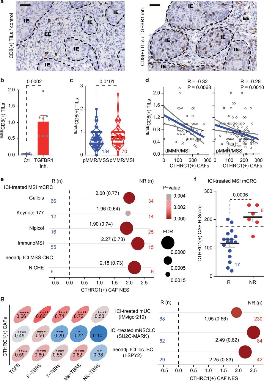
In patient tumors, increased CTHRC1-positive CAF abundance also correlated with TGF-β-associated gene expression programs in fibroblasts and immune populations, including macrophages, natural killer cells, and T lymphocytes.
Notably, these relationships were observed in both pMMR/MSS and dMMR/MSI colorectal cancers.
This finding is important because it suggests that immunosuppressive stromal signaling is not restricted to tumors traditionally classified as poorly immunogenic. Even dMMR/MSI tumors, which frequently contain abundant immune cells, may exhibit a TGF-β-associated stromal environment capable of limiting effective antitumor immunity.
CTHRC1-Positive Fibroblasts and the Exclusion of CD8+ T Cells
The presence of CD8+ cytotoxic T lymphocytes is generally associated with favorable immune activity in colorectal cancer. However, the number of T cells within a tumor does not necessarily indicate whether they can directly interact with malignant cells.
To explore this distinction, the investigators evaluated the spatial distribution of CD8+ tumor-infiltrating lymphocytes, separating them into two populations: intraepithelial T cells located within cancer-cell compartments and extraepithelial T cells confined to the surrounding tumor microenvironment.
They then calculated the ratio between these populations to assess how effectively CD8+ T cells penetrated tumor-cell regions.
As expected, dMMR/MSI tumors generally demonstrated greater intraepithelial CD8+ T-cell infiltration than pMMR/MSS tumors. However, substantial variation was observed within both groups. Some MSI tumors had relatively low levels of intraepithelial infiltration, while certain MSS tumors demonstrated more favorable T-cell distribution.
The investigators found that higher CTHRC1-positive CAF abundance was associated with a lower ratio of intraepithelial to extraepithelial CD8+ T cells, indicating greater immune exclusion.
This relationship was observed independently of mismatch repair status, suggesting that CTHRC1-positive fibroblasts may identify tumors in which cytotoxic T cells are less able to reach cancer-cell compartments.
Experimental findings provided additional support for this relationship. In mouse models treated with a TGF-β receptor inhibitor, the proportion of CD8+ T cells located within cancer-cell regions increased substantially compared with untreated controls.
Together, these observations connect CTHRC1-positive fibroblasts with a biologically relevant feature of immunotherapy resistance: the spatial separation of cytotoxic T cells from their tumor-cell targets.
Although these findings support an association between stromal TGF-β signaling and immune exclusion, they do not establish that CTHRC1 itself directly prevents T-cell infiltration.
Can CTHRC1 Predict Resistance to Immune Checkpoint Inhibitors?
The investigators next examined whether CTHRC1-positive fibroblasts were associated with treatment outcomes in patients receiving immune checkpoint inhibitors.
They analyzed transcriptomic data from patients with MSI metastatic colorectal cancer enrolled in or represented by several immunotherapy studies, including KEYNOTE-177, NIPICOL, ImmunoMSI, and the Gallois cohort.
Across these datasets, higher expression of CTHRC1-positive CAF-associated genes was observed in patients classified as non-responders compared with responders. Response was defined according to RECIST 1.1, with complete and partial responses grouped separately from stable and progressive disease.
The investigators also evaluated CTHRC1 expression at the protein level in tumor samples from 23 patients with dMMR/MSI metastatic colorectal cancer treated with immune checkpoint inhibitors.
In this cohort, non-responders demonstrated higher CTHRC1-positive fibroblast abundance than patients who achieved an objective response.
Receiver operating characteristic analysis identified a CTHRC1 immunohistochemical H-score threshold of 175 that distinguished responders from non-responders, with an area under the curve of 0.936, sensitivity of 88.2%, and specificity of 83.3%.
These findings suggest that CTHRC1 expression may help identify MSI colorectal cancers with reduced likelihood of objective response to checkpoint inhibition.
However, the threshold was derived from a small retrospective cohort and has not been prospectively validated. Its apparent performance therefore should not be interpreted as sufficient evidence for routine clinical decision-making.
The study also evaluated a cohort of 15 patients with localized pMMR/MSS colorectal cancer who received neoadjuvant immune checkpoint inhibition in the exploratory NICHE study.
Similar associations were observed between CTHRC1-positive fibroblasts, increased TGF-β signaling, and lack of response to treatment.
This finding is particularly interesting because checkpoint inhibitors are not standard treatment for unselected pMMR/MSS colorectal cancer. The results suggest that stromal profiling might eventually help distinguish immune-inflamed MSS tumors from those with a more immunosuppressive microenvironment.
Nevertheless, the small number of patients and retrospective nature of the analyses mean that these findings remain hypothesis-generating.
Could the Findings Extend Beyond Colorectal Cancer?
To investigate whether the relationship between CTHRC1-positive fibroblasts and immunotherapy response might be relevant to other malignancies, the researchers examined additional cohorts involving metastatic urothelial carcinoma, metastatic non-small cell lung cancer, and high-risk localized HER2-negative breast cancer.
These analyses included patients treated with immune checkpoint inhibitor-based regimens in the IMvigor210, SU2C-MARK, and I-SPY2 studies.
Across these tumor types, CTHRC1-positive CAF-associated signatures correlated with increased TGF-β signaling in stromal and immune compartments. The same signatures were enriched among patients who did not achieve an objective response to immunotherapy.
Although the findings suggest that CTHRC1-associated stromal biology may be relevant beyond colorectal cancer, differences in tumor biology, treatment regimens, and patient populations limit their direct clinical interpretation.
The results therefore provide a rationale for additional tumor-specific investigation rather than evidence supporting a universal predictive biomarker across cancers.
Could Targeting TGF-β Improve Immunotherapy Response?
The association between CTHRC1-positive fibroblasts, TGF-β activity, and immune exclusion raises an important therapeutic question: could inhibiting TGF-β signaling make resistant tumors more susceptible to immune checkpoint blockade?
TGF-β contributes to multiple aspects of the tumor microenvironment, including fibroblast activation, extracellular matrix remodeling, and regulation of immune-cell function. In tumors with prominent stromal activation, these processes may create conditions that restrict effective T-cell infiltration and antitumor activity.
Previous preclinical research has demonstrated that combining TGF-β pathway inhibition with immune checkpoint blockade can produce synergistic antitumor effects in experimental models of MSS colorectal cancer.
The present study adds a potentially useful biomarker to this therapeutic concept. Because CTHRC1-positive fibroblast abundance closely reflects TGF-β activity, assessing this population may help identify tumors in which stromal pathway inhibition could be biologically relevant.
However, no TGF-β inhibitor is currently approved for cancer treatment, and the study did not demonstrate that selecting patients according to CTHRC1 expression improves outcomes with TGF-β-targeted combinations.
Prospective clinical trials will therefore be required to determine whether CTHRC1 can identify patients who benefit from combining TGF-β inhibition with immune checkpoint blockade.
Could CTHRC1 Become a Practical Biomarker in Clinical Pathology?
One of the most clinically relevant aspects of the study is the potential feasibility of evaluating CTHRC1 through routine immunohistochemistry.
Unlike complex molecular signatures that require extensive transcriptomic analysis, CTHRC1-positive fibroblasts can be assessed in formalin-fixed, paraffin-embedded tumor tissue using protein-based staining.
The investigators demonstrated that immunohistochemical assessment of CTHRC1 was associated with disease outcomes across independent patient cohorts, supporting its potential use as a practical marker of stromal biology.
Such an approach could eventually complement established colorectal cancer biomarkers, including mismatch repair status, by providing information about the immune and stromal characteristics of the tumor microenvironment.
However, important questions remain regarding the standardization of staining methods, scoring thresholds, reproducibility between pathology laboratories, and the ability of CTHRC1 assessment to improve clinical decisions.
The investigators also reported variation in optimal CTHRC1 cutoffs across patient subgroups, highlighting the need for prospective validation before implementing a standardized clinical assay.
What Do These Findings Mean for Immuno-Oncology?
This study highlights the importance of looking beyond tumor-cell genetics when evaluating immunotherapy sensitivity in colorectal cancer.
Mismatch repair status remains a central biomarker, but it does not fully capture the biological complexity of the tumor microenvironment. The presence of CTHRC1-positive fibroblasts may help explain why some dMMR/MSI tumors demonstrate limited response to checkpoint inhibitors despite their generally immunogenic characteristics.
At the same time, the identification of immune-inflamed features in a subset of pMMR/MSS tumors suggests that additional microenvironmental biomarkers may eventually contribute to more refined patient selection.
The study also connects a potentially accessible histopathological marker with a biologically relevant therapeutic pathway. By reflecting TGF-β-associated stromal activation and immune exclusion, CTHRC1-positive fibroblasts may provide a foundation for investigating biomarker-guided combinations of immune checkpoint inhibitors and stromal-targeted therapies.
However, the evidence remains predominantly retrospective, and the findings do not establish CTHRC1 as a validated predictive biomarker or justify changing current immunotherapy selection practices.
Conclusion
The study by Iglesias Coma and colleagues identifies CTHRC1-positive cancer-associated fibroblasts as a potentially important stromal biomarker in colorectal cancer, linking increased TGF-β signaling with immune exclusion, unfavorable clinical outcomes, and limited response to immune checkpoint inhibitors.
Importantly, these associations were observed across both pMMR/MSS and dMMR/MSI tumors, suggesting that stromal biology may provide clinically relevant information beyond mismatch repair status and conventional molecular classification.
The possibility of detecting CTHRC1-positive fibroblasts through routine immunohistochemistry makes this approach particularly interesting for future clinical translation. However, prospective validation is needed to establish reliable thresholds, confirm predictive performance, and determine whether biomarker-guided treatment strategies can improve patient outcomes.
As immuno-oncology continues to move toward more personalized treatment approaches, understanding not only whether immune cells are present but also whether the surrounding stroma allows them to reach and attack cancer cells may become increasingly important.
Read Full Article Here