
The retinoblastoma protein, or Rb, is one of the best-known tumor suppressors in cancer biology. In hormone receptor-positive breast cancer, its activation is also central to how CDK4/6 inhibitors stop tumor-cell proliferation.
But new research published in Nature shows that activated Rb has a second, unexpected function.
After CDK4/6 inhibition, hypophosphorylated Rb does not simply repress cell-cycle genes. It is redistributed across chromatin and becomes incorporated into estrogen receptor-rich transcriptional networks, where it can increase expression of estrogen-responsive genes, including pro-proliferative genes such as CCND1.
In endocrine-sensitive breast cancer, anti-estrogen therapy can suppress this Rb-driven feedback, helping explain the strong therapeutic interaction between endocrine therapy and CDK4/6 inhibition. In ESR1-mutant disease, however, estrogen receptor signaling can remain active despite estrogen deprivation, allowing the compensatory transcriptional program to persist and potentially limiting the depth of CDK4/6-mediated cell-cycle arrest (Watt et al., 2026).
The study therefore challenges the traditional view of Rb as an exclusively transcriptionally repressive tumor suppressor and provides a mechanistic explanation for why CDK4/6 inhibition and effective ER suppression need to work together in HR-positive breast cancer.
Why Is Rb So Important to CDK4/6 Inhibition?
Rb normally limits cell-cycle progression by binding E2F transcription factors and suppressing genes required for entry into S phase.
During cell-cycle progression, CDK4, CDK6 and CDK2 phosphorylate Rb. Hyperphosphorylated Rb becomes functionally inactive, releasing E2F and allowing cells to progress through the G1–S transition.
CDK4/6 inhibitors reverse this process. By preventing Rb phosphorylation, drugs such as abemaciclib and palbociclib maintain Rb in its hypophosphorylated, active state, suppress E2F-regulated transcription and induce G1 arrest.
This mechanism is fundamental to the activity of CDK4/6 inhibitors in HR-positive breast cancer.
Until now, however, considerably less was known about what activated Rb does elsewhere in the genome after prolonged pharmacological CDK4/6 inhibition (Watt et al., 2026).
What Did the Researchers Discover?
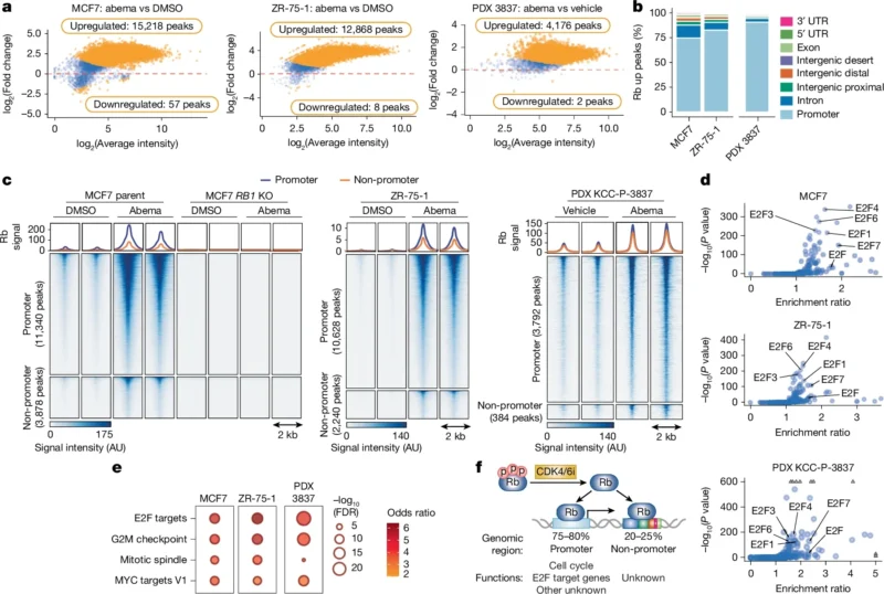
Watt and colleagues used high-resolution chromatin profiling to map endogenous Rb across the genome after CDK4/6 inhibition.
In MCF7 breast cancer cells treated with abemaciclib, the investigators identified:
15,218 regions with increased Rb binding
compared with only:
- 57 regions with decreased binding.
- In ZR-75-1 cells, they observed:
- 12,868 increased Rb-binding regions
and only:
- 8 decreased regions.
Approximately 75%–80% of newly occupied Rb sites were located at promoters, where Rb behaved as expected: it was enriched at E2F-associated cell-cycle genes and accompanied by reduced transcriptional activity.
But approximately 20%–25% occurred outside promoters, including enhancer and other regulatory regions. It was this second group that revealed the unexpected biology (Watt et al., 2026).
The dramatic redistribution is shown in Figure 1 on page 2, where the chromatin profiling demonstrates thousands of newly gained Rb-binding sites after abemaciclib treatment in both breast cancer cell lines and a patient-derived xenograft model.
Rb Was Not Only Repressing Genes
At classical cell-cycle promoters, activated Rb performed its familiar tumor-suppressive function.
It accumulated near E2F-regulated genes, reduced H3K27 acetylation and suppressed cell-cycle transcription.
At non-promoter regions, however, the pattern was almost the opposite.
These newly occupied Rb sites were associated with increased H3K27 acetylation, a chromatin mark associated with active transcription, and with increased expression of nearby genes.
Gene-set analyses repeatedly identified estrogen-response pathways among the transcriptional programs associated with these sites (Watt et al., 2026).
This suggested that pharmacologically activated Rb could have two simultaneous functions:
At E2F promoters: suppress cell-cycle genes.
At ER-associated regulatory regions: enhance estrogen-responsive transcription.
That duality is the central discovery of the study.
How Does Rb Become Connected to Estrogen Receptor Signaling?
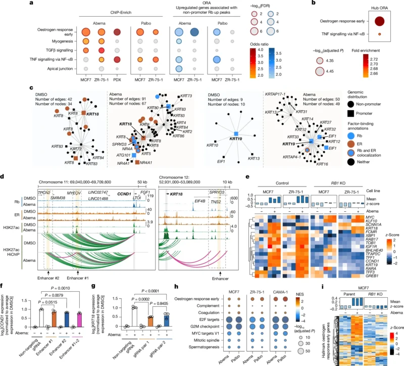
The investigators examined three-dimensional chromatin organization and found that Rb-binding regions frequently occurred within complex transcriptional hubs enriched for estrogen receptor activity.
Among 345 transcriptional hubs containing non-promoter Rb peaks, 188, or approximately 54%, also contained ER-bound regions.
The investigators then identified 405 non-promoter Rb-bound elements that physically looped to gene promoters.
These promoter-connected genes were again strongly enriched for estrogen-response pathways.
Seventeen belonged directly to the Hallmark Estrogen Response Early gene set, including:
- CCND1
- TFF1
- KRT18
- KRT19
- GREB1
and other canonical luminal or estrogen-responsive genes (Watt et al., 2026).
The transcriptional hubs and enhancer–promoter interactions are illustrated in Figure 2 on page 4, where Rb recruitment after abemaciclib is shown at regulatory regions surrounding genes including CCND1 and KRT18.
Did Rb Actually Cause These Genes to Increase?
The investigators went beyond demonstrating correlation.
Using CRISPR–Cas9, they disrupted Rb-bound regulatory elements that interacted with the promoters of CCND1 and KRT18.
Disrupting these enhancers had little effect on baseline expression. But after abemaciclib treatment, the expected increase in CCND1 or KRT18 expression was substantially impaired.
The effect was also demonstrated at the protein level for cyclin D1.
These experiments provide functional evidence that the Rb-bound regulatory elements were involved directly in the transcriptional activation occurring during CDK4/6 inhibition (Watt et al., 2026).
Why Is CCND1 Particularly Important?
CCND1 encodes cyclin D1, a major regulator of the cell cycle and an established component of CDK4/6 signaling.
This creates a biological paradox.
CDK4/6 inhibition activates Rb to suppress proliferation.
But activated Rb can simultaneously contribute to increased transcription of CCND1, generating a compensatory pro-proliferative signal.
When the investigators experimentally reduced CCND1 expression during CDK4/6 inhibition, suppression of E2F target genes became deeper.
The findings therefore suggest that cyclin D1 is one component of the transcriptional feedback that can partially counteract the tumor-suppressive consequences of Rb activation (Watt et al., 2026).
Was the Estrogen Response Seen Only in Cell Lines?
No.
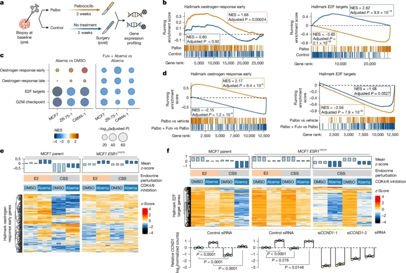
The investigators examined breast cancer samples from the POP trial, in which patients with primary ER-positive breast cancer received two weeks of preoperative palbociclib monotherapy or no treatment before surgery.
Transcriptomic data were available from:
- 44 paired palbociclib-treated samples
and:
- 18 paired untreated controls.
As expected, palbociclib markedly suppressed E2F target genes.
But at the same time, estrogen-response gene sets increased significantly after palbociclib treatment.
The same pattern was not observed in untreated controls (Watt et al., 2026).
These clinical-sample data are particularly important because they demonstrate that the transcriptional phenomenon identified experimentally can also occur in human HR-positive breast cancers exposed to CDK4/6 inhibition.
The paired clinical analysis is presented in Figure 3 on page 5.
Why Does Endocrine Therapy Make CDK4/6 Inhibition More Effective?
CDK4/6 inhibitor monotherapy has considerably less clinical activity than treatment combining CDK4/6 inhibition with endocrine therapy.
The new study offers a mechanistic explanation.
If activating Rb simultaneously increases ER-responsive transcription, then effective suppression of ER signaling would remove the compensatory pathway while allowing Rb to continue repressing E2F-driven cell-cycle progression.
The investigators tested this directly using fulvestrant, a selective estrogen receptor degrader.
Across MCF7, ZR-75-1 and CAMA-1 cells, abemaciclib increased estrogen-response gene expression.
Adding fulvestrant markedly reduced this response.
Combination therapy also produced deeper suppression of E2F targets and more sustained inhibition of tumor-cell proliferation than CDK4/6 inhibition alone (Watt et al., 2026).
The same principle was observed in the patient-derived xenograft model, where palbociclib monotherapy induced estrogen-responsive genes while the combination of palbociclib and fulvestrant produced stronger suppression of E2F signaling and tumor growth.
Does Rb Increase Estrogen Receptor Expression?
Not substantially.
One important distinction is that the increased estrogen-response program was not explained simply by increased ESR1 expression.
Across the ER-positive breast cancer models, CDK4/6 inhibition consistently increased expression of estrogen-responsive genes without a major corresponding increase in ER itself.
Instead, the data point toward changes in chromatin organization and transcriptional activity surrounding existing ER-regulated loci (Watt et al., 2026).
That means CDK4/6 inhibition appears to change how ER-regulated transcription operates, rather than merely increasing the amount of estrogen receptor.
What Happens in ESR1-Mutant Breast Cancer?
This is where the findings become particularly relevant to endocrine resistance.
The investigators studied MCF7 cells carrying the ESR1 Y537S mutation, which produces constitutive, ligand-independent ER activity and is associated with resistance to estrogen-deprivation strategies such as aromatase inhibition.
CDK4/6 inhibition again increased estrogen-responsive genes, including CCND1.
But unlike endocrine-sensitive cells, this transcriptional response persisted even when estrogen was removed from the culture environment.
As a result, suppression of E2F target genes was less complete (Watt et al., 2026).
In simplified terms:
- CDK4/6 inhibition → Rb activation → ER-responsive genes increase
In endocrine-sensitive disease:
- effective ER suppression → feedback blocked → deeper cell-cycle arrest
In ESR1-mutant disease:
- ligand-independent ER remains active → feedback persists → cell-cycle suppression becomes less complete
This provides a potential mechanistic explanation for how ESR1 mutations can limit the effectiveness of CDK4/6 inhibition when ER signaling is not adequately suppressed.
Could SERDs Overcome This Problem?
The experimental findings suggest that direct degradation of ER can suppress this pathway.
When the researchers replaced estrogen deprivation with a selective estrogen receptor degrader, suppression of E2F targets improved and tumor-cell proliferation was more effectively controlled in ESR1-mutant models.
Both fulvestrant and the oral SERD imlunestrant were evaluated experimentally.
The authors therefore propose a mechanistic framework consistent with clinical evidence supporting SERD-based strategies when ESR1-mutant disease emerges (Watt et al., 2026).
Importantly, the study itself was not a clinical trial comparing SERDs with aromatase inhibitors and does not independently establish a new treatment sequence.
Its contribution is mechanistic: it provides a biological explanation for why stronger ER suppression could become particularly important once ligand-independent ESR1 signaling develops.
Where Does KDM5A Enter the Story?
The investigators next asked how activated Rb could convert from a classical transcriptional repressor into a facilitator of ER-driven gene expression.
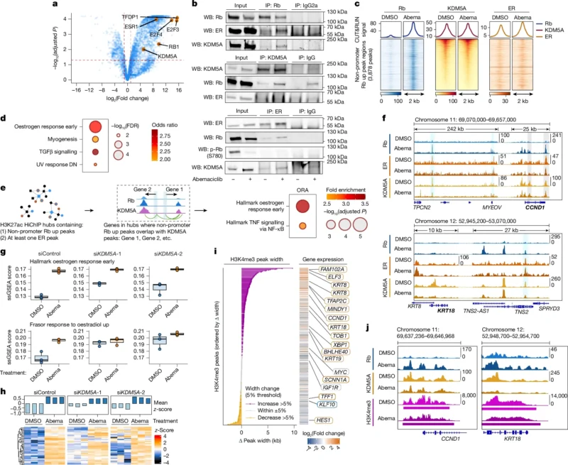
Proteomic analysis identified interactions between Rb, ER and KDM5A, a histone demethylase involved in chromatin regulation.
Three-way co-immunoprecipitation showed that pulling down any one of these proteins could co-precipitate the other two, supporting the existence of a shared nuclear complex.
Chromatin profiling showed that newly recruited Rb frequently localized to regulatory regions already occupied by ER and KDM5A.
The data therefore suggest that Rb is recruited into pre-existing ER-associated transcriptional environments rather than creating these structures from scratch (Watt et al., 2026).
The Rb–ER–KDM5A relationship is shown in Figure 4 on page 7, including co-immunoprecipitation experiments and chromatin occupancy around estrogen-responsive genes such as CCND1 and KRT18.
How Does Rb Affect KDM5A?
KDM5A normally removes trimethylation from histone H3 lysine 4, or H3K4me3, a chromatin mark associated with transcriptionally active promoters.
The investigators found evidence that Rb recruitment can inhibit KDM5A-mediated repression.
When KDM5A was experimentally depleted, estrogen-responsive genes increased even without CDK4/6 inhibition.
Adding abemaciclib after KDM5A depletion produced little additional transcriptional induction.
At the same time, CDK4/6 inhibition increased the breadth of H3K4me3 around several estrogen-responsive genes (Watt et al., 2026).
The proposed mechanism is therefore:
- CDK4/6 inhibition
→ hypophosphorylated Rb
→ Rb enters ER/KDM5A-associated transcriptional hubs
→ KDM5A-mediated repression is relieved
→ H3K4me3 increases
→ ER-responsive transcription increases.
This pathway provides a chromatin-level explanation for the paradoxical gene activation induced by a classical tumor suppressor.
Does This Mean Rb Promotes Breast Cancer?
That would be an oversimplification.
Rb remains a major tumor suppressor.
The dominant and well-established consequence of pharmacologically activating Rb is repression of E2F-driven proliferation and cell-cycle arrest.
The new study instead shows that Rb has context-dependent dual functionality.
At one set of genomic regions it suppresses transcription.
At another set, particularly within ER-rich regulatory networks, it can facilitate transcription.
Some of those ER-responsive genes, including CCND1, can support proliferation and partially counteract the depth of cell-cycle suppression.
The biological effect therefore depends on the transcriptional context surrounding Rb activation (Watt et al., 2026).
Why Could This Explain the Success of CDK4/6 Plus Endocrine Therapy?
The findings provide an elegant mechanistic interpretation of a clinically established treatment principle.
CDK4/6 inhibition activates Rb and suppresses E2F.
At the same time, Rb increases ER-dependent transcription.
If ER is simultaneously inhibited or degraded, that compensatory response is suppressed.
The result is deeper cell-cycle repression than either mechanism achieves alone.
The study therefore suggests that endocrine therapy does more than independently block an oncogenic pathway alongside CDK4/6 inhibition.
It also appears to neutralize a transcriptional feedback program created by CDK4/6-mediated Rb activation itself (Watt et al., 2026).
Could This Help Explain CDK4/6 Resistance?
Potentially.
Several mechanisms of CDK4/6 inhibitor resistance are already recognized, including loss of RB1, alterations affecting cyclin E–CDK2 signaling and increased cyclin D activity.
The current study adds a different concept: incomplete suppression of Rb-induced ER transcription.
This may be especially relevant when ER signaling becomes constitutively active through ESR1 mutation.
The researchers found that ESR1-mutant cells continued to express ER-responsive genes during estrogen deprivation and CDK4/6 inhibition, thereby limiting complete E2F suppression.
Direct ER degradation reversed this effect experimentally (Watt et al., 2026).
The study therefore suggests that some apparent resistance to CDK4/6 inhibition could reflect not failure to activate Rb, but rather an unintended transcriptional consequence of successfully activating it.
Does the Study Change Clinical Practice?
Not directly.
This is primarily a mechanistic study integrating breast cancer cell lines, patient-derived xenografts, chromatin profiling, CRISPR experiments and transcriptomic analysis of human breast cancer samples.
It does not prospectively compare clinical treatment strategies based on Rb-driven transcription, KDM5A activity or the identified enhancer program.
It also does not establish a validated biomarker for deciding which patient should receive a particular endocrine partner.
Its clinical importance lies in providing a molecular framework that can help explain established and emerging therapeutic observations.
In particular, it reinforces why effective ER suppression remains essential during CDK4/6 treatment and why direct ER degradation could be particularly relevant when ESR1 mutations produce ligand-independent ER activity.
What Are the Main Limitations?
Several limitations are important.
Much of the detailed mechanistic work was performed in established HR-positive breast cancer cell lines.
Although the investigators validated major findings using a patient-derived xenograft and human breast cancer samples from the POP trial, the clinical transcriptomic analysis involved relatively short exposure to palbociclib, two weeks of monotherapy, rather than the long-term combination regimens used routinely in metastatic disease.
Some integrated chromatin analyses also combined datasets generated after different treatment durations. The authors specifically note that Rb profiling was performed after two days of treatment, whereas the previously generated H3K27ac HiChIP data reflected seven days of abemaciclib exposure.
The study also does not establish that ER itself recruits Rb to chromatin. Other transcription factors and chromatin regulators may participate.
Finally, the ESR1-mutant findings are predominantly mechanistic and do not independently prove that one clinical endocrine strategy is superior to another (Watt et al., 2026).
The Bottom Line
The study substantially expands the understanding of how CDK4/6 inhibitors affect HR-positive breast cancer.
After CDK4/6 inhibition, activated Rb undergoes extensive genome-wide redistribution.
In MCF7 cells, abemaciclib produced 15,218 newly increased Rb-binding regions, and in ZR-75-1 cells, 12,868.
At classical cell-cycle promoters, Rb suppresses E2F transcription as expected.
But at non-promoter regulatory regions, Rb enters ER-rich transcriptional hubs, interacts with ER and KDM5A, and promotes estrogen-responsive gene expression—including CCND1, a pro-proliferative mediator capable of limiting complete cell-cycle repression.
In endocrine-sensitive models, ER-directed therapy neutralized this transcriptional feedback and deepened CDK4/6-mediated growth suppression.
In ESR1-mutant models, the Rb-driven ER program persisted during estrogen deprivation, while pharmacologic ER degradation restored stronger cell-cycle suppression.
The findings therefore reveal an unexpected duality:
Rb can simultaneously enforce cell-cycle arrest and activate a transcriptional program capable of weakening that arrest.
For HR-positive breast cancer, that paradox provides a new explanation for why CDK4/6 inhibition works most effectively when estrogen receptor signaling is also adequately suppressed.
References
- Watt AC, Ahn A, Blyth C, Dixon-Douglas JR, Ambani K, Coulson R, Taylor M, Chan KT, Dietrich C, Russ BE, Ramm S, Mahendra CA, Lu K-H, Pires N, Garcia-Sannicolas J, Voulgaris O, Nunag S, Ang C-S, Dawson MA, Lim E, Arnedos M, Chandarlapaty S, André F, Goel S. Rb-driven transcription limits its tumour-suppressive effects in breast cancer. Nature. 2026. doi:10.1038/s41586-026-10886-w.
- Arnedos M, et al. Modulation of Rb phosphorylation and antiproliferative response to palbociclib: the preoperative-palbociclib (POP) randomized clinical trial. Annals of Oncology. 2018;29:1755–1762.
- Watt AC, et al. CDK4/6 inhibition reprograms the breast cancer enhancer landscape by stimulating AP-1 transcriptional activity. Nature Cancer. 2021;2:34–48.
- Toy W, et al. ESR1 ligand-binding domain mutations in hormone-resistant breast cancer. Nature Genetics. 2013;45:1439–1445.
- Bidard FC, et al. First-line camizestrant for emerging ESR1-mutated advanced breast cancer. New England Journal of Medicine. 2025. doi:10.1056/NEJMoa2502929.