Could molecular changes associated with breast cancer be detectable in blood years before a tumor becomes clinically apparent?
A new study published in Cell Genomics suggests that the answer may be yes, but with important limitations for clinical translation. Nicholas Cheng, Tom W. Ouellette, Kimberly Skead, and colleagues analyzed genome-wide cell-free DNA methylation patterns in prospectively collected plasma from individuals who were cancer-free at blood collection but subsequently developed breast or prostate cancer. The study identified regulatory-element methylation changes that could be detected years before diagnosis and explored whether these signals could be used to stratify future cancer risk.
For breast cancer, the most informative signals arose from hypermethylated enhancer regions. These signatures distinguished future breast cancer cases from controls only modestly, but they were associated with future risk, varied according to tumor subtype and age, and became substantially more detectable in established late-stage disease. The results therefore support cfDNA methylation as a potentially complementary risk-stratification tool rather than a replacement for mammography or a stand-alone early-detection test.
The study is particularly relevant because most liquid-biopsy development has relied on blood collected after cancer has already been diagnosed. By contrast, this analysis used pre-diagnosis plasma, providing a rare opportunity to examine the molecular changes that precede clinical detection.
A Prospective Window Into Cancer Before Diagnosis
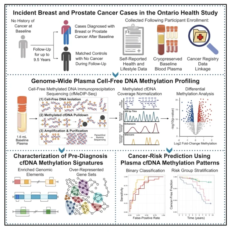
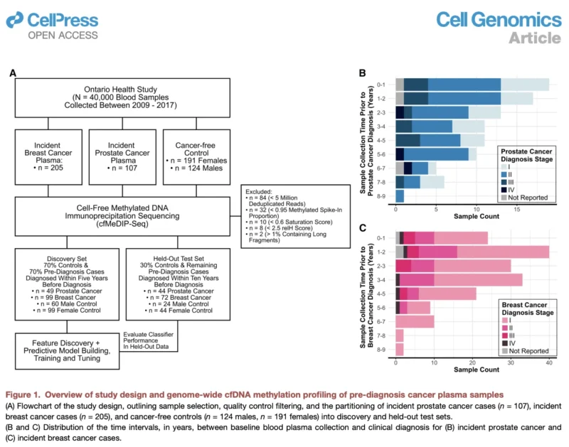
The investigators used biospecimens from the Ontario Health Study, a prospective population cohort that collected biological samples and health information from more than 40,000 participants.
Participants were cancer-free when blood was collected. By linking the cohort with the Canadian Cancer Registry, investigators subsequently identified individuals who developed breast or prostate cancer from a few weeks to as long as nine years after plasma collection. Cancer-free controls were matched according to factors including age, sample timing, smoking, and alcohol consumption.
Genome-wide plasma cfDNA methylation was characterized using cell-free methylated DNA immunoprecipitation sequencing, or cfMeDIP-seq.
Following quality control, the analysis included 491 plasma samples, comprising 171 incident breast cancer cases, 93 incident prostate cancer cases, and 227 cancer-free controls. Samples from future cancer cases had been obtained between two weeks and nine years before clinical diagnosis.
The breast cancer population was particularly representative of screen-detected early disease: 67.8% of incident breast cancers were stage I, and nearly 89% of cases and controls had undergone mammography before blood collection. This feature is important because early-stage breast cancer is one of the most difficult settings for blood-based cancer detection. Small localized tumors often release very little tumor-derived DNA into the circulation.
Early cfDNA Changes Were Concentrated in Regulatory Regions
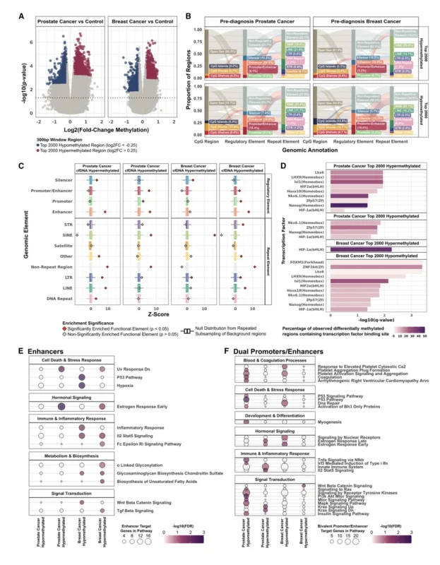
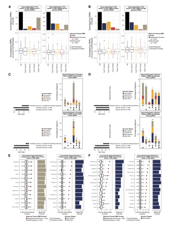
Rather than focusing only on individual cancer-associated genes, the investigators analyzed methylation patterns across the genome. Differentially methylated regions identified before diagnosis were frequently located in promoters, enhancers, silencers, and repetitive genomic elements. Approximately 37.5%–51.2% of the leading regions mapped to promoters, enhancers, or silencers.
The biological pathways associated with these regulatory changes were also notable. In breast cancer, differentially methylated regions were associated with pathways involving DNA repair, hypoxia, P53 signaling, hormonal regulation, cell growth, and immune-related processes.
The authors interpret these findings as evidence that pre-diagnosis cfDNA may contain information originating not only from emerging tumor cells but also from systemic host and immune changes associated with cancer development. This distinction matters. At very early stages, the amount of DNA directly shed by a tumor may be extremely small. A useful blood-based signal may therefore need to capture both tumor-derived and host-derived biology.
Enhancer Methylation Provided the Strongest Breast Cancer Signal
For breast cancer, the investigators found that methylation changes within enhancer regions provided the strongest predictive performance. A penalized logistic-regression classifier incorporating the top 90 hypermethylated enhancer regions was developed in a discovery cohort of 99 breast cancer cases and 99 controls.
The resulting model achieved a cross-validated:
- AUROC of 0.62
- C-index of 0.61
across breast cancer subtypes, ages, and pre-diagnosis intervals extending up to five years. Performance was weaker in the independent held-out cohort of 72 cases and 44 controls:
- AUROC 0.58
- C-index 0.59.
These values are modest and are central to interpreting the study correctly. The results demonstrate biological detectability and potential for risk stratification, but they do not indicate that this assay is ready to function as an independent breast cancer screening test.
The authors explicitly acknowledge this point, concluding that cfDNA methylation signatures may arise years before clinical detection but currently have limited stand-alone diagnostic utility in breast cancer.
Risk Groups Began to Separate Approximately 1.5 Years Before Diagnosis
Although discrimination between individual cases and controls was limited, the methylation score was able to separate groups with different future breast cancer risk. Using a prespecified classifier threshold, women categorized as high risk had a:
- 3.6% five-year breast cancer incidence in the discovery set
compared with:
- 1.2% among women classified as low risk.
In the held-out cohort, eight-year breast cancer incidence was:
- 6.2% in the high-risk group
versus:
- 1.8% in the low-risk group.
The high- and low-risk curves began to separate approximately 1.5 years before diagnosis. In the held-out cohort, the estimated hazard ratio for high versus low methylation risk was 2.3, although the confidence interval was wide and the comparison was not statistically significant:
- HR 2.3; 95% CI, 0.9–6.0; P=0.09.
This nuance is important. The observed separation is biologically and clinically interesting, but the study does not yet establish a sufficiently precise individual risk model for clinical decision-making.
Breast Cancer Subtype Appeared to Influence Detectability
One of the more provocative observations was that cfDNA methylation signals differed by breast cancer subtype. Among the relatively small number of patients with available receptor information, pre-diagnosis classifier performance appeared higher in triple-negative and HR-positive/HER2-positive disease than in some other subgroups.
In the held-out dataset, triple-negative and HR-positive/HER2-positive tumors had C-index values of approximately 0.73 and 0.78, respectively. However, these estimates were based on extremely small numbers, only three cases in each subgroup in the test set, and therefore had very wide confidence intervals. They should not be interpreted as validated subtype-specific performance estimates.
The biological pattern nevertheless raises an important hypothesis. The authors observed that triple-negative cancers appeared more detectable within approximately four years of diagnosis, potentially reflecting the faster evolution of aggressive tumors. By comparison, slower-growing HR-positive/HER2-negative disease showed more consistent methylation signals across time.
If confirmed, this could mean that different breast cancer subtypes have different molecular detection windows before clinical diagnosis.
Performance Also Varied According to Age
Age appeared to influence classifier performance. In the held-out pre-diagnosis cohort, the highest C-index was observed among women diagnosed between ages 30 and 50, at 0.67, compared with 0.49 among women diagnosed after age 70. A similar pattern was observed in the external late-stage breast cancer cohort.
The explanation remains uncertain, and the subgroup numbers were limited. However, the finding suggests that age-related biological or epigenetic differences may influence the performance of methylation-based cancer detection. This will require larger validation studies before it can inform screening strategies.
The Relationship With Mammography Is Particularly Interesting
The study also examined the timing of the most recent mammogram before blood collection. Among women in the test cohort, classifier performance was lowest when mammography had been performed within six months of the blood draw:
- C-index 0.44
Performance increased when the last mammogram had occurred one to two years earlier:
- C-index 0.64
and more than two years earlier:
- C-index 0.70.
The finding does not establish a causal relationship between mammography timing and methylation signal. It may instead reflect differences in disease biology, screening interval, stage, or the timing of emerging cancer relative to imaging. Nevertheless, it supports the idea that blood-based biomarkers may eventually provide information that is complementary to imaging rather than competing with it.
The Signal Became Much Stronger in Established Advanced Breast Cancer
The same classifier developed from pre-diagnosis samples was also tested in an external cohort of patients with established late-stage breast cancer from the OCTANE study.
Here, discriminatory performance increased substantially. The classifier achieved AUROC 0.87 when late-stage breast cancer cases were compared with Ontario Health Study cancer-free controls, and AUROC 0.94 when compared with external non-breast cancer samples. This contrast illustrates one of the central challenges of liquid biopsy. Detecting advanced cancer is comparatively easier because tumor burden is larger and more tumor-derived material enters the circulation.
The real challenge is identifying a biologically meaningful signal before a small, asymptomatic cancer becomes detectable by conventional methods. The modest performance observed in the pre-diagnosis cohort reflects precisely that challenge.
cfDNA Methylation Is Unlikely to Replace Mammography
The study does not support replacing mammographic screening with cfDNA methylation testing. The authors are explicit that the current sensitivity of cfDNA-based approaches in early-stage breast cancer remains insufficient for this purpose. Instead, a more plausible future application could involve risk-adapted screening.
A sufficiently validated blood-based methylation score might eventually identify women whose molecular risk appears higher than expected from conventional clinical factors, potentially prompting earlier or more intensive surveillance. Conversely, a robust low-risk result could theoretically contribute to decisions about screening intensity.
Those possibilities remain investigational. The present study establishes biological feasibility rather than clinical utility. Prospective trials would need to demonstrate that acting on the blood test improves cancer detection, reduces advanced-stage diagnoses, or produces another clinically meaningful outcome without unacceptable false-positive testing.
Sample Quality Had a Major Effect on Model Performance
An important technical finding was the sensitivity of the classifier to assay quality. When samples failing predefined cfMeDIP-seq quality-control thresholds were included, breast cancer classifier performance fell to:
- AUROC 0.54
- C-index 0.55.
The investigators found that variation in immunoprecipitation efficiency, sequencing complexity, and other laboratory parameters could shift predicted risk scores and potentially generate false-positive or false-negative classifications. This is highly relevant to eventual clinical implementation.
A biomarker intended for population screening must perform reproducibly across laboratories, sample-processing conditions, storage intervals, and patient populations. Analytical standardization is therefore not a secondary technical issue, it is fundamental to whether a methylation-based screening strategy can ever become clinically reliable.
Several Limitations Prevent Immediate Clinical Translation
The study has important limitations. Only 1.6 mL of archived plasma was available per participant. Larger plasma volumes could increase the amount of tumor-derived cfDNA captured and potentially improve sensitivity. The exact interval between blood collection and cryopreservation was also unavailable, and archived EDTA samples may not behave identically to samples processed immediately or collected using contemporary stabilizing tubes.
Some clinically important breast cancer subgroups were very small, particularly triple-negative disease, producing wide confidence intervals and preventing reliable conclusions about subtype-specific performance. Follow-up among cancer-free controls was also not uniform. Some apparently false-positive results could therefore represent individuals with occult cancer that had not yet been diagnosed.
Most importantly, the investigators did not have an independent external population-based pre-diagnosis cohort profiled using the same assay. Although they used held-out validation within the Ontario Health Study, true external validation will be necessary before the model’s generalizability can be established.
What This Study Adds to Breast Cancer Early Detection
The importance of the study lies less in the current diagnostic accuracy than in its design. Most cancer liquid-biopsy studies ask whether molecular signatures can distinguish patients after cancer is already present clinically from healthy controls. Cheng and colleagues instead asked what the blood looks like before diagnosis. That distinction is essential for developing genuine early-detection biomarkers.
The study demonstrates that epigenetic changes associated with future breast cancer can be detected years before diagnosis and that these signals contain information related to regulatory biology, immune processes, tumor subtype, age, and subsequent disease progression.
At the same time, it shows why early detection remains difficult: the molecular signal at this stage is subtle, heterogeneous, and substantially weaker than in established advanced disease. That combination of biological promise and limited current sensitivity makes the study particularly informative.
The Bottom Line
Genome-wide cfDNA methylation profiling identified breast cancer-associated enhancer signals in blood collected years before clinical diagnosis. Among women who later developed breast cancer, a classifier based on 90 hypermethylated enhancer regions achieved modest discrimination:
- Discovery AUROC: 0.62
- Held-out AUROC: 0.58.
Nevertheless, methylation-based risk scores separated groups with different future breast cancer incidence, with high-risk individuals in the held-out cohort showing an 8-year incidence of 6.2% versus 1.8% in the low-risk group. The signals also varied by breast cancer subtype, age, and mammography interval and became considerably stronger in established late-stage disease.
The clinical message should therefore remain measured. This is not yet a blood test for breast cancer screening, and its performance is insufficient to replace mammography.
But it provides compelling evidence that the molecular evolution of breast cancer may leave detectable epigenetic traces in plasma years before clinical diagnosis. The future opportunity may not be to replace conventional screening, but to combine imaging, clinical risk, and molecular information to identify who should be screened more closely, and when.
Reference
- Cheng N, Ouellette TW, Skead K, Singhawansa A, Elliott M, Cescon DW, Bratman SV, De Carvalho DD, Soave D, Awadalla P. Pre-diagnosis plasma cell-free DNA reveals early signatures of prostate and breast cancer risk up to eight years prior to clinical detection. Cell Genomics. 2026;6:101364. doi:10.1016/j.xgen.2026.101364.