
Breast cancer treatment decisions rely on an accurate estimate of recurrence risk.
Clinical stage, nodal involvement, tumor grade, hormone receptor status, HER2 status, and patient characteristics remain central to this process. In selected hormone receptor-positive, HER2-negative cancers, genomic assays can provide additional information and help guide decisions about adjuvant chemotherapy.
However, these tools are not available or guideline-supported across every breast cancer subtype. Their role is also more limited in triple-negative and HER2-positive disease, where treatment decisions often depend on broader clinical risk factors and response to systemic therapy.
A study published in Nature Communications evaluated whether artificial intelligence could extract prognostic information from routine H&E pathology slides and combine it with standard clinical data to estimate recurrence risk across invasive breast cancer subtypes.
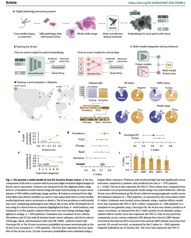
The study developed a multimodal AI test using data from 8,161 patients across 15 cohorts in seven countries. The model combined digital pathology features with routinely available clinical variables and was externally evaluated in 3,502 patients.
The findings suggest that routine pathology images may contain clinically meaningful information beyond established clinicopathological factors. However, the test remains prognostic rather than predictive. It has not yet been shown to identify which patients benefit from a specific treatment or to replace established genomic assays in clinical practice.
Why Current Risk Assessment Has Limits
Risk stratification in breast cancer is designed to distinguish patients with a lower likelihood of recurrence from those who may need more intensive systemic treatment.
For hormone receptor-positive, HER2-negative disease, genomic assays such as Oncotype DX, MammaPrint, and Prosigna have become important tools in selected clinical settings. These assays measure gene-expression patterns associated with recurrence risk and, in some settings, chemotherapy benefit. [2–4]
Still, genomic assays require tissue processing, may take time to return results, and are not designed for every subtype. They also do not fully capture all morphological features within the tumor microenvironment.
Pathologists already evaluate features such as tumor grade, histological subtype, tumor-infiltrating lymphocytes, and proliferation markers. Yet these conventional measurements may not capture the full biological complexity visible in a standard pathology slide.
The investigators asked whether a digital model could identify additional patterns from H&E images that are not routinely measured or reported.
How the AI Test Was Built
The model used digitized H&E-stained pathology slides obtained from core needle biopsies or surgical specimens.
A pathology foundation model called Kestrel extracted image-based features from the whole-slide images. Kestrel had been trained using self-supervised learning on 400 million pathology image patches from a large pan-cancer dataset.
The pathology-derived information was combined with eight routinely collected clinical variables: age, ER status, PR status, HER2 status, T stage, N stage, invasive ductal histology, and invasive lobular histology.
The final model generated a continuous risk score between 0 and 1. Higher scores were associated with a higher estimated risk of recurrence.
The study design is illustrated on page 3 of the paper. Standard pathology slides were processed through the AI model, then integrated with clinical information to produce one multimodal recurrence-risk score.
External Validation Across Different Populations
The model was developed in 4,659 patients from 10 cohorts and evaluated in 3,502 patients from five independent external cohorts.
These evaluation cohorts included broad populations of invasive breast cancer, as well as three hormone receptor-positive, HER2-negative cohorts in which patients had undergone Oncotype DX testing.
For the primary endpoint of disease-free interval, the pooled external analysis reported a C-index of 0.71 (95% CI, 0.68–0.75). A C-index closer to 1 reflects better ability to rank patients according to recurrence risk.
For every 0.2-unit increase in the AI score, the pooled hazard ratio for disease-free interval was 3.63 (95% CI, 3.02–4.37; p<0.001).
The model was also prognostic for distant recurrence-free interval, recurrence-free survival, distant recurrence-free survival, and overall survival.
These findings support the idea that the model could separate lower-risk and higher-risk patients across multiple external datasets. They do not show that acting on the score improves outcomes.
How Did the Test Compare With Oncotype DX?
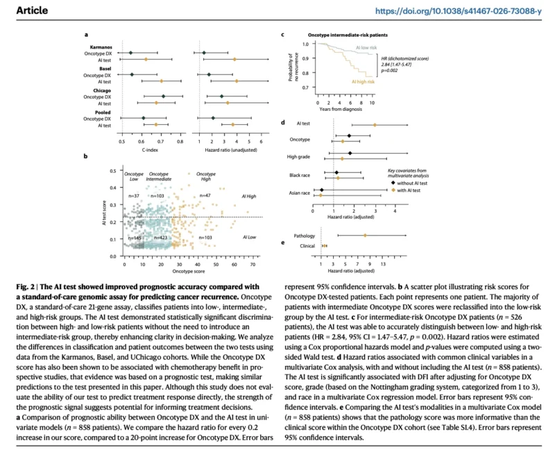
The study included 858 patients from three external cohorts who had previously received Oncotype DX testing in routine care.
In the pooled comparison, the AI test showed a numerically higher C-index for disease-free interval than Oncotype DX: 0.67 versus 0.61.
The AI score also remained independently associated with disease-free interval after adjustment for Oncotype DX score, Nottingham grade, race, and dataset. The adjusted hazard ratio was 2.95 (95% CI, 1.82–4.79; p<0.001).
However, this result should not be interpreted as proof that the AI test is clinically superior to Oncotype DX.
The comparison was retrospective, the confidence intervals overlapped, and the study was not designed to compare treatment-guidance strategies. Oncotype DX has prospective evidence supporting its role in selected chemotherapy decisions, whereas the AI model has not yet been tested for chemotherapy prediction.
The study instead suggests that digital pathology may capture prognostic information that is not fully represented by conventional clinical variables or a genomic recurrence score.
A Possible Way to Refine Intermediate Risk
One of the more clinically interesting analyses involved patients with intermediate Oncotype DX scores.
Among 526 patients classified as intermediate risk by Oncotype DX, the AI model reclassified 423 patients as low risk and 103 as high risk.
Within this intermediate-risk group, the continuous AI score remained associated with recurrence risk, with a hazard ratio of 3.45 (95% CI, 1.85–6.42; p<0.001).
This does not mean that patients classified as low risk by the AI test can safely avoid chemotherapy. It also does not mean that high-risk patients should automatically receive more intensive treatment.
The study did not evaluate treatment benefit. It evaluated prognosis.
Still, this finding raises an important future question: could digital pathology help clarify recurrence risk in patients whose genomic result does not offer a simple treatment direction?
Why the TNBC Findings Matter
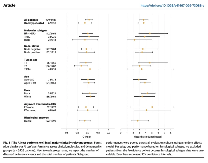
The model was also tested in triple-negative breast cancer, a subtype for which the authors noted there are no NCCN guideline-supported prognostic assays for treatment selection.
Among 230 patients with TNBC, the AI test achieved a C-index of 0.71 (95% CI, 0.62–0.81). The associated hazard ratio for disease-free interval was 3.81 (95% CI, 2.35–6.17; p=0.02).
This is relevant because treatment decisions in early TNBC are increasingly complex.
For many patients with stage II–III disease, neoadjuvant pembrolizumab plus chemotherapy is now central to treatment. At the same time, ongoing studies are examining whether patients with favorable response patterns, including pathological complete response, could receive less intensive therapy in the future. [5–7]
A reliable prognostic tool could eventually contribute to these discussions. But the current AI score has not been validated to predict benefit from pembrolizumab, platinum chemotherapy, capecitabine, PARP inhibition, or treatment de-escalation.
It should not yet be used to select or omit therapy in TNBC.
Could Digital Pathology Improve Access to Prognostic Testing?
The proposed model uses standard H&E slides, which are already generated in routine pathology workflows.
The authors estimate that slide accessioning, scanning, and model inference could be completed in less than one hour in a digital pathology environment. They also note that this approach could preserve tumor tissue for later genomic profiling if disease recurs.
This potential advantage is practical rather than proven.
Widespread use would require high-quality digital pathology infrastructure, harmonized staining and scanning procedures, prospective validation, regulatory review, and integration into multidisciplinary clinical decision-making.
The reliability of the score would also need to be confirmed across laboratories, scanners, specimen types, racial and ethnic groups, and global health-care settings.
Important Limitations
This was a retrospective observational study. The investigators were not blinded during experiments or data analysis, and the test was developed using pre-existing cohort data.
The model predicts recurrence risk. It does not predict whether a patient benefits from a specific treatment.
A high-risk result does not establish that treatment escalation will improve outcomes. A low-risk result does not establish that treatment can be safely reduced or omitted.
The study also used proprietary model code. Several authors hold equity in Ataraxis AI, and New York University reported financial and intellectual-property interests related to the research.
These disclosures do not invalidate the results. They do reinforce the importance of independent validation and prospective clinical studies before the score is incorporated into routine treatment decisions.
The Bottom Line
This large international study suggests that AI applied to routine pathology slides, combined with clinical data, may improve recurrence-risk stratification across invasive breast cancer subtypes.
The model showed prognostic activity in external cohorts, demonstrated numerical advantages over Oncotype DX in a retrospective comparison, and showed promising results in TNBC and HER2-positive disease.
The most important message is not that AI is ready to replace genomic assays.
It is that standard pathology images may contain prognostic information that current clinical and molecular tools do not fully capture.
Before this approach can influence treatment selection, it will need prospective validation, standardized risk thresholds, independent testing, regulatory review, and evidence that use of the score improves patient outcomes.
References
- Witowski J, Zeng KG, Cappadona J, et al. Multi-modal AI for comprehensive breast cancer prognostication. Nature Communications. 2026;17:5879. doi:10.1038/s41467-026-73088-y.
- Paik S, Shak S, Tang G, et al. A multigene assay to predict recurrence of tamoxifen-treated, node-negative breast cancer. New England Journal of Medicine. 2004;351:2817–2826.
- Sparano JA, Gray RJ, Makower DF, et al. Adjuvant chemotherapy guided by a 21-gene expression assay in breast cancer. New England Journal of Medicine. 2018;379:111–121.
- Kalinsky K, Barlow WE, Gralow JR, et al. 21-gene assay to inform chemotherapy benefit in node-positive breast cancer. New England Journal of Medicine. 2021;385:2336–2347.
- Schmid P, Cortés J, Pusztai L, et al. Event-free survival with pembrolizumab in early-stage triple-negative breast cancer. New England Journal of Medicine. 2022;386:556–567.
- Schmid P, Cortés J, Dent R, et al. Overall survival with pembrolizumab in early-stage triple-negative breast cancer. New England Journal of Medicine. 2024;391:1981–1991.
- Tolaney SM, et al. OptimICE-pCR: de-escalation of therapy in early-stage triple-negative breast cancer patients who achieve pathological complete response after neoadjuvant chemotherapy with checkpoint inhibitor therapy. San Antonio Breast Cancer Symposium. 2023.