Siba El Hussein, Assistant Professor of Pathology and Laboratory Medicine at The University of Vermont Medical Center, shared a post on X:
“Acute promyelocytic leukemia (APL) in the setting of primary or secondary MPO deficiency can create diagnostic challenges, as bright MPO expression by flow cytometry analysis is a key feature of APL.
Six cases of APL with reduced MPO expression (2 cases) and absent MPO expression (4 cases) have been described so far: Four of the microgranular variant and two classic APL cases. One of the MPO negative microgranular APL has been extensively analyzed and found to have a germline heterozygous MPO loss of function mutation (MPO c.2031 -2A > C) with somatic uniparental disomy of 17q, resulting in homozygous MPO deficiency and subsequent MPO negativity in the myeloblasts.
Three different flow cytometric patterns of immunoreactivity with the MPO protein have been described in the setting of MPO deficiency:
- (a) Dim/absent MPO expression, characteristic of patients with primary complete MPO deficiency.
- (b) Moderate MPO expression, typical of patients with primary partial MPO deficiency.
- (c) Bright MPO expression, typical of patients with secondary MPO deficiency (a functionally inactive form of the enzyme is present at normal levels).
MPO deficiency was previously thought to be extremely rare; However, recent studies have suggested that the incidence might be much higher, with a reported frequency of 1 per 200 to 4,000 in the US. The prevalence of heterozygotes for MPO c.2031-2A > C may close to 1% in the population.
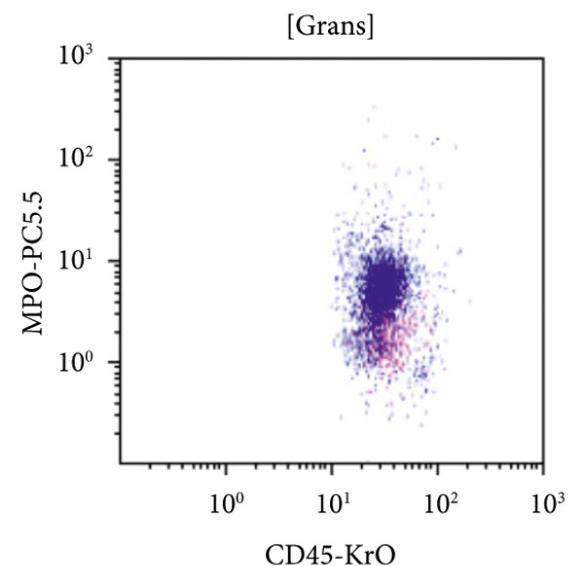
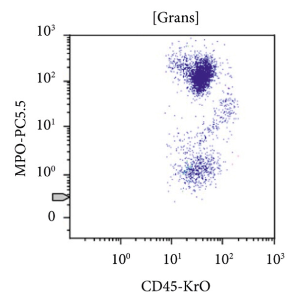
In the setting of an acute leukemia with morphologic and immunophenotypic features typical of APL, with dim to absent MPO expression, evaluating MPO expression in background granulocytes is very helpful to highlight the possibility of a primary (hereditary) form of MPO deficiency (partial or complete):
- Figure 1 below illustrates a case of primary MPO deficiency with dim/negative MPO expression in granulocytes,
- while Figure 2 illustrates bright MPO expression in granulocytes in normal control.
For more on this + figure credits, check this article by Kritika Krishnamurthy, Jui Choudhuri and Yanhua Wang.”
MPO Expression of Background Neutrophils in MPO Negative Acute Promyelocytic Leukemia, An Easy Clue to Corroborate a Challenging Diagnosis: A Case Report and Review of Literature.
Authors: Kritika Krishnamurthy, et al.
She continued:
“More on rare presentations of APL.
APL with circulating myeloblasts, and “aleukemic” promyelocytes confined to the marrow may cause diagnostic challenges:
If diagnosis relied only on peripheral blood findings, the promyelocytic component hidden in the marrow may be missed, particulalry that the typical myeloblastic morphology identified in the blood (as seen in the top left figure below) and phenotype (bottom left figure) may not alerting to perform STAT FISH for PML::RARA, delaying appropriate treatment.
The top right figure shows promyelocytes in the bone marrow aspirate smear, with a phenotype classic of APL by flow cytometry analysis (negative CD34 and bright MPO expression).
Look out for more on this and figure credits.”
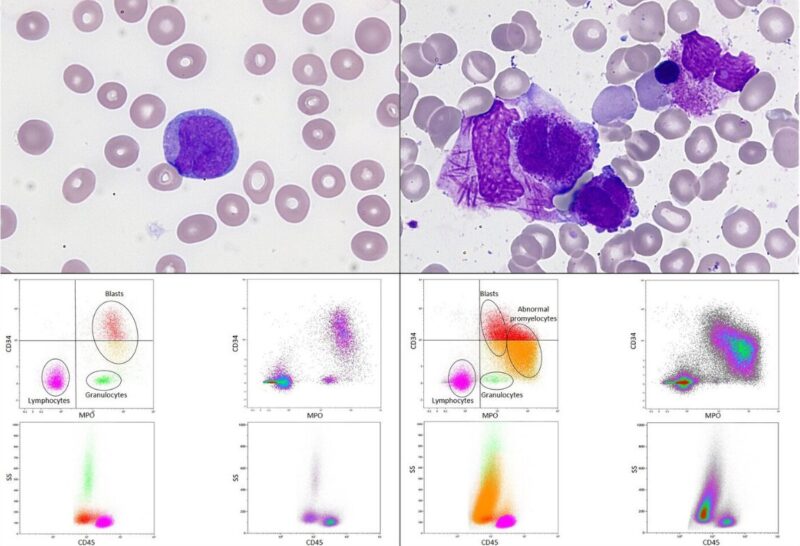
When acute promyelocytic leukaemia changes its face in the peripheral blood.
Authors: Sophie Le Grand, et al.
