Systemic mastocytosis often enters clinical practice through deceptively familiar symptoms, leading patients first to allergists or other specialists rather than hematologists. Systemic mastocytosis develops not simply from excessive mast-cell activation, but because one of the immune system’s oldest defense mechanisms becomes incorporated into a persistent hematopoietic clone.
The resulting clonal mast cells retain the capacity for rapid mediator release while persisting and progressively accumulating within the bone marrow and other tissues. These processes do not necessarily evolve in parallel, nor do symptom burden, disease extent, and prognosis.
Why Mast Cells Are Meant to React So Quickly
Mature mast cells reside immediately beneath the epithelial surfaces of the skin, respiratory and gastrointestinal tracts, around blood vessels, peripheral nerves, and connective tissues. These locations are not accidental. They place mast cells at the interface between the host and potential environmental threats.
Unlike neutrophils or monocytes, which are recruited from the circulation during inflammation, mast cells are long-lived tissue residents. They originate from hematopoietic progenitors in the bone marrow and complete their maturation only after entering peripheral tissues. Although they share several mediators and receptors with basophils, they represent a distinct cellular lineage.
Their principal advantage is speed. The cytoplasm of mature mast cells is packed with preformed mediators, including histamine, tryptase, and heparin. Activation also rapidly induces the synthesis of prostaglandins, leukotrienes, cytokines, chemokines, and growth factors, allowing to initiate an immediate response while sustaining inflammation when needed.
The best-characterized activation pathway involves the high-affinity IgE receptor (FcεRI), which triggers rapid degranulation following allergen-induced cross-linking of receptor-bound IgE. They also respond to microbial products, complement, physical stimuli, alcohol, medications, exercise, emotional stress, and tissue injury, an important reason why mast-cell disorders often lack an obvious allergic trigger.
How a Normal Mast Cell Becomes Neoplastic
In SM mast cells become descendants of a common clone that persists independently of the stimulus that originally brought the patient to medical attention.
In most adults, this transformation is driven by the KIT D816V mutation. KIT is a receptor tyrosine kinase that normally requires stem cell factor to regulate mast-cell survival and maturation. The D816V mutation constitutively activates the receptor, enabling ligand-independent signaling. Rather than causing rapid proliferation, it primarily enhances mast-cell survival, allowing a slowly expanding clone to accumulate over years or decades.
The Shared Origin of Systemic Mastocytosis and Myeloid Neoplasms
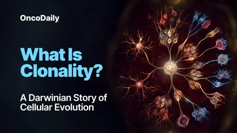
Increasing use of next-generation sequencing has shown that many patients, particularly those with advanced systemic mastocytosis, harbor additional somatic mutations. Perhaps more importantly, accumulating evidence suggests that, in some patients, mutations affecting epigenetic regulation or RNA splicing occur before KIT D816V is acquired by one branch of the evolving clone.
This evolutionary model explains why systemic mastocytosis may coexist with myelodysplastic syndromes, chronic myelomonocytic leukemia, myeloproliferative neoplasms, or acute myeloid leukemia: these disorders may represent divergent descendants of the same ancestral hematopoietic clone rather than entirely independent diseases.
A Multisystem Disease in Disguise
The delay of diagnosis rarely reflects a lack of symptoms. A patient may first seek medical attention because of recurrent flushing after meals. Another because of anaphylaxis following a wasp sting. Others undergo years of investigation for chronic diarrhea, abdominal pain, dyspepsia, osteoporosis, recurrent fractures, palpitations, presyncope, or persistent fatigue. Each complaint has a broad differential diagnosis, and each is frequently evaluated in isolation.
Only when these apparently unrelated manifestations are considered together does a characteristic pattern begin to emerge. Because mast cells reside throughout barrier tissues, the consequences of abnormal mediator release are inherently multisystemic. It presents as a disease of one cell distributed throughout many organs.
Demonstrating Clonality Rather Than Activation
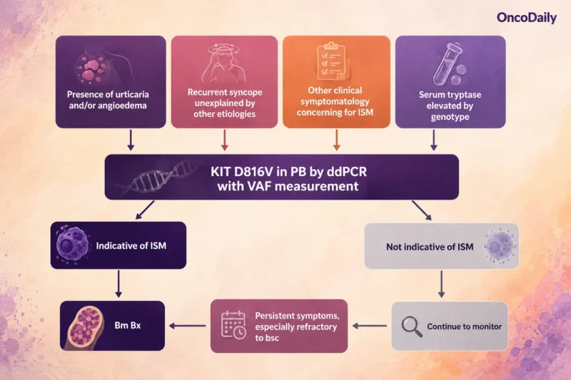
Because mast cells constitutively release small amounts of tryptase, expansion of the mast-cell population generally increases basal serum levels. A persistently elevated basal tryptase concentration above 20 ng/mL is therefore included as a minor diagnostic criterion in both the WHO and ICC classifications.
Yet tryptase is only a clue. Some patients with indolent systemic mastocytosis have lower basal levels, whereas hereditary alpha-tryptasemia, chronic kidney disease, other myeloid neoplasms, and several non-neoplastic conditions can also increase serum tryptase. Its main value is identifying patients who warrant further evaluation.
Definitive evidence usually comes from the bone marrow. Instead of scattered individual mast cells, systemic mastocytosis is characterized by dense multifocal aggregates of at least 15 mast cells, often displaying the spindle-shaped morphology typical of neoplastic cells. Morphology is complemented by immunophenotyping. In addition to KIT (CD117), neoplastic mast cells characteristically express CD25 and often CD2 and/or CD30.
The final layer of evidence comes from molecular testing. Detection of KIT D816V strongly supports the diagnosis. Because mast cells represent only a small fraction of bone marrow cells, highly sensitive techniques such as allele-specific PCR or digital droplet PCR are often required, particularly in indolent disease.
Each investigation answers a different question: serum tryptase estimates mast-cell burden, histology demonstrates tissue infiltration, immunophenotyping identifies an aberrant phenotype, and molecular testing confirms clonality.
The Overlap With Mast Cell Activation Syndrome
Patients with systemic mastocytosis may fulfill diagnostic criteria for MCAS because their neoplastic mast cells continue to release inflammatory mediators. Conversely, many individuals with secondary or idiopathic MCAS have no evidence of a clonal mast-cell disorder despite experiencing recurrent flushing, gastrointestinal symptoms, hypotension, or even anaphylaxis. It is therfore primarily a disorder of mast-cell function.
Why Disease Course Varies
Once systemic mastocytosis has been established, another question follows. If most patients share the same defining feature, a KIT-driven mast-cell clone, why does the disease range from an almost normal life expectancy in one individual to rapidly progressive organ failure in another?
Modern classifications recognize systemic mastocytosis as a spectrum of distinct entities rather than a single disease with varying severity.
Indolent Systemic Mastocytosis: A Stable Clone With an Unstable Clinical Picture
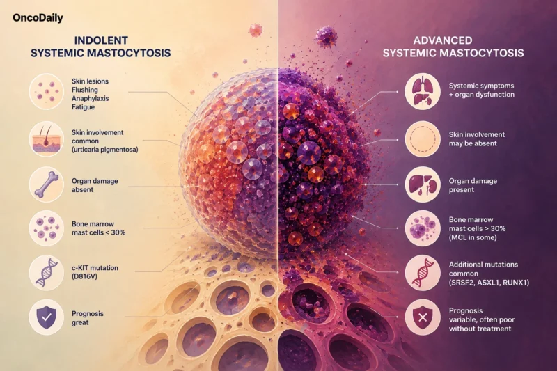
Indolent systemic mastocytosis (ISM) accounts for approximately 80-90% of adult cases. Despite its name, indolent describes the behavior of the neoplasm rather than the severity of symptoms.
Patients may experience recurrent flushing, gastrointestinal symptoms, osteoporosis, fatigue, or even anaphylaxis, yet the mast-cell clone remains biologically stable. Organ function is preserved, progression is uncommon, and life expectancy is close to that of the general population in most patients.
Smoldering Systemic Mastocytosis: When the Clone Begins to Expand
Some patients develop a substantially greater mast-cell burden without evidence of organ damage. These individuals are classified as having smoldering systemic mastocytosis (SSM), an intermediate category between indolent and advanced disease.
SSM is defined by one or more B-findings, which reflect a high disease burden without irreversible organ dysfunction. These include extensive bone marrow infiltration, markedly elevated serum tryptase, organomegaly without functional impairment, and evidence of dysregulated but preserved hematopoiesis. Many patients remain stable for years, but SSM carries a higher risk of progression than ISM and therefore requires closer follow-up.
Advanced Systemic Mastocytosis: When Infiltration Causes Organ Damage
Advanced systemic mastocytosis comprises aggressive systemic mastocytosis (ASM), systemic mastocytosis with an associated hematologic neoplasm (SM-AHN), and mast-cell leukemia (MCL). These entities differ clinically but share one defining feature: mast-cell infiltration causes clinically significant organ dysfunction.
Progressive infiltration of the bone marrow, liver, spleen, gastrointestinal tract, and skeleton may result in cytopenias, portal hypertension, malabsorption, osteolytic lesions, or pathological fractures. These manifestations are recognized as C-findings, indicating organ damage that requires disease-directed therapy rather than symptom control alone.
Why Some Patients Experience Anaphylaxis While Others Never Do
Anaphylaxis is one of the most feared complications of systemic mastocytosis, yet it does not occur uniformly. Its risk depends on the interaction between clonal mast cells, environmental triggers, coexisting IgE-mediated allergy, and individual susceptibility to mediator release.
This relationship is particularly evident in Hymenoptera venom allergy, where severe sting-induced anaphylaxis, often characterized by profound hypotension with relatively limited cutaneous manifestations, is a well-recognized presentation of systemic mastocytosis. Coexisting hereditary alpha-tryptasemia can amplify mediator-related symptoms and increase susceptibility to severe anaphylaxis.
The key clinical message is that anaphylaxis reflects the propensity of mast cells to degranulate rather than the extent of the disease.
What Should Be Treated?
Mast cells release a broad range of mediators that influence vascular permeability, inflammation, tissue remodeling, and bone metabolism. Some manifestations result primarily from mediator release, others reflect progressive tissue infiltration.
These processes also operate on different timescales. Mediator release produces symptoms within seconds or minutes, while persistent inflammation and clonal expansion remodel tissues over months or years.
Bone illustrates this well. Mast-cell mediators disrupt the balance between bone resorption and formation, leading to osteopenia, osteoporosis, and vertebral fractures, even in patients with otherwise indolent disease. In advanced systemic mastocytosis, extensive infiltration may additionally produce osteolytic or osteosclerotic lesions.
Recognizing which process predominates helps determine whether treatment should target mediators, the neoplastic clone, or both.
Managing Consequences of Mast-Cell Activation
When mediator-related symptoms predominate, treatment focuses on preventing mast-cell activation and blocking the effects of released mediators. Antihistamines, leukotriene antagonists, proton pump inhibitors, corticosteroids, trigger avoidance, and, when indicated, venom immunotherapy or anti-IgE therapy can substantially reduce symptoms and the risk of anaphylaxis.
Bone disease should be assessed independently, as it may require antiresorptive therapy such as bisphosphonates regardless of overall disease extent. Patients at risk of anaphylaxis require self-injectable epinephrine and education regarding its prompt use.
The Evolution of KIT Inhibition: What Worked, and What Worked Better
Imatinib was the first logical candidate, given its success in chronic myeloid leukemia and gastrointestinal stromal tumors. Because imatinib preferentially binds the inactive conformation of KIT, the D816V mutation, which stabilizes the receptor in its active state, renders most adult cases resistant. Imatinib therefore retains a role only in the uncommon subgroup lacking D816V or harboring imatinib-sensitive KIT variants.
Midostaurin marked the first major advance. It inhibits both wild-type KIT and KIT D816V, producing meaningful reductions in mast-cell burden, serum tryptase, splenic volume, and organ damage in patients with advanced systemic mastocytosis. For the first time, treatment directly interfered with the molecular driver but the gastrointestinal toxicity and variable efficacy in genetically complex disease, have raised the need for better selectivity.
Avapritinib represented the next stage in this evolution, designed specifically to inhibit activation-loop mutant kinases. The EXPLORER and PATHFINDER studies demonstrated that deep molecular and pathological responses were achievable.
Perhaps even more importantly, the phase III PIONEER trial extended this concept to indolent systemic mastocytosis in patients with persistent symptoms despite optimized supportive care.
Next-generation inhibitors such as bezuclastinib and elenestinib, aim to maintain potent inhibition of KIT D816V while improving long-term tolerability.
Precision Medicine Requires Precision Patient Selection
The availability of effective KIT inhibitors does not mean that every patient requires it. Patients with well-controlled indolent disease generally derive little benefit from long-term KIT inhibition. Treatment decisions therefore remain individualized, balancing expected efficacy against toxicity, comorbidities, quality of life, and patient preference.
Moreover, KIT inhibition addresses only one component. In patients with additional high-risk mutations (SRSF2, ASXL1, RUNX1) or in SM associated with another hematologic neoplasm, prognosis is determined by broader factors than the mast-cell component alone.
Allogeneic hematopoietic stem-cell transplantation remains the only potentially curative treatment. Owing to its considerable risks, it is reserved for selected patients with advanced systemic mastocytosis, especially those with high-risk disease or SM-AHN, often after disease control with KIT-directed therapy.
Living With Systemic Mastocytosis
Though systemic mastocytosis is a lifelong disease, many patients achieve good symptom control and maintain an excellent quality of life with appropriate treatment and self-management.
Ultimately, living well with systemic mastocytosis depends not only on effective therapy but also on recognizing individual triggers (medications, insect stings, physical exertion, temperature changes, or emotional stress…), and maintaining long-term follow-up with a multidisciplinary team.
(Soare D., et al., Syal A., et al., Ustun C., et al., Degboé Y., et al., Rüfer A., et al., Akin C et al., Madigan L.M., et al., 2025)
You can also read: Chronic Myelomonocytic Leukemia (CMML): Beyond the Overlap, Toward the Bigger Picture
Written by Susanna Mikayelyan, MD
FAQ
Can systemic mastocytosis develop without skin lesions?
Yes. Although many people associate mastocytosis with urticaria pigmentosa or other cutaneous lesions, a substantial proportion of adults with systemic mastocytosis have little or no skin involvement. In some cases, recurrent anaphylaxis, unexplained osteoporosis, gastrointestinal symptoms, or persistently elevated serum tryptase may be the first clues. The absence of skin lesions should therefore not exclude the diagnosis when other clinical features raise suspicion.
Does having a KIT D816V mutation always mean a person has systemic mastocytosis?
No. Although KIT D816V is the hallmark driver mutation in most adults with systemic mastocytosis, its detection alone does not establish the diagnosis. The mutation must be interpreted alongside clinical findings, bone marrow morphology, immunophenotyping, and other diagnostic criteria. Conversely, a small minority of patients with systemic mastocytosis do not harbor KIT D816V and may carry alternative KIT mutations.
Can systemic mastocytosis affect life expectancy?
It depends on the disease subtype. Most patients with indolent systemic mastocytosis have a life expectancy close to that of the general population, although symptoms may significantly affect quality of life. In contrast, advanced forms, particularly aggressive systemic mastocytosis, SM-AHN, and mast-cell leukemia, are associated with progressive organ damage and substantially worse outcomes.
Are vaccinations, surgery, or anesthesia safe for people with systemic mastocytosis?
In most cases, yes. Patients with systemic mastocytosis can safely undergo surgery and receive routine vaccinations, but careful planning may be required because certain medications, anesthetic agents, or physical stress can trigger mast-cell mediator release. Informing the treating team in advance allows appropriate precautions, including continuation of regular anti-mediator therapy and, in selected cases, premedication before procedures.
Can systemic mastocytosis coexist with other blood cancers?
Yes. In some patients, systemic mastocytosis shares a common hematopoietic ancestor with another myeloid neoplasm, resulting in systemic mastocytosis with an associated hematologic neoplasm (SM-AHN). The associated disorder may include myelodysplastic syndromes, chronic myelomonocytic leukemia, myeloproliferative neoplasms, or acute myeloid leukemia. This overlap has important implications for prognosis and treatment selection.