
Flow cytometry can characterize thousands to millions of individual cells within minutes. In hematology, that makes it particularly useful for diseases defined by abnormal cell populations and measurable residual disease.
Flow cytometry determines whether an abnormal population exists and, if so, what lineage it belongs to, how mature it is, and whether its antigen pattern differs from normal cells. The diagnosis comes from that pattern, not from a list of positive and negative CD (Cluster of Differentiation) markers.
Take CD5. Its expression on T cells is normal. On a clonal B-cell population, it raises a differential, particularly chronic lymphocytic leukemia/small lymphocytic lymphoma and mantle cell lymphoma. The same marker can have very different significance depending on which cells express it.
How Does Flow Cytometry Work?
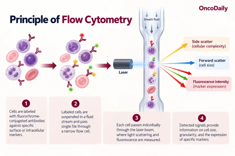
Cells from blood, bone marrow, lymph node, cerebrospinal fluid, or another specimen are suspended in fluid and passed individually through one or more lasers. The instrument records two broad types of information.
Light scatter provides physical information about the cell. Forward scatter roughly correlates with cell size, and side scatter reflects internal complexity or granularity. These properties help separate lymphocytes, monocytes, granulocytes, blasts, and debris, but scatter alone cannot identify a malignancy.
Fluorescence provides the immunophenotype. Before analysis, the cell suspension is incubated with antibodies against selected cellular antigens. Each antibody is linked to a fluorochrome. As the labeled cells pass through the laser, the fluorochromes emit light at characteristic wavelengths. Detectors capture these signals and convert them into electronic data, allowing the expression of multiple antigens to be measured on each individual cell.
Modern multiparameter flow cytometry measures many antigens on the same cell at the same time, so flow can show that one abnormal B-cell population coexpresses CD19 and CD5, grade the intensity of each antigen, and test the same cells for CD23, CD200, κ/λ light chains, and other markers.
What Does Gating Mean?
A marrow or blood specimen contains many different cells. Gating defines the population that will be analyzed. An initial gate may use CD45 and side scatter to separate broad leukocyte populations.
Additional marker combinations then progressively define the cells of interest. Gating prevents a common interpretive mistake: reading percentages without asking what the denominator is. “CD10: 35%” means little by itself. Thirty-five percent of all acquired cells, lymphocytes, B cells, or an abnormal gated population are very different findings.
Types of Flow Cytometry Panels
A screening or orientation panel determines what kind of abnormal population is present and guides further testing. In suspected acute leukemia, the EuroFlow Acute Leukemia Orientation Tube uses a compact combination of markers to distinguish major differentiation pathways before lineage-specific characterization.
A diagnostic characterization panel goes further. Once a B-cell, T-cell, myeloid, or plasma-cell abnormality has been identified, additional markers define its phenotype and narrow the differential diagnosis.
When the differential is already narrow, the panel can be tailored to the suspected disease, using markers selected to confirm its characteristic immunophenotype and distinguish it from its closest mimics. An MRD panel searches for very small residual abnormal populations after treatment and is built for sensitivity.
Acute Leukemia
In acute leukemia, flow cytometry first identifies the immature blast population. CD34, CD117, TdT, and HLA-DR help characterize its maturation but do not independently establish lineage.
Leukemic blasts frequently express antigens outside their expected lineage: AML may aberrantly express CD7 or CD19, which does not automatically make it mixed-phenotype acute leukemia. Lineage assignment follows defined lineage-associated criteria, regardless of how many markers happen to be positive.
After lineage is established, the phenotype becomes more specific. AML commonly includes combinations of CD13, CD33, CD117, and myeloperoxidase, with variable CD34 and HLA-DR.
B-ALL commonly shows CD19, CD22, and CD79a, with variable CD10, CD20, CD34, and TdT depending on the maturation stage and molecular subtype. T-ALL centers on cytoplasmic or surface CD3 with combinations of CD7, CD5, CD2, CD1a, and CD4/CD8.
The exact phenotype can also direct the next test. Flow may strongly suggest acute promyelocytic leukemia, for example, but detection of the PML::RARA fusion by FISH or RT-PCR establishes the diagnosis.
Mature B-Cell Neoplasms
For a mature B-cell population, surface κ and λ light chains are commonly used to assess clonality. A marked predominance of one light chain within an abnormal B-cell population supports a clonal process. Classification then depends on the rest of the phenotype.
In a CD5-positive clonal population, CD23, CD200, and FMC7 help separate CLL/SLL from mantle cell lymphoma. Flow may strongly favor one diagnosis, but mantle cell lymphoma may still require cyclin D1, SOX11, or CCND1 rearrangement assessment.
A CD10-positive clonal B-cell population suggests germinal-center differentiation. Follicular lymphoma becomes an important consideration, but CD10 expression doesn’t establish it. The CD5-negative/CD10-negative group is broader. Marginal zone lymphoma, lymphoplasmacytic lymphoma, and other mature B-cell neoplasms. In these cases, flow often narrows the differential.
Antigen intensity matters as much as positivity. Dim CD20 and weak surface immunoglobulin support a different pattern from bright CD20 and strong surface IG, even when both populations are technically CD20-positive.
The Limits of Flow Cytometry in Lymphoma Diagnosis
Flow cytometry removes cells from their tissue architecture, which limits what it can determine about lymphoma. A clonal population can establish that abnormal B cells are present and define their immunophenotype. It cannot show a follicular versus diffuse growth pattern or fully assess the relationship between malignant cells and surrounding tissue.
Histology and immunohistochemistry are therefore central to many lymphoma diagnoses. A lymph node may contain lymphoma even when flow is negative if malignant cells are sparse, fragile, lost during processing, or poorly represented in the submitted material.
Mature T-Cell Neoplasms
B-cell clonality can often be approached through κ/λ restriction. Mature T-cell neoplasms are less straightforward. Flow instead looks for an abnormal T-cell population through antigen loss, altered intensity, unusual combinations, skewed CD4/CD8 patterns, and, where available, restricted T-cell receptor-associated expression. Loss or dim expression of CD2, CD3, CD5, or CD7 may indicate an aberrant phenotype.
None is independently proof of lymphoma. Reactive T-cell populations can show unusual phenotypes, and malignant T cells may retain apparently normal pan-T-cell markers. Molecular clonality studies and tissue findings may therefore be required.
Flow Cytometry at the MRD Level
AML MRD commonly combines two approaches. A leukemia-associated immunophenotype (LAIP) identified at diagnosis can be tracked after treatment. A different-from-normal (DfN) approach searches for populations whose maturation pattern differs from normal or regenerating marrow.
AML immunophenotypes can change during treatment: a residual clone may lose or gain antigen expression, making exclusive reliance on the original diagnostic phenotype unsafe. MRD interpretation also depends on how many cells were acquired, assay sensitivity, and specimen quality. A negative MRD result is only as informative as the assay’s detection limit.
Bone marrow quality is particularly important. Hemodilution can reduce the apparent disease burden because the aspirate increasingly resembles peripheral blood. A technically “negative” result from a poor-quality specimen provides less reassurance than the word suggests.
You can also read:

Flow Cytometry vs FISH, PCR, and NGS
These tests examine different levels of the disease and their results are complementary:
- Flow cytometry identifies abnormal cells through phenotype.
- FISH searches for selected chromosomal abnormalities.
- PCR detects defined molecular targets with high analytical sensitivity.
- Next-generation sequencing (NGS) interrogates sequence alterations across the genes included in the assay.
After treatment, flow MRD and molecular MRD may follow different targets and have different sensitivities. Discordant results require biological and technical interpretation before one assay is assumed to be wrong.
Could AI Change Flow Cytometry Interpretation?
Flow cytometry data interpretation still relies heavily on manual gating. AI can analyze relationships across multiple markers simultaneously, recognize abnormal cell clusters, and reduce operator-dependent variation. In a 2024 study of 1,820 samples, a deep-learning model identified acute leukemia with an AUROC of 0.961 and differentiated AML from B- and T-lymphoblastic leukemia with an AUROC of 0.965.
MRD may be particularly suited to AI-assisted analysis because residual malignant cells can be a tiny fraction of millions of acquired events. In the B-ALL study, AI-assisted analysis agreed completely with conventional interpretation for MRD ≥0.01%.
AI can find suspicious populations faster and more consistently. Algorithms still have to contend with specimen quality, hemodilution, treatment-related phenotypic shifts, different antibody panels, and variation between laboratories.
How to Read the Final Flow Cytometry Impression
The most useful part of the report is usually the description of the abnormal population.
For diagnostic studies: What population was found? What lineage and maturation does it show? What makes it abnormal or clonal? Which diagnosis or differential does that phenotype support?
Then look for what flow cannot resolve and which additional tests are recommended.
For MRD: Was the specimen adequate? How much abnormal population was detected? What sensitivity did the assay achieve?
You can also read:

FAQ
Can flow cytometry diagnose leukemia from a blood sample?
Yes, in many cases. If abnormal cells are circulating in sufficient numbers, peripheral blood flow cytometry can identify their lineage, maturation, clonality, and aberrant antigen expression. Bone marrow examination may still be required for complete classification, disease assessment, cytogenetic or molecular testing, and treatment planning.
Can leukemia be present even if flow cytometry is negative?
Yes. A negative result does not exclude leukemia when abnormal cells are below the assay's detection limit, absent from the sampled compartment, or poorly represented because of specimen quality. Interpretation should always consider morphology, clinical findings, and genetic or molecular studies.
Why are so many CD markers needed in flow cytometry?
Few markers are specific enough to define a hematologic malignancy alone. Diagnostic information comes from combinations of markers expressed on the same cell population, including their intensity and patterns of coexpression. Multiparameter analysis can distinguish malignant cells from normal populations that share individual markers.
Why can a leukemia's flow cytometry pattern change after treatment?
Leukemic populations can undergo immunophenotypic shifts under treatment pressure. Residual cells may gain, lose, or change the intensity of antigens present at diagnosis. This is one reason AML MRD assessment can combine tracking of the original leukemia-associated immunophenotype with a different-from-normal approach.
How sensitive is flow cytometry for MRD?
Sensitivity depends on the assay, disease, number of cells acquired, specimen quality, and laboratory methodology. A negative MRD result therefore needs to be interpreted alongside the assay's reported detection capability; “MRD negative” does not mean that absolutely no malignant cells remain.