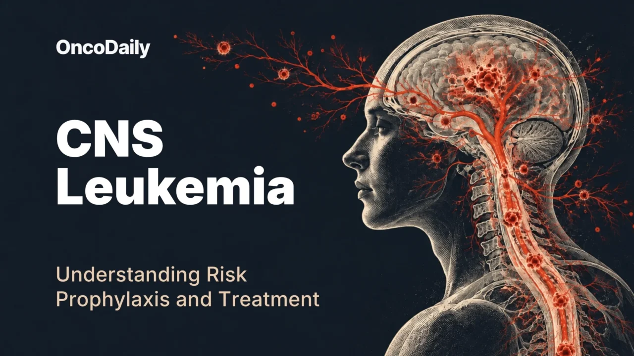
CNS leukemia refers to leukemic involvement of the central nervous system, most commonly the cerebrospinal fluid (CSF) and leptomeninges surrounding the brain and spinal cord. This is most clinically important in acute lymphoblastic leukemia, where the CNS has long been recognized as a site of treatment failure and relapse.
Without CNS-directed prophylaxis, historical CNS relapse rates reached 30-40% in adults with ALL, with modern prophylaxis, reported rates are roughly 4-15%. Those numbers explain an otherwise counterintuitive part of ALL treatment. A patient can have no neurologic symptoms and no detectable leukemia in the CSF yet still receive repeated intrathecal chemotherapy.
How Does Leukemia Reach the Central Nervous System?
The traditional explanation centered on the blood-brain barrier: leukemic cells crossed it, systemic chemotherapy penetrated poorly, and the CNS became a protected “sanctuary.” Current biology is more interesting.
Leukemic cells appear to use several routes into CNS-associated tissues. Experimental studies have implicated migration along the outside of vessels connecting bone marrow and meningeal structures, including emissary vessels. Chemokine and adhesion pathways, including CXCR4-CXCL12 and CCR7-CCL19 signaling, as well as integrin-mediated interactions, can promote leukemic cell migration, retention, and survival within these niches. Research has also identified direct connections between skull and vertebral marrow and the adjacent meninges.
CSF is a metabolically austere environment compared with blood and bone marrow, so ALL cells must adapt. Experimental models suggest that contact with meningeal cells activates survival programs, such as PI3K-AKT signaling and can reduce sensitivity to chemotherapy. Leukemic cells may also alter their metabolism to survive, using increased fatty-acid oxidation.
Future CNS-directed treatment may target those dependencies. Several candidate pathways are under investigation, but for now, established CNS-directed treatment relies on intrathecal chemotherapy and systemic CNS-active therapy.
CNS Disease in ALL vs AML?
CNS involvement is disproportionately common in ALL, with particularly strong experimental evidence for CCR7- and CXCR4-mediated trafficking in T-ALL. In adults, CNS disease is detected at diagnosis in ~5-11% of ALL, compared with ~1-3% of AML. Risk is associated with features including:
- T-cell lineage
- elevated lactate dehydrogenase (often >3 times the upper limit of normal)
- hyperleukocytosis (WBC >100 × 10⁹/L is an independent risk factor)
- adverse genomic abnormalities (KMT2A-rearranged, BCR::ABL1-positive, TCF3::PBX1-positive ALL)
No clinical risk model can reliably identify every patient harboring or destined to develop CNS disease. Modern ALL therapy therefore treats biological risk before overt disease becomes measurable, and the strategy has worked.
In AML, certain situations, including monocytic differentiation, hyperleukocytosis and extramedullary disease, may increase concern, but available evidence does not support routine intrathecal prophylaxis. It would expose a large population with a relatively low baseline event rate to repeated invasive procedures and treatment without population-level benefit.
You can also read:

Early Signs and Red Flags of CNS Involvement in Leukemia
CNS leukemia often causes no symptoms at all, especially when it’s confined to the CSF or meninges. When symptoms do show up, what matters is a pattern that’s new, persistent, progressive, or points to a specific site.
Early or subtle signs: new or persistent headache, unexplained nausea or vomiting, visual changes, facial numbness or weakness, hearing changes, paresthesias, subtle gait or balance issues. More diffuse involvement can show up as cognitive or behavioral change.
Cranial neuropathies are worth watching closely because they pinpoint a nerve: diplopia, facial weakness or numbness, hearing loss, vision changes, or trouble swallowing. In ALL, clear cranial nerve involvement counts as CNS disease on its own, even if the CSF cell count doesn’t meet the CNS3 threshold.
Spinal cord, cauda equina, or nerve-root involvement looks different early on: persistent back or radicular pain, limb weakness, sensory loss, or gait trouble, with bladder and bowel function still intact.
Red flags: progressive headache with repeated vomiting or papilledema (raised intracranial pressure), new seizures, altered consciousness, focal weakness, fast-worsening cranial nerve deficits, or marked gait decline. For spinal or cauda equina disease, the red flag is progression, worsening weakness or sensory loss, and especially new bladder or bowel dysfunction, which means significant compression and needs urgent evaluation.
Infection, bleeding, hyperleukocytosis, metabolic disturbance, and treatment-related neurotoxicity can also look similar, so diagnosis rests on the clinical picture plus CSF testing and, when needed, imaging.
How Is CNS Involvement in Leukemia Detected?
Lumbar puncture remains the foundation of CNS assessment in ALL. CSF is evaluated for leukemic blasts, traditionally by cytology and increasingly with multiparameter flow cytometry. Flow cytometry can detect leukemic cell populations below the sensitivity of conventional cytology.
According to ALL protocols, CNS1 indicates no detectable blasts. CNS2 describes detectable blasts with fewer than 5 white blood cells/µL, while CNS3 generally requires blasts with at least 5 white blood cells/µL or clinical evidence of CNS leukemia (cranial nerve involvement, for instance).
A traumatic lumbar puncture complicates this classification because circulating blasts introduced with peripheral blood can mimic true CNS disease. The Steinherz/Bleyer criteria and similar algorithms use the relationship between blood and CSF cell counts to help distinguish contamination from real involvement.
Contrast-enhanced MRI is useful when neurologic symptoms suggest cranial nerve, leptomeningeal, parenchymal or other structural involvement, but its sensitivity is insufficient to exclude microscopic CNS leukemia.
How Is CNS Prophylaxis Delivered?
CNS prophylaxis usually combines intrathecal chemotherapy, delivered directly into the CSF, with systemic drugs that have meaningful CNS activity. Methotrexate is the most widely used intrathecal agent, cytarabine and corticosteroids are also used, either alone, alternated, or in combinations depending on the protocol. Systemic methotrexate and cytarabine contribute additional CNS activity in many regimens.
In pediatric high-risk ALL, triple intrathecal therapy with methotrexate, cytarabine and a corticosteroid did not improve 5-year disease-free survival compared with intrathecal methotrexate alone, and older data raised concerns about inferior overall survival with the triple approach. The optimal CNS strategy therefore depends on the entire systemic regimen.
The same principle comes from systemic methotrexate studies. In children and young adults with B-ALL enrolled in AALL0232, high-dose methotrexate produced favorable event-free survival with CNS relapse below 5%. Yet in T-ALL, AALL0434 found fewer isolated CNS relapses with Capizzi escalating-dose methotrexate plus pegaspargase than with high-dose methotrexate, 0.4% versus 3.0%.
Why Is Cranial Radiation Rarely Used for Routine Prophylaxis?
Radiation can control leukemia in the CNS, but long-term exposure carries risks that are especially consequential for children and young adults, including neurocognitive effects, endocrinopathy and secondary malignancies. As systemic and intrathecal chemotherapy improved, studies showed that prophylactic radiation could be omitted from many regimens without unacceptable increases in CNS relapse.
Radiotherapy has therefore moved from routine prophylaxis toward selected use, including some cases of overt CNS leukemia, cranial nerve or focal involvement, persistent disease and relapse. Treatment must also account for planned hematopoietic cell transplantation because conditioning regimens may add further radiation exposure.
You can also read:
What Changes When CNS Leukemia Is Already Present?
Once leukemia is demonstrated in the CNS, intrathecal therapy is generally intensified and repeated until CSF clearance, alongside treatment of the systemic leukemia. Radiation may enter the strategy for selected patients. Allogeneic hematopoietic cell transplantation may also be considered according to the leukemia subtype, response, MRD, prior therapy, age, and transplant fitness.
In two large French adult ALL studies summarized by Kopmar and Cassaday, approximately 75% of CNS relapses occurred with leukemia at another site, only about 25% were isolated to the CNS. Historically, median overall survival after CNS relapse in adults has been less than one year.
The approach in children is more risk-adapted. Early isolated CNS relapse and CNS relapse with marrow involvement frequently lead to consideration of allogeneic transplantation, whereas some children with late isolated CNS relapse can be cured with intensive systemic and intrathecal therapy, historically combined with cranial irradiation, without transplantation. In UKALL R3, late isolated CNS relapse was associated with 5-year EFS of 81% and OS of 85%.
CNS Control With Modern ALL Therapies
Blinatumomab, inotuzumab ozogamicin, potent BCR::ABL1 tyrosine kinase inhibitors and CAR T-cell therapy have strengthened systemic disease control. Some modern regimens can achieve deep molecular remissions with far less conventional chemotherapy.
For Philadelphia chromosome-positive ALL, imatinib has poor CNS penetration, while dasatinib has demonstrated that activity. In a pediatric trial comparing chemotherapy plus dasatinib with chemotherapy plus imatinib, the 4-year cumulative risk of CNS relapse was 2.7% with dasatinib versus 8.4% with imatinib.
Blinatumomab can achieve responses in CNS ALL, but prospective evidence is limited. In AALL1331, 4-year disease-free survival was 24% with blinatumomab and chemotherapy in isolated CNS relapse. Inotuzumab ozogamicin has limited CNS activity and is inadequate for established CNS disease.
Chemotherapy-free or chemotherapy-light regimens raise an even broader concern: what is now providing CNS protection? Intrathecal prophylaxis therefore remains an intentional component of contemporary ALL treatment while researchers determine how much conventional CNS-directed therapy newer systemic approaches can safely replace.
CAR T cells can traffic into the CNS and have demonstrated activity in patients with CNS ALL, showing that the compartment is not immunologically or therapeutically sealed. Evidence is still evolving, particularly regarding optimal patient selection and the distinction between active CNS leukemia and treatment-related neurotoxicity.
You can also read
FAQ
Can leukemia affect the brain even when the blood or bone marrow is in remission?
Yes. CNS relapse can occur without simultaneous marrow relapse, although isolated CNS relapse is less common than CNS relapse accompanied by leukemia elsewhere. In two large adult ALL studies, about 25% of CNS relapses were isolated to the CNS.
Why does ALL spread to the CNS more often than AML?
ALL has a stronger biological tendency to traffic into and survive within CNS-associated tissues. Pathways involving CXCR4–CXCL12 and CCR7–CCL19, particularly studied in T-ALL, appear to facilitate leukemic cell migration and retention. At diagnosis, CNS involvement is reported in roughly 5–11% of adults with ALL compared with about 1–3% with AML.
Can CNS leukemia be present without neurological symptoms?
Yes. Microscopic CNS leukemia may produce no headache, weakness, seizures, or other neurological symptoms. This is one reason lumbar puncture and CNS-directed prophylaxis are built into ALL treatment even when patients feel neurologically well.
Can a normal brain MRI rule out CNS leukemia?
No. Contrast-enhanced MRI is useful when clinicians suspect leptomeningeal, cranial nerve, parenchymal, or other structural involvement, but it cannot reliably exclude microscopic CNS disease. Cerebrospinal fluid analysis remains central to assessment.
Does more intrathecal chemotherapy always provide better CNS protection?
No. CNS control depends on the entire treatment regimen. In pediatric high-risk ALL, triple intrathecal therapy with methotrexate, cytarabine, and a corticosteroid did not improve 5-year disease-free survival compared with intrathecal methotrexate alone. Systemic agents with CNS activity also contribute substantially to CNS control.
Could future ALL treatment eliminate repeated lumbar punctures and intrathecal chemotherapy?
Possibly, but current evidence does not support doing so routinely. Research is investigating leukemic trafficking, meningeal adhesion, metabolic adaptation, and biomarkers that could identify patients at very low or high risk of CNS disease. As systemic ALL therapy becomes increasingly targeted and chemotherapy-light, determining who still needs CNS prophylaxis, how much, and for how long is becoming an important research question.