
Whole-body HER2 imaging may provide information about trastuzumab deruxtecan sensitivity that a single tumor biopsy cannot capture.
Updated results from the phase II PREDICT-HER trial, presented at ESMO Congress 2026, showed that baseline HER2-PET status was strongly associated with progression-free survival in patients with advanced breast cancer receiving trastuzumab deruxtecan (T-DXd), independently of conventional pathological HER2 classification.
The central finding was particularly striking in patients whose tumors were classified as HER2-low or HER2-zero by pathology.
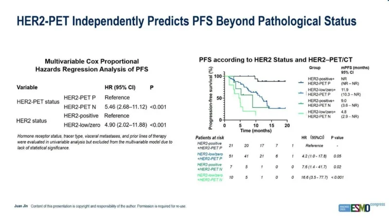
Patients with HER2-low/zero disease but positive HER2-PET had a median PFS of 11.9 months, whereas those with HER2-low/zero disease and negative HER2-PET had median PFS of only 4.8 months.
Even among conventionally HER2-positive cancers, HER2-PET separated patients into markedly different outcome groups. Median PFS was not reached in patients with HER2-positive, HER2-PET-positive disease compared with 9.0 months in those whose tumors were pathologically HER2-positive but whose whole-body HER2-PET was negative.
In multivariable analysis, HER2-PET-negative status was associated with a more than five-fold higher hazard of progression or death compared with HER2-PET positivity, with an HR of 5.46 (95% CI 2.68–11.12; P<0.001).
The findings suggest that the clinically relevant question may increasingly be not simply whether a biopsy is labeled HER2-positive, HER2-low or HER2-zero, but whether enough accessible HER2 target is present across a patient’s metastatic disease for a HER2-directed ADC to bind effectively.
Why Conventional HER2 Testing May Not Tell the Whole Story
T-DXd has dramatically expanded the therapeutic importance of HER2 expression.
The drug was initially established in HER2-positive metastatic breast cancer but subsequently demonstrated activity in HER2-low disease and, more recently, HER2-ultralow HR-positive disease. In the United States, T-DXd is approved for HR-positive HER2-low or HER2-ultralow metastatic breast cancer after endocrine therapy, illustrating how increasingly small degrees of HER2 expression can carry therapeutic relevance.
Yet conventional HER2 testing remains based largely on tissue obtained from one tumor site at one point in time. That creates an important limitation in metastatic disease. HER2 expression can vary between the primary tumor and metastatic lesions, between different metastases in the same patient and over time following systemic treatment.
A single biopsy may therefore classify a tumor as HER2-low while other lesions contain little or no accessible HER2, or conversely underestimate HER2 expression elsewhere in the body.
This spatial heterogeneity becomes especially important for an ADC such as T-DXd, because efficacy depends not only on histopathological classification but also on sufficient target availability, antibody binding, internalization and delivery of the cytotoxic payload.
PREDICT-HER explores whether molecular imaging can provide a whole-body measurement of that target landscape.
 PREDICT-HER Used HER2-Directed PET Before T-DXd
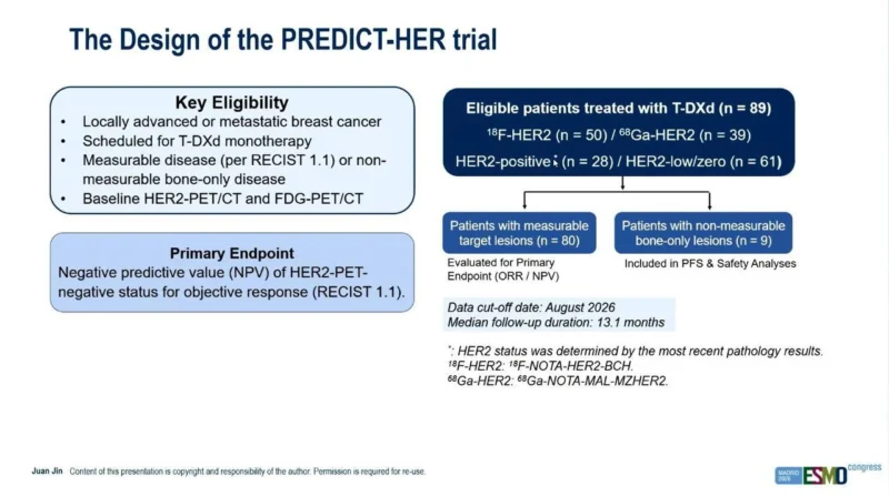
The phase II study enrolled patients with locally advanced or metastatic breast cancer who were already scheduled to receive T-DXd monotherapy.
All participants underwent both conventional FDG-PET/CT and HER2-directed PET/CT before starting treatment. Two HER2-directed tracers were used: 18F-NOTA-HER2-BCH in 50 patients and 68Ga-NOTA-MAL-MZHER2 in 39 patients.
Both are affibody-based imaging agents designed to bind HER2 and allow visualization of HER2 target availability throughout the body. The final ESMO presentation included 89 treated patients, of whom 28 had HER2-positive disease and 61 had HER2-low or HER2-zero disease according to the most recent pathology results.
Eighty patients had measurable target lesions and were evaluable for the primary response-related analysis, while nine patients had non-measurable bone-only disease and were included in progression-free survival and safety analyses.
The data cutoff was August 2026, with a median follow-up of 13.1 months.
HER2-PET Was Designed as a Rule-Out Biomarker
PREDICT-HER was not primarily designed to prove that HER2-PET-positive tumors would always respond. Instead, its primary objective focused on the negative predictive value of HER2-PET-negative status. That distinction is clinically important.
A useful rule-out biomarker could identify patients whose cancers appear eligible for T-DXd based on conventional pathology but have insufficient whole-body HER2 target availability to derive meaningful benefit.
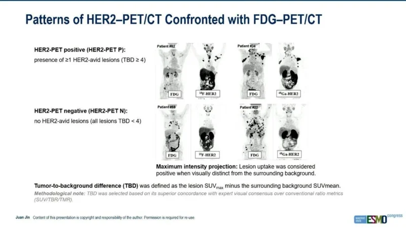
In the imaging analysis, a patient was considered HER2-PET positive if at least one lesion demonstrated clearly HER2-avid uptake, using a tumor-to-background difference threshold of at least 4.
HER2-PET-negative disease was defined by the absence of HER2-avid lesions, with all evaluated lesions below that threshold.
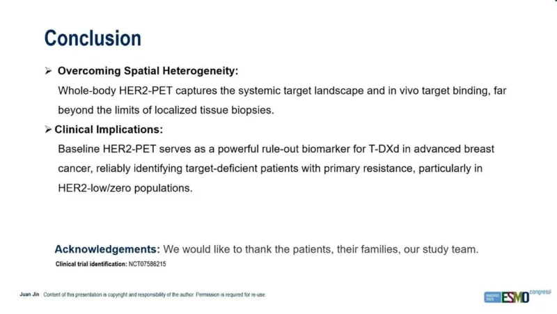
The updated presentation concluded that baseline HER2-PET performed as a strong rule-out biomarker, particularly in HER2-low/zero cancers.
HER2-PET Predicted PFS Independently of Tissue HER2 Status
One of the strongest results came from the multivariable analysis. When HER2-PET status and pathological HER2 classification were considered simultaneously, both remained independently associated with PFS.
Compared with patients whose tumors were HER2-PET positive, HER2-PET-negative patients had an HR for progression or death of:
- 5.46 (95% CI 2.68–11.12; P<0.001)
Separately, patients with pathological HER2-low/zero disease had an HR of:
- 4.90 (95% CI 2.02–11.88; P<0.001)
compared with HER2-positive disease.
Hormone receptor status, tracer type, visceral metastases and number of prior treatment lines were evaluated but were not retained as statistically significant variables in the multivariable model.
The implication is important: HER2-PET was not merely reproducing information already available from pathology. It added independent prognostic information about response durability during T-DXd therapy.
Four Groups Revealed a Clear Biological Gradient
When patients were divided by both pathological HER2 status and HER2-PET findings, a striking gradient emerged. Patients with HER2-positive/HER2-PET-positive disease had the most favorable outcome, with median PFS not reached at the time of analysis.
Patients with HER2-low/zero but HER2-PET-positive disease had median PFS of 11.9 months. Patients with HER2-positive but HER2-PET-negative disease had median PFS of 9.0 months. The worst outcome occurred in patients with HER2-low/zero and HER2-PET-negative disease, where median PFS was only 4.8 months.
Using HER2-positive/HER2-PET-positive disease as the reference group, the hazard ratio for progression was approximately 4.2 for HER2-low/zero PET-positive disease, 7.6 for HER2-positive PET-negative disease and 16.6 for HER2-low/zero PET-negative disease.
The confidence intervals are wide because some groups were small, but the directional pattern is highly provocative.
A HER2-Positive Biopsy Did Not Guarantee Strong T-DXd Sensitivity
Perhaps the most interesting group consisted of patients whose pathology said HER2-positive but whose PET imaging was HER2-negative. Conventional thinking would generally consider these patients strong candidates for HER2-directed therapy.
Yet their median PFS was substantially shorter than that of HER2-positive/HER2-PET-positive patients. This discordance may reflect spatial heterogeneity. A biopsy could sample one HER2-rich lesion while much of the remaining metastatic burden contains little accessible HER2.
Alternatively, HER2 may be present histologically but insufficiently accessible to the imaging tracer, and potentially to therapeutic antibody binding in vivo.
PREDICT-HER does not yet establish which biological explanation predominates, but it demonstrates that tissue expression and whole-body target availability are not always equivalent.
HER2-Low Disease May Be Where PET Adds the Most Value
The potential clinical value appears particularly high in HER2-low and HER2-zero disease. This population encompasses tumors with relatively limited HER2 expression, where small differences in receptor density may meaningfully influence ADC delivery. A HER2-low label based on IHC 1+ or 2+/ISH-negative status describes a broad biological spectrum.
Whole-body HER2-PET could potentially divide that spectrum into cancers with enough accessible target to support T-DXd activity and cancers with effectively target-poor disease despite an eligible biopsy result.
That possibility is consistent with the large PFS difference between HER2-low/zero PET-positive and PET-negative patients.
Importantly, this should not yet be interpreted as evidence that HER2-PET-negative patients should routinely be denied T-DXd.
PREDICT-HER is a relatively small, single-arm phase II biomarker study. Prospective validation is necessary before imaging can replace or modify regulatory tissue-based eligibility criteria.
Whole-Body Imaging Addresses Spatial Heterogeneity
The conceptual strength of HER2-PET is straightforward: metastatic cancer exists throughout the body, whereas a biopsy samples millimeters of one lesion. HER2-PET provides a systemic view. FDG-PET identifies metabolically active disease, while HER2-PET can show which of those lesions actually demonstrate HER2-specific tracer uptake.
A patient can therefore have extensive FDG-avid metastatic disease but very little HER2-targetable signal. Conversely, HER2-directed imaging may reveal strong target uptake across lesions even when pathology categorizes a tumor in a lower HER2-expression group.
This is particularly relevant in the ADC era, where treatment selection increasingly depends on quantitative or heterogeneous target expression rather than simple binary positive-versus-negative classification.
PREDICT-HER Is a Biomarker Study, Not Yet a Treatment-Selection Standard
The results are compelling, but there are several reasons for caution. First, the study was single-arm. Every patient received T-DXd, so HER2-PET currently functions as a marker of outcome during T-DXd rather than a definitively validated predictive biomarker demonstrating interaction with treatment versus another therapy.
Second, the cohort was modest, with only 89 patients and relatively small numbers in some PET/pathology combinations.
Third, two different radiotracers were used. Although tracer type did not emerge as an important variable in the multivariable analysis, standardization will be necessary if HER2-PET is to become broadly reproducible.
Fourth, accessibility matters. Molecular HER2 imaging requires specialized tracers, nuclear medicine infrastructure and standardized interpretation that are not currently available in routine breast oncology practice.
Finally, thresholds will need external validation before treatment decisions can reliably depend on them.
Could HER2-PET Prevent Futile ADC Treatment?
This is ultimately the clinically attractive possibility. T-DXd is highly active but is not a trivial treatment. It carries meaningful risks, including interstitial lung disease/pneumonitis and gastrointestinal and hematologic toxicities, and requires repeated intravenous treatment.
If a baseline imaging test could identify patients with a very low probability of meaningful response, clinicians might eventually redirect them toward another effective therapy rather than exposing them to ineffective ADC treatment.
That concept becomes particularly relevant as the number of breast cancer ADCs grows. Treatment selection may increasingly involve choosing among agents targeting HER2, TROP2, HER3 and other proteins.
Whole-body target imaging could potentially become part of a broader strategy for selecting the ADC whose target is most accessible across an individual patient’s metastatic disease.
That remains a future possibility rather than current standard practice.
OncoDaily Takeaway
PREDICT-HER challenges the assumption that one tissue biopsy fully describes whether metastatic breast cancer is biologically suited for T-DXd. In the updated ESMO 2026 presentation, baseline HER2-PET status independently predicted progression-free survival even after accounting for conventional pathological HER2 status.
HER2-PET-negative disease was associated with a 5.46-fold higher hazard of progression or death compared with PET-positive disease. The most favorable patients had both HER2-positive pathology and positive HER2-PET, with median PFS not reached.
At the opposite extreme, patients with HER2-low/zero, HER2-PET-negative disease had median PFS of only 4.8 months, compared with 11.9 months when HER2-low/zero disease remained PET positive.
Perhaps most provocatively, even pathologically HER2-positive patients had shorter PFS when HER2-PET was negative. The study therefore suggests that whole-body in vivo target availability may matter independently of what a single biopsy reports.
PREDICT-HER is not yet sufficient to make HER2-PET a routine gatekeeper for T-DXd. The study is small, single-arm and requires external validation. But the principle could become increasingly important in the ADC era.
The future of biomarker selection may move beyond asking whether a tumor expresses a target somewhere. The more clinically meaningful question may be:
How much target is available across the entire disease, and can the drug actually reach it?
Source
- Jin J, et al. HER2-PET imaging to predict trastuzumab deruxtecan efficacy in advanced breast cancer: The phase II PREDICT-HER trial. Presented at ESMO Congress 2026, October 24, 2026. Clinical trial identifier NCT07586215.