





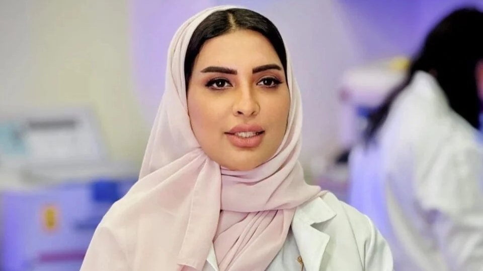
Rawia Mohamed, Head of the Anatomical Pathology Department at Burjeel Medical City, Regional Ambassador of the Asian society of Digital Pathology and Associate Professor at RAK Medical and Health Sciences University, shared a post on LinkedIn:
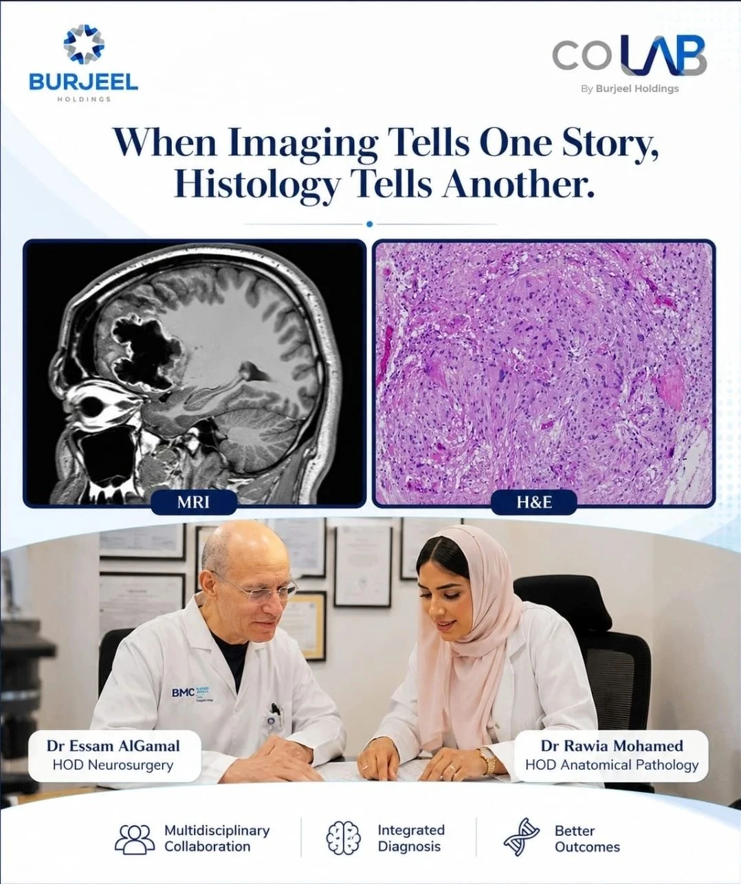
“When Imaging Tells One Story, Histology Tells Another
One of the most rewarding aspects of neuropathology is seeing how multidisciplinary collaboration changes patient care.
Recently, we encountered a ‘challenging brain tumor in a young Emirati male patient. Based on the ‘clinical presentation, radiological findings, and intraoperative impression,’ the lesion raised a ‘strong suspicion for a high-grade malignant glioma.’
However, careful ‘histopathological evaluation painted a different picture’. The microscopic features suggested pleomorphic xanthoastrocytoma (PXA), and subsequent molecular testing confirmed the diagnosis by demonstrating a BRAF V600E mutation.
This case is a valuable reminder that even lesions with highly aggressive clinical and radiological features ‘may ultimately represent tumors with a different biological behavior’ and therapeutic implication. It also highlights the importance of integrating clinical information, imaging, histopathology, immunohistochemistry, and molecular diagnostics before reaching a final diagnosis.
These are the cases that reinforce why pathology remains the cornerstone of precision medicine—transforming uncertainty into evidence-based diagnosis and helping guide the most appropriate patient management.
A sincere thank you to our neurosurgical colleagues for the excellent collaboration and open discussion throughout this case as well as Molecular Team from our Oncohelix lab for the fast PCR results. It is through teamwork that we achieve the best outcomes for our patients.”
Other posts featuring Rawia Mohamed on OncoDaily.