


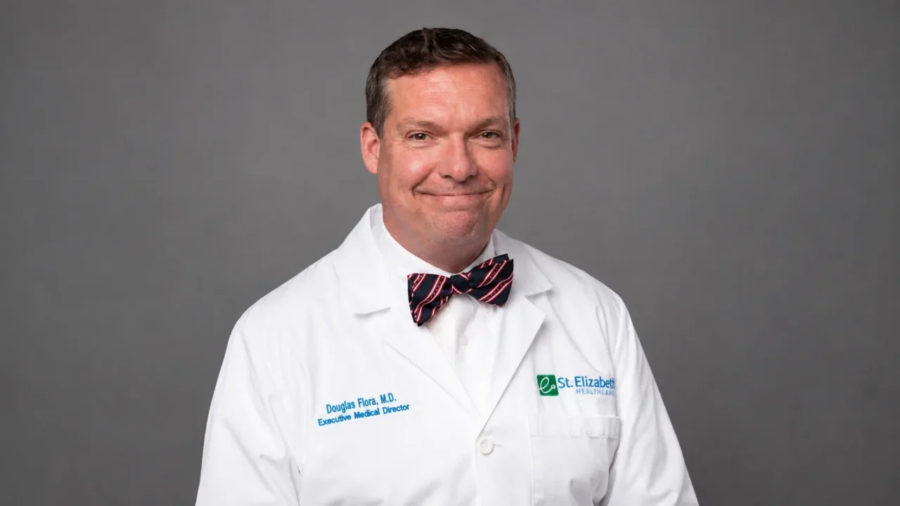
Douglas Flora, President-Elect at Association of Cancer Care Centers, Executive Medical Director of Yung Family Cancer Center at St. Elizabeth Healthcare, shared on LinkedIn:
“As multiomics, radiomics, and liquid biopsies detect signs of malignancy before a tumor can be measured on routine imaging, oncology has to reckon with a new clinical stage: Molecular Only Disease.
‘It is the theory which decides what we can observe.’ – Albert Einstein, in conversation with Werner Heisenberg, 1926
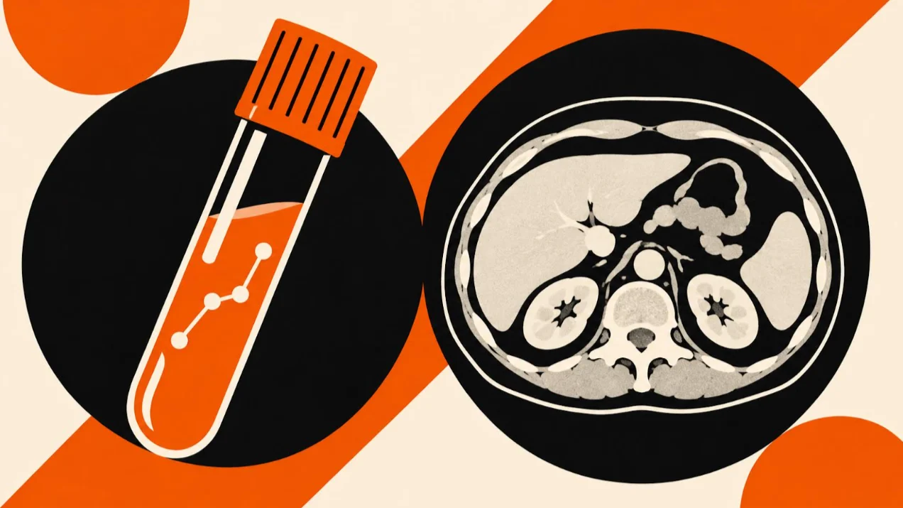
In an examination room on a Wednesday morning, a woman in her late fifties sits across from her oncologist, waiting to hear what happens next. On the screen between them are two results that contradict each other. A multiomic panel reports circulating tumor DNA, aberrant methylation patterns, and circulating chromosome arm alterations in her bloodstream—molecular and phenomic signatures indicating that malignancy is active somewhere in her body. Beside that report sits a complete series of cross-sectional scans of her chest, abdomen, and pelvis. Every slice is clean.
She feels completely well. She walked in from the parking lot without a cough, without pain, without a pound of unexplained weight loss. She asks a very reasonable question: what is the plan?
In most clinics across the country, nobody has a good answer. Our diagnostic tools have produced a result that our clinical workflows do not know how to handle.
Patients are not waiting for us to sort this out before ordering the tests. Driven by a desire to catch disease early, people with the means to pay out of pocket are signing up for preventive whole-body MRI scans, advanced radiomics profiling, and multi-cancer blood tests at wellness visits or their executive physicals. They are walking into our clinics holding folders of incidental findings and molecular blood tests that primary care physicians and oncologists were never trained to interpret. The consumer appetite for early detection has outrun our clinical readiness.
For generations, oncology managed to bypass this dilemma simply because we work with physical lumps. A clinician palpated a mass, or a screening film caught a shadow. A radiologist stuck a needle into the lesion, and a pathologist read the tissue on a glass slide. That biopsy told us what the disease was, where it belonged, and how far it had traveled.
Nearly everything downstream in modern cancer care was built on the premise that a physical mass would be there to find. Our staging manuals, the reimbursement codes, the response criteria that regulators use to judge whether an experimental drug works, and the physical layout of cancer centers divided into breast clinics and gastrointestinal floors all depend on a visible lesion.
A growing population of patients now fall outside that premise. They carry multiomic, genomic, proteomic, and radiomic warning signs in their blood and imaging data, yet show no measurable tumor anywhere in their bodies. I have previously used the word “pre-patient” to describe people in this position, though the term does not capture what is actually happening biologically. As these assays improve and the American Joint Committee on Cancer (AJCC) eventually updates its staging manuals to reflect modern biology, we are going to need a formal clinical designation for this state: Molecular Only Disease (MOD). Right now, patients with MOD exist in a sort of medical no-man’s-land.
A category without a code
The first thing that happens to someone with Molecular Only Disease is that they fall straight through the healthcare system’s administrative categories.
Medical billing runs on the Tenth Revision of the International Classification of Diseases, ICD-10, which requires a diagnosis, and a diagnosis in solid tumor oncology conventionally needs tissue. Without tissue, there is no diagnostic code that establishes cancer. Without that code, there is frequently no approved route to the high-resolution imaging that might locate an occult lesion, or to the systemic therapy that might treat it. Prior authorization software checks whether clinical documentation matches an established rule. When the documentation is a statistical probability in the blood or phenomic profile of a person with clean routine scans, the software issues an autopmatic denial.
Insurance of other kinds poses an even sharper problem. The Genetic Information Nondiscrimination Act of 2008, GINA, bars discrimination on the basis of genetic information in health coverage and in employment. It does not extend to life insurance, disability insurance, or long-term care insurance. Whether a positive multi-cancer blood test must be disclosed on those underwriting applications remains unsettled in law, and the people rolling up their sleeves for the blood draw are rarely warned of the financial risk they are taking on.
Which leaves us with surveillance. Repeat the blood draw in three months, repeat the imaging in six, and wait for the disease to grow large enough to show up on a scan. This makes no sense to me from a cancer kinetics standpoint. Hence my passion around this idea of ‘Cancer Interception.’ Lets do trials that let us intervene in these patients, and measure if we can change the trajectory of these cancers.
Decades ago, the sociologist Talcott Parsons described the social contract normally granted to an ill person as the “sick role”: a patient accepts the obligation to follow medical guidance in exchange for permission to be ill, exemption from ordinary duties, and the organized care of the clinic. But when you are handed a positive molecular result with clean scans, you inherit all the heavy psychological duties of being sick without any of the institutional permissions or relief. You carry the quiet panic of a cancer diagnosis while moving through daily life as a healthy person, trapped in a holding pattern that can drag on for years.
Why oncology was built around the lump
We built our system around the physical tumor for good reason. The requirement for tissue was one of medicine’s greatest breakthroughs, which is why oncology is so reluctant to let it go.
Before Rudolf Virchow, disease was understood as a systemic imbalance: humors, miasma, constitutional weakness, conditions of the whole body that had no physical address. When Virchow published his lectures on cellular pathology in 1858, he traced disease directly to the cell and gave illness a physical location. For oncology, this changed everything. Cancer became something a physician could point to on a scan and a surgeon could cut out. Whether a person had cancer depended on whether a pathologist could identify malignant cells on a slide.
More than a century later, when medical oncology needed a consistent way to judge whether a chemotherapy drug was working, it reached for the same ruler. Measure the mass across its longest diameter, give the drug, and measure the mass again. The Response Evaluation Criteria in Solid Tumors, RECIST, first published in 2000, turned this into the rulebook by which most solid tumor therapies are still judged. Regulators still largely approve cancer drugs based on what happens to a measurable lesion on a radiologist’s monitor, or how long a patient lives after that lesion is found.
Hematologists moved past this limitation twenty years ago because blood was always their medium. In chronic myeloid leukemia and acute lymphoblastic leukemia, clinicians learned to measure minimal residual disease, MRD, counting malignant transcripts by polymerase chain reaction at levels far too low to show up on a bone marrow biopsy. They began treating the molecular signal directly, before a clinical relapse could take hold.
What the clinical trials actually show
Two recent randomized trials show where the science stands today. One proves how powerful treating a blood signal can be after surgery; the other shows how complicated it gets when we test healthy people.
The first is IMvigor011, published in the New England Journal of Medicine in 2025 by Thomas Powles and colleagues. Investigators enrolled 761 patients who had undergone bladder removal for muscle-invasive bladder cancer and whose postoperative scans were completely clear. Rather than treating everyone blindly or waiting for tumors to show up on imaging, the study team drew blood on a regular schedule to look for tumor-informed circulating tumor DNA.
Roughly four in ten patients eventually turned positive in their blood while their scans stayed clean, facing the same dilemma as the woman in my exam room. Among those patients with Molecular Only Disease, 250 were randomly assigned in a two-to-one ratio to receive the immunotherapy atezolizumab (167 patients) or placebo (83 patients). Median disease-free survival in the treated group was 9.9 months versus 4.8 months with placebo. Median overall survival reached 32.8 months compared to 21.1 months, giving treated patients nearly an extra year of life, with grade 3 or 4 side effects occurring in 28 percent versus 22 percent of patients.
What makes IMvigor011 so important is what happened before it. A previous trial had tested the exact same drug in postoperative bladder cancer patients without using a blood test to select them, and the drug failed to show a benefit. The medicine did not change; our ability to see who needed it did. The blood test turned a failed treatment strategy into a life-extending one because it found the patients who still had microscopic cancer hiding out when their scans looked normal.
The second trial is NHS-Galleri, led by Richard Neal, Charles Swanton, Peter Sasieni, and colleagues, which tested a cell-free DNA methylation blood assay in 142,250 asymptomatic adults aged 50 to 77 in England, randomized one-to-one to annual blood testing plus usual care or usual care alone. The trial asked whether screening a general population over three years could reduce the combined number of stage III and stage IV diagnoses across 12 deadly cancers.
When results were reported in 2026 alongside diagnostic performance data in Nature Medicine, the trial missed its primary endpoint. The combined count of stage III and stage IV diagnoses across those 12 cancers was nearly identical between the screened and unscreened groups.
Looking beyond that top-line miss, the rest of the data tells a more complicated story. Adding the blood test caught 1,173 cancers compared with 290 in standard care alone, roughly quadrupling the number of cancers found. Stage IV diagnoses fell by about 14 percent overall, with drops of more than 20 percent in the second and third years of testing. Even better, the number of cancers diagnosed only after a patient showed up in crisis at an emergency department fell by 25 percent.
The primary endpoint failed because stage III diagnoses rose enough in the screened group to cancel out the drop in stage IV disease. Clinicians looking at those numbers see two very different possibilities. The hopeful view is stage migration: aggressive cancers caught one step earlier at stage III, where surgery and cure are still possible, rather than at stage IV. The skeptical view is overdiagnosis: picking up slow-growing tumors that never would have harmed the patient. We will not know which interpretation is right until we see whether fewer people in the screened group actually die of cancer over the next several years.
The gap between postoperative tracking and general screening
Some caveats here I need to point out:
Every patient in IMvigor011 had already gone through surgery for a known, proven cancer. Their blood test was custom-built in a lab using the exact mutational signature of their resected tumor, hunting for one specific fingerprint and nothing else.
Screening a healthy person during a routine wellness visit is an entirely different deal. Here, we are looking for a needle in a haystack without knowing what the needle looks like in advance, in a population where cancer is thankfully uncommon. Proving we can intercept residual disease after a bladder is removed does not automatically mean we know how to interpret a stray methylation signal in an otherwise healthy executive.
We also have to be candid about what population screening misses. Across the three rounds of NHS-Galleri, specificity was 99.5 to 99.6 percent, with positive predictive value moving from 58.0 percent down to 45.8 percent. That near-perfect specificity is a real achievement; when the test turns positive, you have to take it seriously. Sensitivity, on the other hand, ran from 26.7 to 37.2 percent across all cancer types, and 47.6 to 63.4 percent across the 12 prespecified lethal cancers. The blood test missed between two-thirds and three-quarters of all cancers that showed up in the study population during a given round. If a patient’s test comes back positive, it deserves an immediate workup. If it comes back negative, it does not mean they are cancer-free, even though most people paying out of pocket for an “early detection” test assume a clean result gives them the all-clear.
Nor do multiomic and liquid biopsy assays catch cancer as early as the marketing sometimes suggests. Stage I tumors are simply too small to shed enough DNA into the bloodstream for current methylation tests to pick up reliably. These platforms do not find cancer when it is a tiny cluster of cells. Their real value is more practical: catching deep-seated cancers of the pancreas, ovary, or liver at two centimeters, when a surgeon can still remove them, rather than at four centimeters when pain or jaundice sends the patient to the emergency room.
Even that window does not exist in every cancer. In the prospective c-TRAK TN trial, published in the Annals of Oncology by Nicholas Turner and colleagues, investigators monitored patients with high-risk triple-negative breast cancer using regular ctDNA blood draws. By 12 months, 27.3 percent of patients had tested positive in their blood. When those patients were brought in for immediate scans before starting treatment, 72 percent already had visible metastases on the screen. In a fast-moving cancer, the disease outran the blood test schedule. Whether we have time to step in depends on how fast the tumor grows and how often we draw blood.
How we prepare the clinic for Molecular Only Disease?
Our ability to read multiomic, genomic, and proteomic signals in the blood has outpaced the clinic’s ability to act on them. Catching up requires four practical changes in how we organize cancer care, none of which requires a new laboratory invention.
First, clinical trials must randomize patients based on their molecular and phenomic signals rather than conventional scans. IMvigor011 and the PADA-1 trial in breast cancer give us the blueprint: find the patients whose blood has turned positive while their scans are still clear, randomize them to early treatment versus standard observation, and see if they live longer. In the screening setting, trials must measure cancer-specific and overall mortality rather than survival timed from the date of diagnosis. Finding a cancer six months earlier with a multiomic blood test automatically makes survival look six months longer on paper, even if the patient dies on the exact same day.
Second, baseline risk should determine who gets screened. When a cancer is rare in the group being tested, even a test with 99.5 percent specificity will produce false alarms alongside real cases. No machine-learning algorithm can repeal Bayes’ theorem, because the math is governed by how common the disease is in the room, not just how good the test is in the lab. Focusing multi-cancer blood tests on higher-risk people first, based on age, inherited polygenic risk scores, smoking history, or family history, makes every positive result much more trustworthy.
Third, we should value using multiomic blood tests to spare patients treatment just as much as we value using them to start treatment early. Look again at the patients in IMvigor011 whose serial blood draws stayed negative after bladder surgery: they skipped adjuvant immunotherapy altogether, including people whose pathology reports looked dangerous under the microscope, and the vast majority stayed cancer-free. When a clean molecular panel gives us permission to withhold six months of punishing chemotherapy and tell someone they can simply go back to living their life, we are offering a profound kind of healing. Knowing who does not need treatment is where liquid biopsy can help the greatest number of patients today.
Fourth, medical societies, staging bodies, and insurers need to build a formal home for these patients. Recognizing Molecular Only Disease as a distinct clinical entity within the AJCC staging system would change everything downstream. A verified molecular signal without a visible mass needs its own ICD-10 diagnostic code, a covered imaging workup, legal protections for life and disability insurance, and dedicated clinics staffed by clinicians who know how to explain what a statistical probability actually means for a patient’s life.
The challenge is about communication as much as infrastructure. Every oncologist using these tests will have to learn a new way of talking with patients, holding honest statistical probability and human uncertainty in the same sentence, and helping someone decide whether to watch or treat an invisible signal without letting fear take over their life.
The woman in the exam chair at the start of this essay will keep showing up in our clinics, in growing numbers, whether she arrived through a postoperative blood draw, a multi-cancer screening panel, or an advanced scan. Nobody can promise her certainty. What oncology can build is a staging system that names her condition, a billing code that covers her workup, and a clinical trial she can enroll in, so that our answer to an early warning in the blood is finally something better than waiting for the tumor to grow large enough to see.
Doug Flora, MD, is a practicing medical oncologist, speaker, and writer on the future of medicine and the power of human possibility within it. He writes about artificial intelligence, precision oncology, and what it means to practice at the inflection point between the science we have and the science we are building. He is the author of Rebooting Cancer Care: Can AI Make Care More Human Again?”
You can also read: Andrea Ferrari: The Rhabdomyosarcoma Paradox – Better Treatment, Persistent Survival Gap