


Cardio-Oncology Bulletin shared a post on LinkedIn:
“Cardio-Oncology Monthly — Issue number 4
Monthly Case Highlight.
Not every baseline cardio-oncology assessment ends with a routine LVEF report.
Sometimes, careful pre-treatment imaging reveals an unexpected structural finding that changes the diagnostic landscape.
This month’s case highlight is especially meaningful for us, as it is based on an accepted case report from our own group, currently in press in EHJ Case Reports.
Case focus:
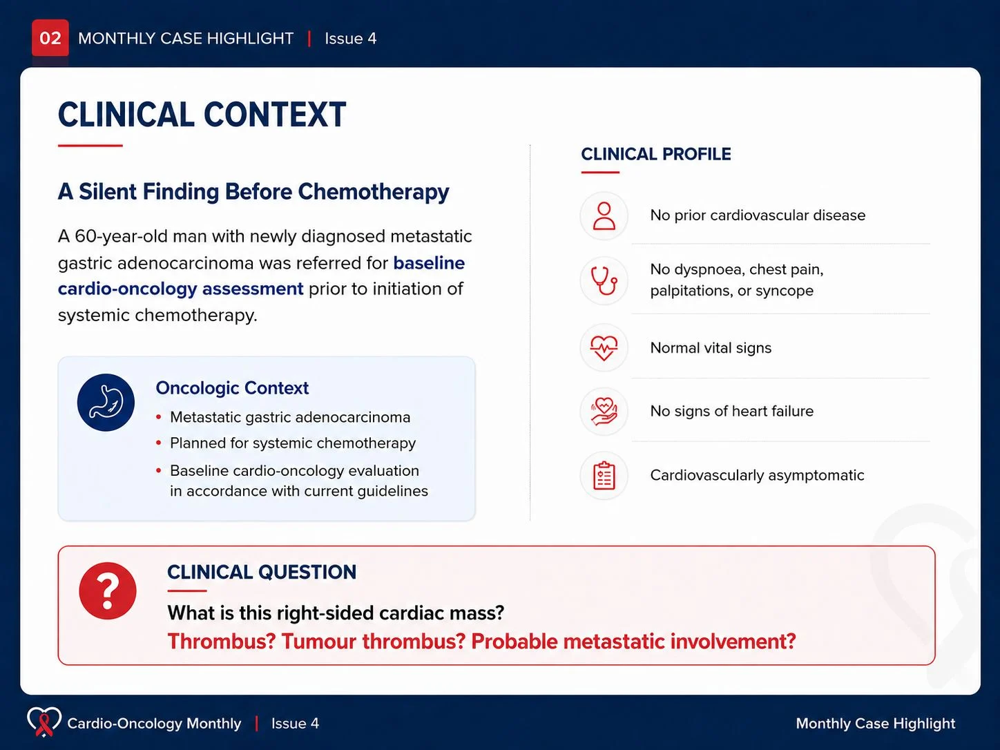
Probable metastatic right ventricular outflow tract mass detected during baseline cardio-oncology assessment.
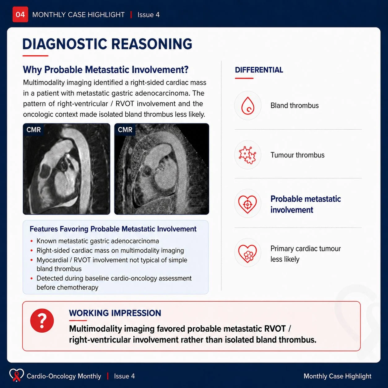
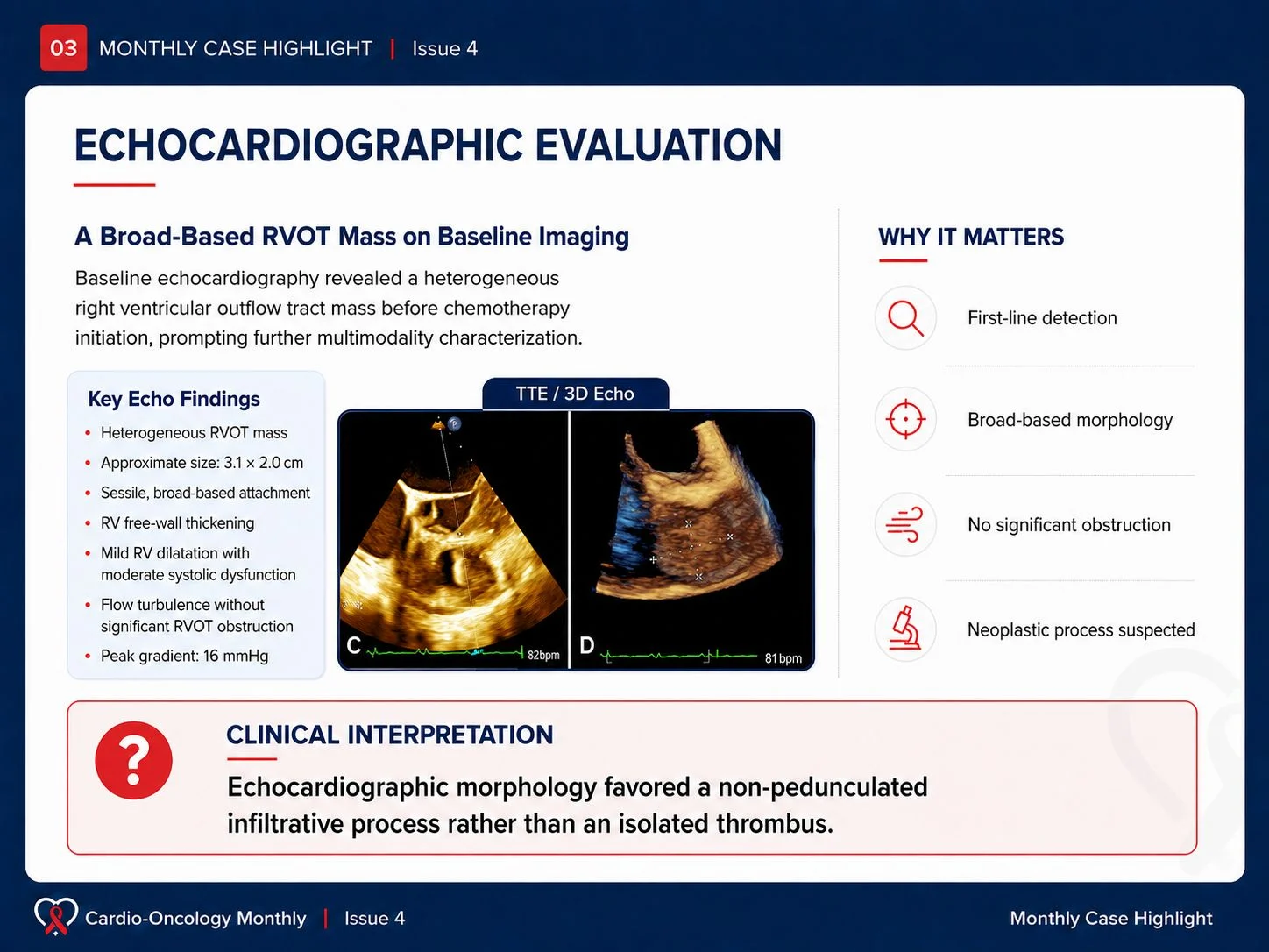
A 60-year-old man with newly diagnosed metastatic gastric adenocarcinoma was referred for baseline cardiovascular evaluation before systemic chemotherapy. Although he was cardiovascularly asymptomatic, echocardiography revealed a large heterogeneous mass involving the right ventricular outflow tract, prompting further multimodality imaging assessment.
Cardiac magnetic resonance imaging supported probable neoplastic infiltration, showing right ventricular free wall involvement, extension into the RVOT, contrast uptake, and heterogeneous late gadolinium enhancement.
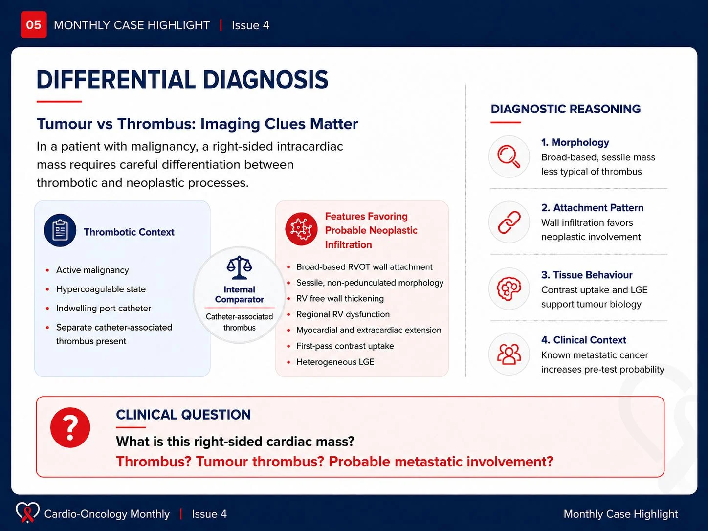
One important aspect of this case was the presence of a separate catheter-associated thrombus, which served as an internal comparator and helped refine the differential diagnosis between thrombotic and probable metastatic involvement.
- Why this case matters
Baseline cardio-oncology imaging is often performed to evaluate cardiovascular risk and ventricular function before cancer therapy. However, this case illustrates that careful pre-treatment imaging may occasionally reveal clinically silent but management-relevant structural cardiac abnormalities. - Key clinical message
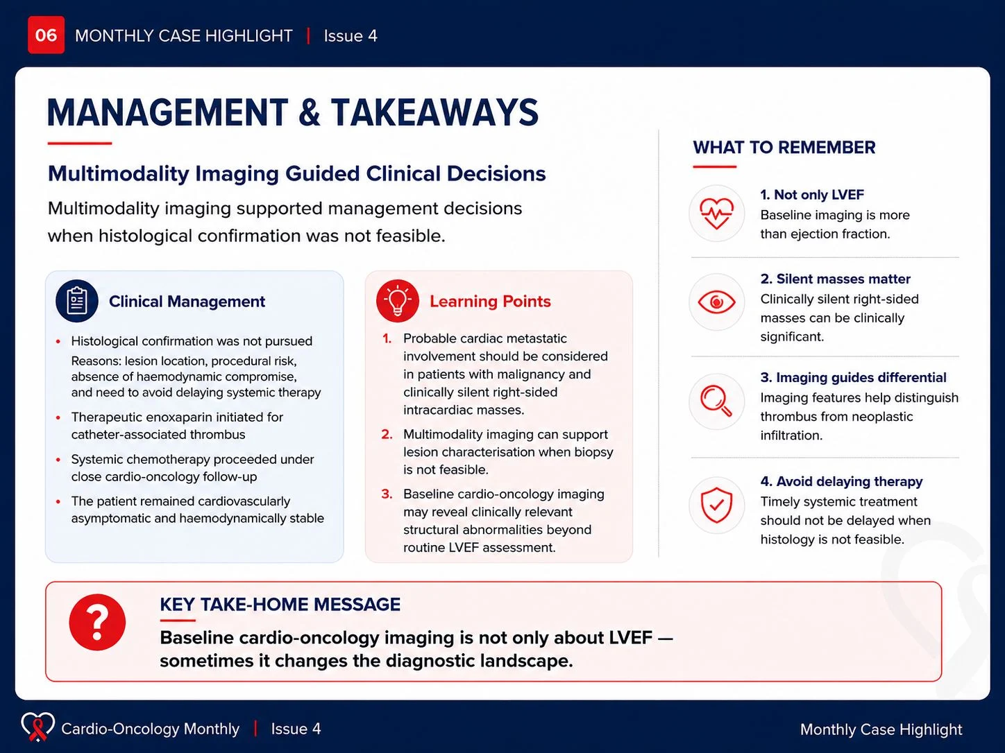
In patients with malignancy, a right-sided intracardiac mass should not automatically be assumed to be thrombus. Multimodality imaging can help characterize lesion morphology, tissue behaviour, and local extension — especially when histological confirmation is not feasible.
Take-home message
Baseline cardio-oncology assessment is not only about LVEF.
Sometimes, it changes the diagnostic landscape.
Accepted in EHJ Case Reports | DOI assigned | Online publication forthcoming.”

Other articles about Cardio-Oncology Bulletin on OncoDaily.