
Ramji Rajendran, Radiation Oncologist at UroPartners LLC, shared on LinkedIn:
“Prostate cancer is inherently a multifocal disease. The Dominiant Intraprostatic Nodule (DIL) is often where radiation failures occur. Providing a boost dose of radiation to the DIL has been shown to decrease recurrence. Before we can boost a prostate tumor precisely, we need to understand where the tumor actually is.
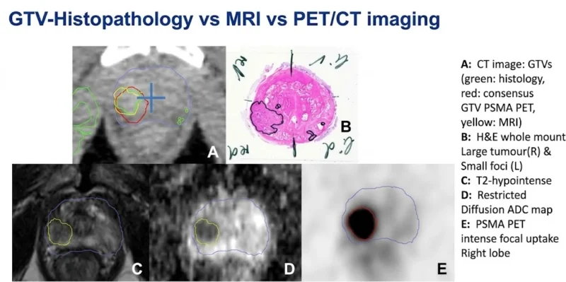
This slide illustrates an important challenge in radiation oncology: the gross tumor volume identified on MRI or PSMA-PET/CT does not necessarily match the full extent of cancer on histopathology.
What are we seeing?
- Panel A overlays histology-derived tumor contours (green), PSMA PET contours (red), and MRI contours (yellow). The boundaries overlap – but are not identical.
- Panel B shows whole-mount histopathology, including a large right-sided tumor and smaller contralateral foci.
- Panels C and D demonstrate the dominant lesion on T2-weighted MRI and the diffusion/ADC map.
- Panel E shows intense focal PSMA uptake corresponding to the dominant right-sided lesion.
Identifying the dominant lesion does not account for every focus of prostate cancer.
MRI – anatomical information. PSMA PET/CT – molecular information.
Histopathology-validates their complementary roles to identify the DIL.
Why does this matter for treatment planning?
- Focal boosting depends on accurate contours.
- Discordant MRI and PET findings deserve careful review.
- Imaging-occult tumor foci are the rationale for whole-gland treatment with selective intensification of visible disease.
- Registration accuracy, contouring methodology, and organ-at-risk constraints are essential when translating imaging into a boost volume.
The randomized FLAME trial demonstrated improved biochemical disease-free survival with an MRI-defined focal boost added to whole-prostate radiotherapy. The role of PSMA PET/CT is still being defined.
_____
Title: Comparison of 68Ga-HBED-CC PSMA-PET/CT and multiparametric MRI for gross tumour volume detection in patients with primary prostate cancer based on slice by slice comparison with histopathology
Authors: Constantinos Zamboglou, Vanessa Drendel, Cordula A Jilg, Hans C Rischke, Teresa I Beck, Wolfgang Schultze-Seemann, Tobias Krauss, Michael Mix, Florian Schiller, Ulrich Wetterauer, Martin Werner, Mathias Langer, Michael Bock, Philipp T Meyer, Anca L Grosu
_____
Title: [68Ga-]PSMA-11 PET/CT and multiparametric MRI for gross tumor volume delineation in a slice by slice analysis with whole mount histopathology as a reference standard – Implications for focal radiotherapy planning in primary prostate cancer
Authors: Alisa S Bettermann, Constantinos Zamboglou, Selina Kiefer, Cordula A Jilg, Simon Spohn, Jasmin Kranz-Rudolph, Thomas F Fassbender, Peter Bronsert, Nils H Nicolay, Christian Gratzke, Michael Bock, Juri Ruf, Matthias Benndorf, Anca L Grosu
_____
Title: Focal Boost to the Intraprostatic Tumor in External Beam Radiotherapy for Patients With Localized Prostate Cancer: Results From the FLAME Randomized Phase III Trial
Authors: Linda G W Kerkmeijer, Veerle H Groen, Floris J Pos, Karin Haustermans, Evelyn M Monninkhof, Robert Jan Smeenk, Martina Kunze-Busch, Johannes C J de Boer, Jochem van der Voort van Zijp, Marco van Vulpen, Cédric Draulans, Laura van den Bergh, Sofie Isebaert, Uulke A van der Heide
This can be interesting:
10 Prostate Cancer Trials to Watch at ESMO 2026