
Kefah Mokbel, Professor of Medicine at Cardiff University School of Medicine, shared on LinkedIn:
“A 60 year old physician , who is under my care, has had a stable 10 mm fibroadenoma in her left breast for the past 15 years. Increasing calcifications were recently noted, prompting an up to date ultrasound-guided core biopsy. This revealed grade 1 infiltrating ductal carcinoma arising within a fibroadenoma. Today, I performed a lumpectomy and sentinel lymph node biopsy. She has been taking HRT and has no family history of breast cancer.
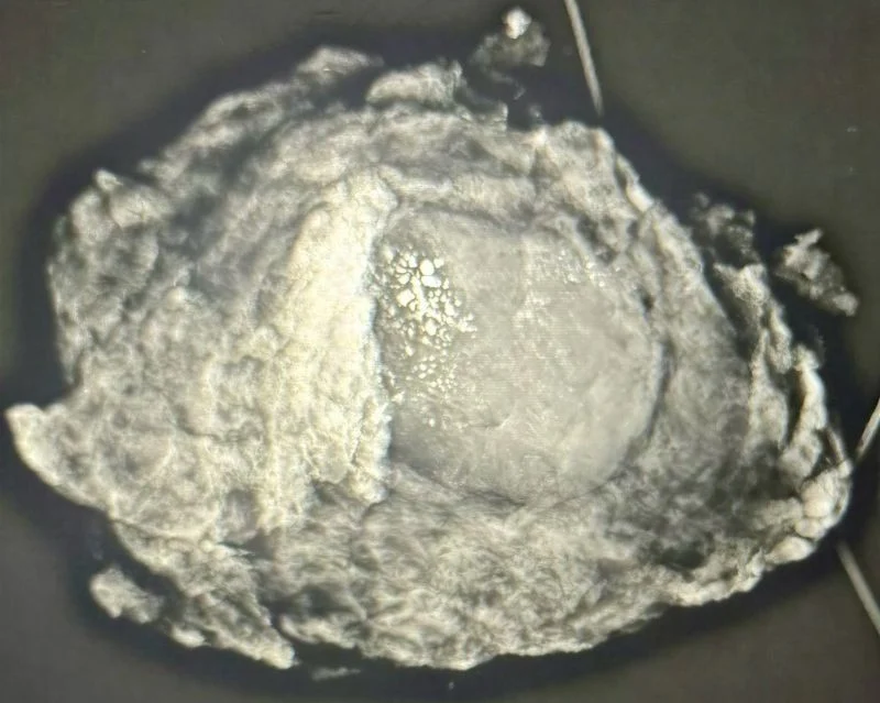
Teaching point
Longstanding stability does not exclude malignancy within a fibroadenoma. New or increasing microcalcifications warrant reassessment and repeat biopsy if their morphology or distribution is suspicious.
Carcinoma arising within a fibroadenoma is exceptionally rare, reported in approximately 0.002–0.125% of fibroadenoma specimens. Most cases are carcinoma in situ; invasive ductal carcinoma is rarer still.
It typically presents in women in their 40s (mean age 43 with the youngest affected is 19) and new suspicious calcifications may be the first indication of malignancy in an otherwise longstanding, stable lesion. Management follows standard breast cancer principles, guided by tumour stage and biology.
This rare occurrence does not justify routine surgical excision of every fibroadenoma. A confirmed fibroadenoma with concordant benign clinical, imaging and, where obtained, biopsy findings can usually be managed conservatively. New suspicious changes warrant reassessment and, where indicated, repeat biopsy – not automatic surgery.”
You can also read: