






Sonali Job, Scientist, Researcher, Nano-Biotechnology Scientist, shared on LinkedIn:
“One Cancer, Five Different Histological Patterns
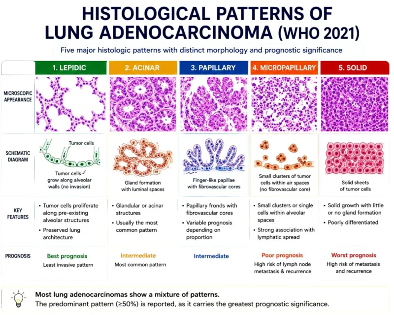
When we hear lung adenocarcinoma, it sounds like a single disease. But under the microscope, pathologists often see five distinct growth patterns-and each tells us something about how the tumor is likely to behave.
- Lepidic Pattern
Tumor cells grow along the existing alveolar walls without destroying the lung architecture. This is the least invasive pattern and is associated with the best prognosis. - Acinar Pattern
The most common subtype, characterized by well-formed gland-like structures. It generally has an intermediate prognosis. - Papillary Pattern
Cancer cells form finger-like projections (papillae) with fibrovascular cores. Prognosis is typically intermediate, though outcomes vary with the proportion of this pattern. - Micropapillary Pattern
Small clusters of tumor cells appear to “float” within the air spaces. Despite its small size, this pattern is highly aggressive and is strongly associated with lymph node metastasis and recurrence. - Solid Pattern
Tumor cells grow in solid sheets with little or no gland formation. This poorly differentiated subtype is often linked to the worst prognosis and a higher likelihood of metastasis.
Why does this matter?
Most lung adenocarcinomas contain more than one histological pattern. Pathologists report the predominant pattern, as it provides valuable prognostic information and helps guide clinical decision-making.
The microscope doesn’t just confirm the diagnosis-it also reveals the tumor’s biological behavior.”
Other articles about lung adenocarcinoma on OncoDaily.