Histologic transformation from lung adenocarcinoma to small-cell lung cancer remains one of the most aggressive and difficult-to-treat mechanisms of resistance in EGFR-mutated non–small cell lung cancer. New multicenter data now provide a more detailed picture of who is at risk, how these tumors evolve genomically, and which treatments may retain activity after transformation.
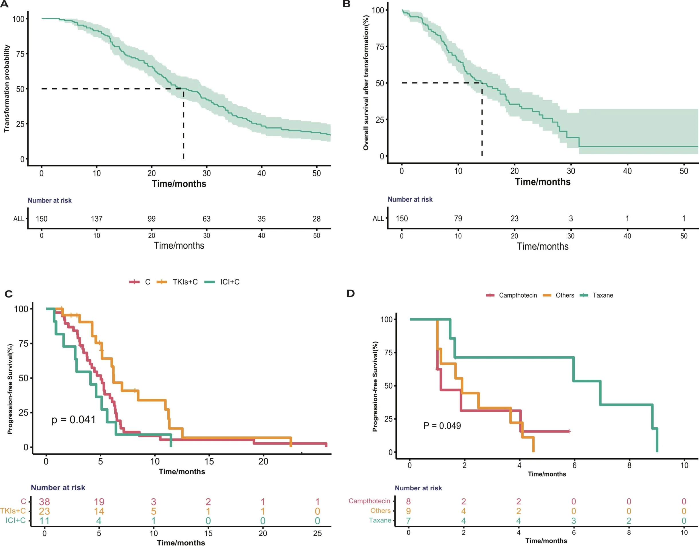
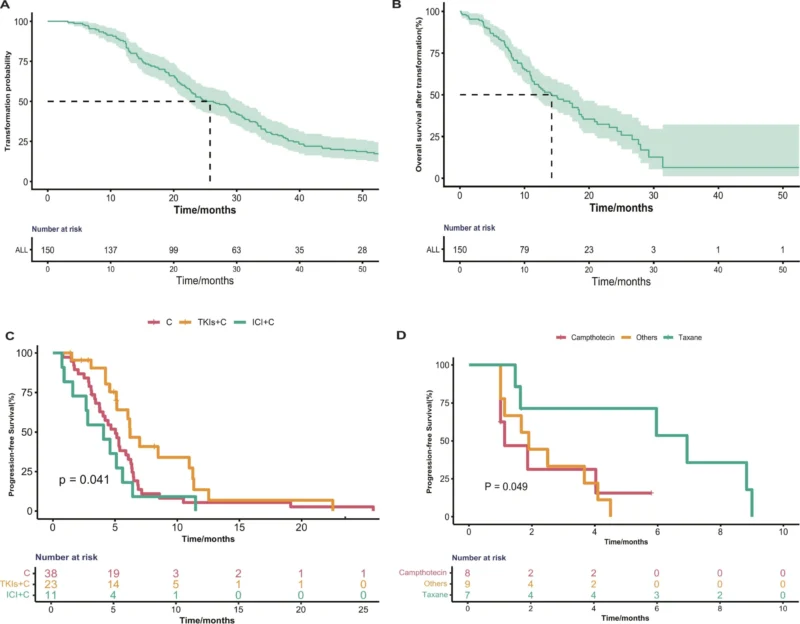
In a 2026 multicenter analysis accepted in Lung Cancer, Gao and colleagues evaluated 163 patients with NSCLC who subsequently developed small-cell lung cancer transformation, including 150 patients with EGFR-mutated disease. The median time from NSCLC diagnosis to transformation in the EGFR-mutated population was 25.8 months, while median overall survival after transformation was 14.2 months.
Among treatment-evaluable patients, EGFR-TKI plus etoposide-platinum chemotherapy produced the longest median first-line progression-free survival after transformation at 6.2 months, compared with 5.3 months for chemotherapy alone and 4.07 months for chemoimmunotherapy. In later lines, taxane-based regimens produced a median PFS of 6.93 months, although this finding came from only seven patients and requires prospective confirmation.
The study also identified extensive genomic remodeling during transformation and developed a machine-learning model incorporating clinical and genomic features that showed high discrimination for transformation risk in internal and external validation cohorts.
Together, the findings reinforce a central message: small-cell transformation is not simply another progression event in EGFR-mutated NSCLC, it represents a major change in tumor lineage, biology and treatment sensitivity.
Why Does Small-Cell Transformation Matter in EGFR-Mutated NSCLC?
EGFR-targeted therapies can produce prolonged disease control in EGFR-mutated lung adenocarcinoma, but acquired resistance eventually develops in most patients.
One recognized mechanism is histologic transformation from adenocarcinoma into a small-cell phenotype.
Previous studies have estimated that approximately 3%–14% of EGFR-mutated NSCLC tumors undergo SCLC transformation during their clinical course. Unlike resistance driven by a single secondary kinase mutation, transformation involves a broader shift in tumor phenotype and lineage state (Gao et al., 2026).
This distinction has important therapeutic consequences.
The transformed tumor can retain the original EGFR alteration while behaving clinically more like small-cell lung cancer, with rapid progression, neuroendocrine biology and reduced dependence on the EGFR pathway.
The result is one of the most difficult treatment scenarios in molecularly driven lung cancer.
How Large Was the New Multicenter Study?
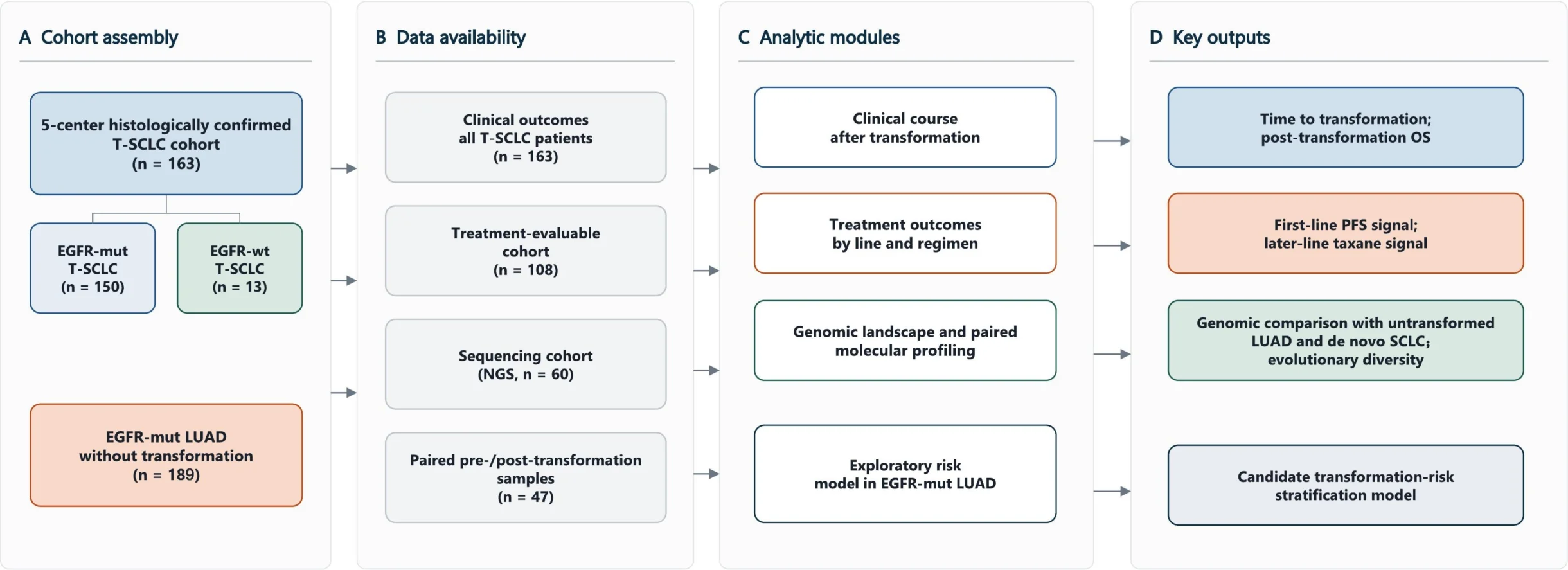
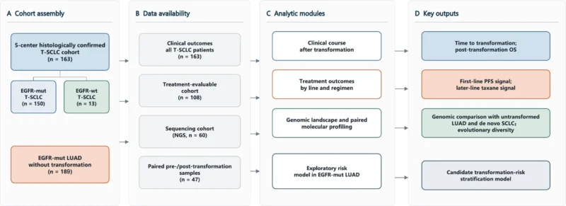
Gao and colleagues retrospectively identified 163 patients who were initially diagnosed with NSCLC and subsequently developed histologically confirmed SCLC transformation across five cancer centers in China between 2011 and 2024.
The median age at initial diagnosis was 56 years, and 52.8% of patients were women.
The cohort was strongly enriched for EGFR-mutated disease:
- 150 patients, or 92.0%, had an EGFR mutation, while 13 patients were EGFR wild type.
Among the EGFR-mutated tumors, exon 19 deletions were most common, occurring in 94 patients. Another 47 patients had L858R, while smaller numbers had L861Q, G719A/G719X or other EGFR alterations.
Most patients with EGFR-mutated disease were never smokers, reflecting the expected clinical phenotype of this molecular subgroup.
When Did SCLC Transformation Occur?
Among the 150 patients with EGFR-mutated NSCLC, the median interval from initial NSCLC diagnosis to SCLC transformation was:
- 25.8 months
Once transformation occurred, prognosis was poor.
Median overall survival from the time of transformation was:
- 14.2 months
- 95% CI, 11.2–18.5 months.
For the small EGFR-wild-type subgroup, median time to transformation was 24.4 months and median post-transformation survival was 10.4 months.
Because only 13 EGFR-wild-type patients were included, the authors appropriately treated this group descriptively rather than drawing comparative conclusions.
One point in the journal pre-proof deserves clarification: the Results and Abstract report post-transformation OS of 14.2 months for the EGFR-mutated population, while one sentence in the Discussion states 10.4 months. The latter corresponds to the EGFR-wild-type result reported elsewhere in the paper. This article therefore uses the 14.2-month value reported consistently in the Abstract and Results section.
What Was the Best First-Line Strategy After Transformation?
Treatment data were available for 108 patients.
Among EGFR-mutated patients receiving first-line therapy after SCLC transformation, investigators compared three main approaches:
EGFR-TKI plus chemotherapy, chemotherapy alone, and immune checkpoint inhibitor plus chemotherapy.
All chemotherapy backbones were based on etoposide plus platinum, reducing some of the treatment heterogeneity.
Median PFS was:
- 6.2 months with EGFR-TKI plus chemotherapy
- 5.30 months with chemotherapy alone
- 4.07 months with ICI plus chemotherapy.
The overall difference reached statistical significance:
- P = 0.041.
The finding suggests that continuing EGFR inhibition alongside chemotherapy could preserve some disease-control benefit in selected patients after transformation.
However, the study was retrospective, and treatment was not randomly assigned. These results therefore cannot establish EGFR-TKI plus chemotherapy as a new standard.
Did Longer PFS Translate Into Longer Overall Survival?
No clear overall survival advantage was demonstrated.
Median OS after initiation of first-line post-transformation treatment was:
- 21.2 months with EGFR-TKI plus chemotherapy
- 27.6 months with chemotherapy alone
- 13.6 months with ICI plus chemotherapy.
The difference was not statistically significant:
- P = 0.193.
Interestingly, chemotherapy alone had the numerically longest median OS despite the shorter PFS.
This discrepancy reinforces the limitations of retrospective treatment comparisons. Subsequent therapies, patient selection, disease burden, treatment timing and other unmeasured factors could all affect overall survival.
The most defensible conclusion is therefore that EGFR-TKI plus chemotherapy was associated with the longest first-line PFS, not that it demonstrated superior overall survival.
Why Continue an EGFR TKI After Transformation?
The biological rationale comes from the evolutionary relationship between the original adenocarcinoma and the transformed SCLC.
Transformed tumors frequently retain the original EGFR driver alteration, indicating that they are descendants of the original EGFR-mutated cancer rather than independent de novo small-cell tumors.
Previous multiomic studies have shown persistence of EGFR-related genomic features after neuroendocrine transformation, although the transformed phenotype can become substantially less dependent on EGFR signaling (Quintanal-Villalonga et al., 2021).
Gao and colleagues therefore suggest that continued EGFR inhibition could retain some activity when combined with chemotherapy.
But the modest PFS and absence of an OS advantage also illustrate the limits of this strategy: once the small-cell phenotype becomes dominant, suppressing EGFR alone is unlikely to reverse the broader lineage transformation.
What Happened With Chemoimmunotherapy?
Chemoimmunotherapy produced the shortest median first-line PFS among the three strategies evaluated:
- 4.07 months.
- Median OS was 13.6 months.
The authors interpret these findings cautiously but note that they are consistent with previous evidence showing limited activity of immune checkpoint inhibitors in oncogene-driven EGFR-mutated tumors.
This is biologically plausible because EGFR-mutated NSCLC generally has a less inflamed tumor immune microenvironment and historically derives less benefit from PD-1/PD-L1 monotherapy than smoking-associated NSCLC.
However, only 11 EGFR-mutated patients in this cohort received ICI plus chemotherapy after transformation. The study therefore cannot definitively determine the value of immunotherapy in this setting.
Could Taxanes Be Important After First-Line Treatment?
One of the most interesting findings emerged in later-line treatment.
Among patients receiving second-line or subsequent therapy after transformation, seven received taxane-based regimens, eight received camptothecin-based treatment, and nine received other therapies.
Median PFS was:
- 6.93 months with taxane-based regimens
compared with:
- 1.13 months with camptothecin-based regimens
and:
- 1.90 months with other therapies.
The overall difference was statistically significant:
- P = 0.049.
This signal is consistent with earlier work by Marcoux and colleagues, in which paclitaxel and nab-paclitaxel showed activity in EGFR-mutated tumors after SCLC transformation.
But the new analysis involved only seven taxane-treated patients.
The result should therefore be viewed as a clinically interesting signal rather than evidence establishing taxanes as the preferred later-line treatment.
Prospective evaluation will be necessary.
What Did Genomic Profiling Reveal Before Transformation?
The molecular analysis was one of the major strengths of the study.
Next-generation sequencing or whole-exome sequencing was performed in 60 EGFR-mutated patients before transformation, and paired pre- and post-transformation samples were available from 47 patients.
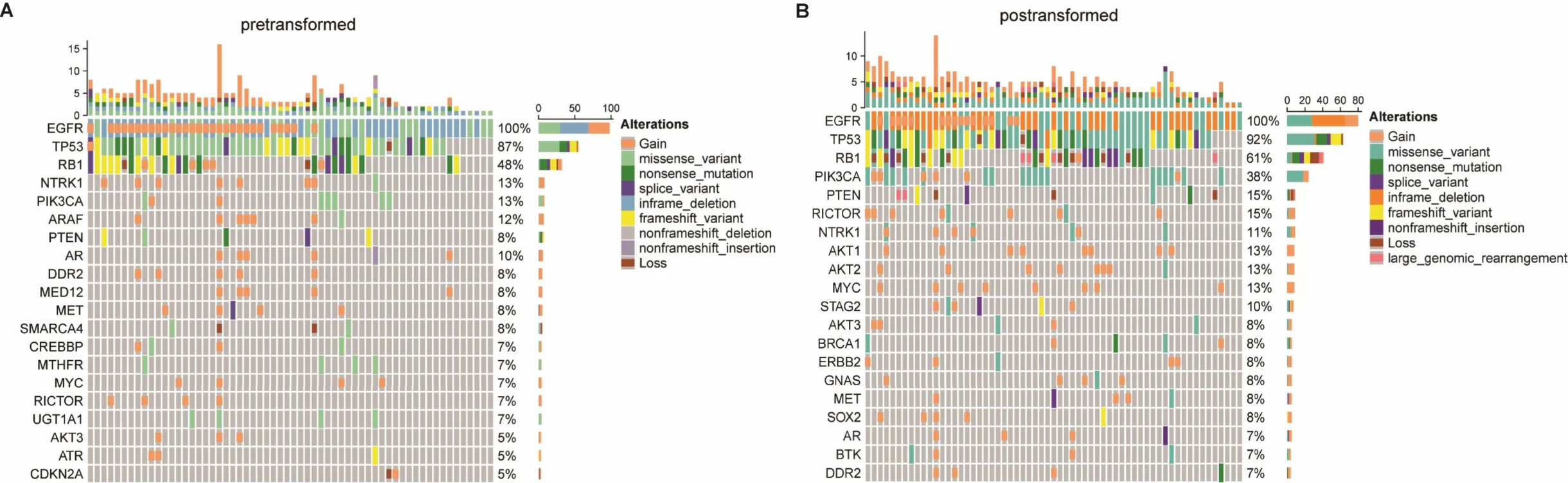
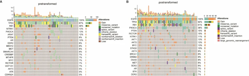
Before transformation, the most frequent co-occurring alterations were:
- TP53: 87%
and:
- RB1: 48%.
These findings reinforce the established relationship between loss of TP53 and RB1 function and susceptibility to small-cell transformation.
When pre-transformation EGFR-mutated tumors were compared with a reference cohort of 189 EGFR-mutated lung adenocarcinomas that had not transformed, several alterations were significantly enriched.
RB1 mutations occurred in:
- 48.33% versus 19.05%
while NTRK1 alterations occurred in:
- 13.33% versus 1.06%.
PMS2 alterations were also more frequent:
- 11.67% versus 0.53%.
All three comparisons remained significant after correction for multiple testing.
How Did the Tumor Genome Change After Transformation?
Paired sequencing demonstrated extensive genomic remodeling.
PIK3CA alterations increased numerically from 8.51% before transformation to 40.43% after transformation, although the adjusted P value of 0.089 did not meet the study’s significance threshold.
When transformed tumors were compared with EGFR-mutated cancers that had progressed but remained adenocarcinoma, several alterations were strikingly enriched in transformed SCLC.
These included:
- RB1: 60.0% vs 14.79%
- TP53: 91.67% vs 52.82%
- PIK3CA: 38.33% vs 5.63%
as well as significant enrichment of NTRK1, AKT1, PTEN and STAG2 alterations.
These differences show that transformation is accompanied by more than a simple histologic change.
It represents a distinct evolutionary route characterized by disruption of tumor-suppressor pathways and additional signaling alterations.
How Different Is Transformed SCLC From De Novo SCLC?
The transformed tumors also remained genomically distinguishable from conventional de novo SCLC.
Compared with a reference cohort of de novo SCLC, transformed tumors had substantially higher frequencies of:
- EGFR: 93.33% vs 3.64%
- PIK3CA: 38.33% vs 3.64%
and higher frequencies of AKT2 and MYC alterations.
Conversely, LRP1B alterations were less frequent in transformed SCLC:
- 5.0% vs 48.18%.
This is an important biological point.
A transformed tumor may look like SCLC under the microscope, but its genome still carries a molecular history inherited from its original EGFR-mutated adenocarcinoma.
Transformed SCLC should therefore not automatically be considered biologically identical to de novo SCLC.
Why Are TP53 and RB1 So Important?
TP53 and RB1 have long been implicated in lineage plasticity and neuroendocrine transformation.
Previous work by Lee and colleagues showed that concurrent loss-of-function alterations involving TP53 and RB1 identified EGFR-mutated lung cancers with dramatically increased transformation risk.
Gao and colleagues again found extremely high frequencies of these alterations.
Before transformation, TP53 mutations were present in 87% and RB1 alterations in 48% of sequenced EGFR-mutated tumors.
After transformation, their frequencies increased to approximately 92% and 60%, respectively.
These findings support the idea that TP53/RB1 disruption establishes a genomic background permissive to lineage plasticity.
However, not every tumor carrying these alterations transforms, meaning additional biological events are required.
Could NTRK1 Have a Role in Transformation?
One of the more novel observations was enrichment of NTRK1 alterations in tumors predisposed to SCLC transformation.
NTRK1 alterations occurred in 13.33% of pre-transformation tumors compared with only 1.06% of baseline EGFR-mutated adenocarcinomas in the non-transformed reference cohort.
The authors emphasize an important caveat: these alterations were mainly copy-number gains and missense mutations, rather than canonical actionable NTRK fusions.
The study therefore does not establish NTRK-directed therapy as a treatment strategy.
Instead, NTRK1 emerged as a possible marker of the biology associated with transformation and requires mechanistic validation.
What Is the Relationship Between T790M and SCLC Transformation?
The relationship between acquired EGFR T790M and small-cell transformation is particularly interesting because the two can represent different evolutionary solutions to EGFR-TKI pressure.
T790M preserves dependence on EGFR signaling while altering sensitivity to earlier-generation EGFR inhibitors.
Small-cell transformation, by contrast, reflects a more fundamental lineage shift away from classical EGFR-dependent adenocarcinoma biology.
The authors note that previous studies have found T790M to be uncommon in transformed tumors and that some tumors previously carrying T790M lose it after SCLC transformation.
In the current dataset, secondary T790M was detected in only 10 of 59 relevant cases discussed in the genomic analysis.
This supports the concept that T790M acquisition and SCLC transformation often represent divergent resistance pathways.
It also reinforces why re-biopsy at progression can be clinically important: the mechanism of resistance cannot always be inferred from the original genomic profile.
What Does “Evolutionary Diversity” Mean?
The investigators went beyond cataloguing individual mutations and reconstructed how tumor clones evolved during transformation.
Using paired pre- and post-transformation samples from 47 patients, mutations were classified as truncal when they persisted across both time points and as branch alterations when they appeared only before or after transformation.
The investigators then quantified the complexity of these evolving tumor populations using measures of clonal heterogeneity and Shannon entropy.
Ten patients were classified as having high evolutionary diversity, while 37 had low diversity.
Median post-transformation OS was:
- 6.77 months in the high-diversity group
versus:
- 11.10 months in the low-diversity group.
The difference was statistically significant:
- P = 0.007.
The finding suggests that tumors undergoing more complex clonal evolution could behave more aggressively after transformation.
However, restricted cubic spline analysis showed only a trend toward an overall relationship and a nonsignificant nonlinear association, and treatment differences could not be fully adjusted for.
The survival association should therefore be considered exploratory.
Can SCLC Transformation Be Predicted Before It Happens?
The investigators also attempted to move beyond retrospective description and build a transformation-risk model.
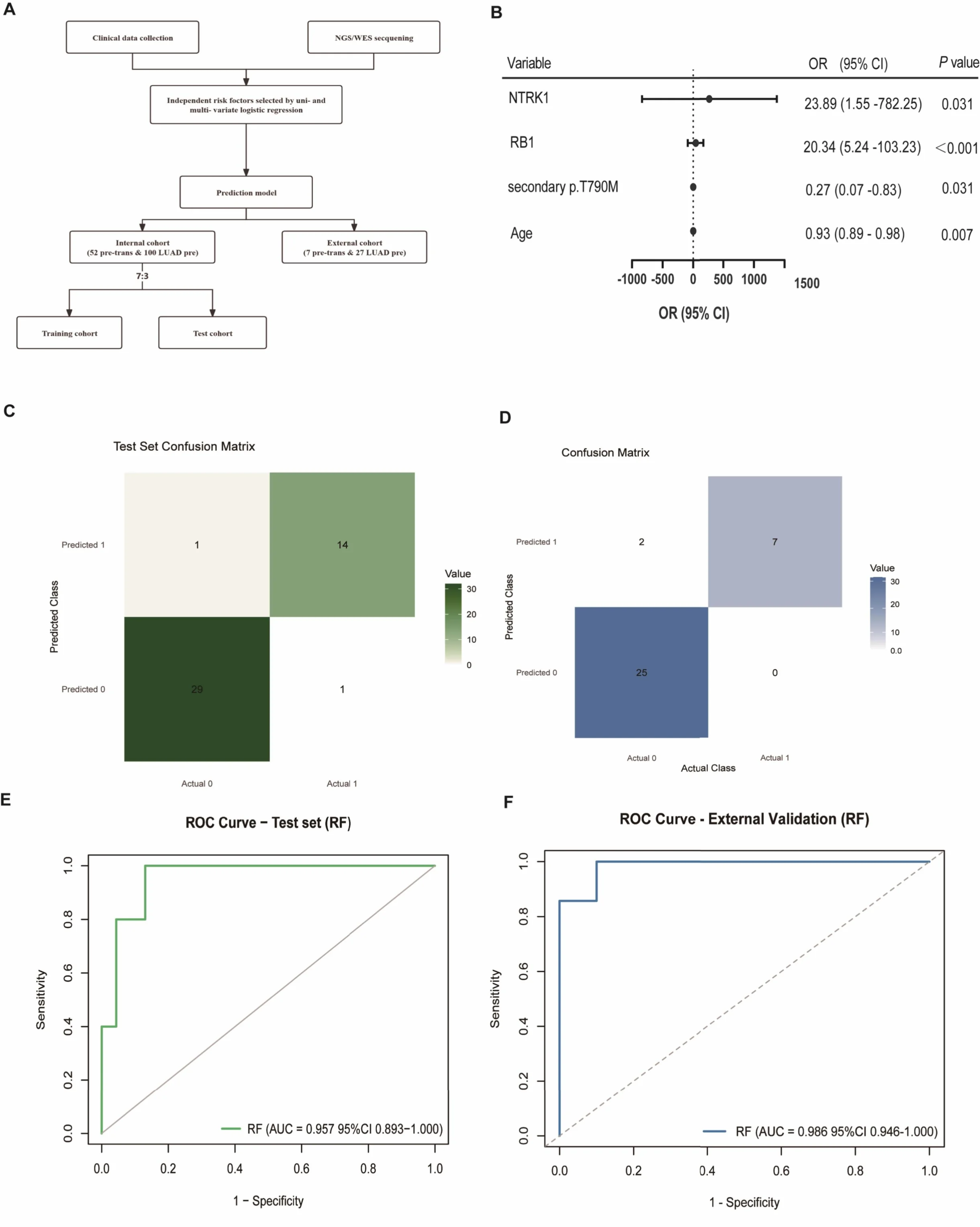
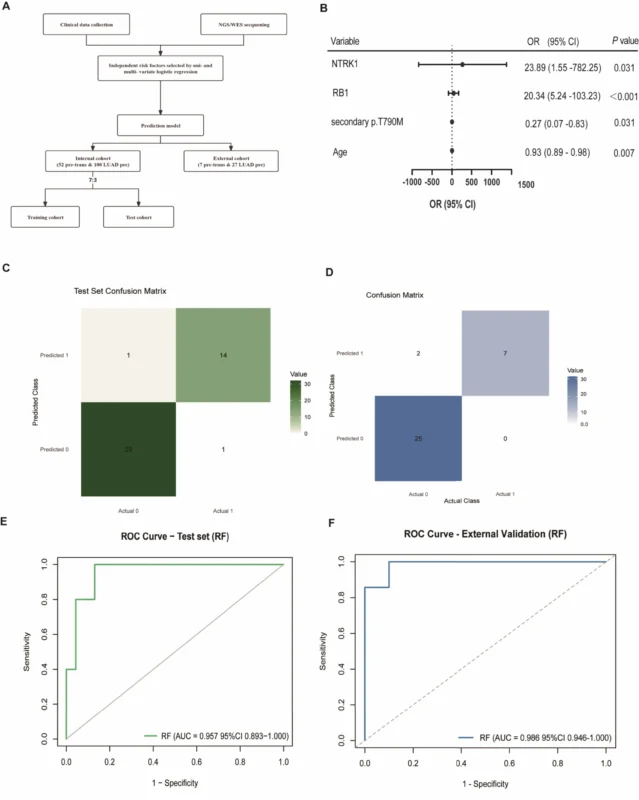
Clinical and molecular variables were first evaluated with logistic regression. The resulting model incorporated:
age, RB1 alteration, NTRK1 alteration and secondary EGFR T790M status.
The main text identifies these variables as independent factors associated with transformation, although the direction and magnitude of each individual association are detailed in supplementary analyses rather than fully reported in the manuscript text.
The investigators then used random forest and support vector machine approaches to construct predictive models.
The internal cohort was divided into a training population of 107 patients and a test population of 45 patients.
An independent external validation cohort included another 34 patients.
How Accurate Was the Prediction Model?
The random forest model demonstrated high discrimination.
In the internal test cohort:
- AUC = 0.957
with:
- 93.3% sensitivity
- 96.7% specificity.
In the external validation cohort:
- AUC = 0.986
with:
- 100% sensitivity
- 92.6% specificity.
These numbers are striking, particularly for a problem as clinically difficult as predicting histologic transformation.
However, they need cautious interpretation.
The external validation cohort contained only seven transformed and 27 non-transformed patients. Large, independent prospective cohorts will therefore be necessary before the model can be considered ready for routine clinical risk stratification.
Could the Model Change Surveillance?
Potentially, but not yet.
A validated transformation-risk model could eventually identify patients who warrant closer clinical monitoring, earlier tissue re-biopsy, or more intensive molecular reassessment when progression occurs.
This could be particularly important because SCLC transformation cannot be reliably diagnosed from imaging alone.
Histologic confirmation remains critical.
The current model therefore provides an important proof of concept: baseline clinical and genomic information might eventually identify tumors with a greater tendency toward lineage transformation.
But prospective clinical utility has not yet been demonstrated.
Why Is Re-Biopsy So Important?
The study strongly reinforces the role of repeat tissue sampling in selected patients with EGFR-mutated NSCLC who develop progression.
A liquid biopsy can detect many acquired genomic alterations, but it cannot demonstrate that adenocarcinoma has transformed morphologically into SCLC.
Histologic transformation is fundamentally a pathology diagnosis.
This matters because treatment following small-cell transformation can differ substantially from treatment used for molecular resistance while adenocarcinoma histology is retained.
The distinction between T790M-mediated resistance and SCLC transformation is a particularly clear example.
A patient who progresses after EGFR-TKI treatment may therefore require more than another genomic test. When clinically feasible, tissue can reveal whether the tumor itself has changed identity.
What Are the Major Limitations?
The study is retrospective.
Treatment allocation was not randomized, meaning that comparisons between EGFR-TKI plus chemotherapy, chemotherapy alone and chemoimmunotherapy can be influenced by patient selection.
Post-transformation treatment data were also available for only a subset of the total cohort.
The later-line taxane result is especially preliminary because only seven patients received taxane-based treatment.
The EGFR-wild-type transformation population included only 13 patients and was too small to support meaningful comparative conclusions.
The evolutionary-diversity analysis involved 47 paired samples, and treatment heterogeneity could not be fully adjusted for.
Finally, the NTRK1 finding remains biologically exploratory, and the predictive model requires broader validation before clinical implementation.
The article is also currently a journal pre-proof, accepted on August 8, 2026, rather than the final Version of Record. Minor changes can therefore still occur during copyediting and production.
The Bottom Line
This multicenter analysis provides one of the most detailed clinical and genomic descriptions to date of small-cell transformation in NSCLC.
Among 150 patients with EGFR-mutated disease, median time to transformation was 25.8 months, and median survival after transformation was 14.2 months.
After transformation, EGFR-TKI plus etoposide-platinum chemotherapy produced the longest first-line median PFS at 6.2 months, although no significant OS advantage was demonstrated.
In later lines, taxane-based treatment produced a median PFS of 6.93 months, but the result came from only seven patients and remains hypothesis-generating.
Genomic analyses reinforced the importance of TP53 and RB1, identified additional enrichment of alterations including NTRK1 and PI3K/AKT pathway changes, and demonstrated substantial clonal remodeling during transformation.
High evolutionary diversity was associated with shorter survival, while a clinicogenomic random forest model achieved AUCs of 0.957 internally and 0.986 in external validation.
The study does not yet establish a new standard of care. But it advances a more precise way of thinking about transformed SCLC: as a biologically distinct evolutionary state that requires early recognition, repeat tissue assessment, genomic interpretation and treatment strategies different from those used for conventional EGFR-TKI resistance (Gao et al., 2026).
References
- Gao R, Li Z, Huang J, Zhao J, Wang Y, Guo Q, Deng G, Bai H, Yang X, Duan J, Wang Z, Wang Y, Wang J, Xu J, Wan R, Sun B, Fei K, Ma Z, Zhong J, Wang J. Genomic Profiling, Risk Stratification, and Post-Transformation Treatment Outcomes in Patients with Transformed Small-Cell Lung Cancer: A Multicenter Analysis. Lung Cancer. 2026. doi:10.1016/j.lungcan.2026.109566.
- Marcoux N, Gettinger SN, O’Kane G, et al. EGFR-mutant adenocarcinomas that transform to small-cell lung cancer and other neuroendocrine carcinomas: clinical outcomes. Journal of Clinical Oncology. 2019;37:278–285.
- Lee JK, Lee J, Kim S, et al. Clonal history and genetic predictors of transformation into small-cell carcinomas from lung adenocarcinomas. Journal of Clinical Oncology. 2017;35:3065–3074.
- Quintanal-Villalonga A, Taniguchi H, Zhan YA, et al. Multiomic analysis of lung tumors defines pathways activated in neuroendocrine transformation. Cancer Discovery. 2021;11:3028–3047.
- Offin M, Chan JM, Tenet M, et al. Concurrent RB1 and TP53 alterations define a subset of EGFR-mutant lung cancers at risk for histologic transformation and inferior clinical outcomes. Journal of Thoracic Oncology. 2019;14:1784–1793.