A patient-derived model of EGFR-mutant non-small cell lung cancer reveals how resistant and sensitive tumor-cell states can coexist despite sharing the same EGFR driver mutation. Published in Lung Cancer, the study highlights CD44-associated phenotypic plasticity, epithelial–mesenchymal programs, and metabolic adaptation as potential contributors to osimertinib resistance, raising new questions about how to target cancer cells that survive despite effective EGFR inhibition.
Acquired resistance to osimertinib remains one of the central challenges in the management of advanced EGFR-mutant non-small cell lung cancer (NSCLC). Although third-generation EGFR tyrosine kinase inhibitors have transformed outcomes for patients with sensitizing EGFR mutations, most patients with advanced disease eventually experience progression.
Historically, resistance research has focused on identifiable molecular alterations, including secondary EGFR mutations, MET amplification, and activation of alternative signaling pathways. However, an increasing body of evidence suggests that treatment failure cannot always be explained by new genomic events.
Tumor cells may also adapt through changes in differentiation, metabolic activity, transcriptional identity, and sensitivity to therapeutic stress. A new study published online on November 5, 2026, in Lung Cancer provides additional evidence supporting this concept.
In the paper, titled Coexistence of Osimertinib-Resistant and Osimertinib-Sensitive Cell States in an EGFR-Mutant NSCLC Model Derived at Osimertinib Resistance, Ilaria C. Chillico, Mariateresa Frascino, Arianna Palladini, and colleagues investigated a cell line established directly from a patient whose cancer had progressed during osimertinib treatment.
Their findings demonstrate that two cellular populations sharing the same EGFR mutation can exhibit profoundly different responses to the same targeted therapy.
The resistant population displayed a CD44-positive, mesenchymal-like and metabolically adapted phenotype, whereas CD44-negative cells retained a more epithelial identity and greater sensitivity to osimertinib. Importantly, osimertinib suppressed EGFR phosphorylation in both populations, suggesting that persistent proximal EGFR activation was not responsible for the resistant state observed in this model.
Acquired Resistance Is More Than a Genetic Problem
Osimertinib is an irreversible third-generation EGFR inhibitor with activity against common sensitizing mutations and the T790M resistance alteration. Its development represented a major advance in EGFR-mutant NSCLC, with the FLAURA trial establishing its efficacy as first-line treatment.
Subsequent studies have expanded treatment options, including combinations of osimertinib with platinum–pemetrexed chemotherapy. Nevertheless, acquired resistance remains a major clinical challenge. Resistance mechanisms can broadly be categorized as EGFR-dependent and EGFR-independent.
EGFR-dependent resistance involves changes that interfere with drug binding or restore receptor function. EGFR-independent resistance includes activation of alternative signaling pathways or adaptive changes allowing tumor cells to survive despite EGFR inhibition.
The latter category increasingly includes non-genetic mechanisms. Epithelial-to-mesenchymal transition (EMT), metabolic reprogramming, altered differentiation, and drug-tolerant persister states may allow a fraction of tumor cells to survive treatment.
These cells can persist under therapeutic pressure and potentially contribute to subsequent disease progression. The new study investigates one such model of heterogeneous resistance.
A Cell Line Established Directly at Disease Progression
The investigators developed a patient-derived NSCLC cell line designated LIBM-ADK-11. The model originated from the malignant pleural effusion of a 65-year-old man with stage IVB EGFR-mutant NSCLC.
At diagnosis, the patient had bilateral pulmonary nodules, a malignant right pleural effusion, and bone and thyroid metastases. Molecular testing identified the EGFR exon 19 alteration p.Leu747_Pro753delinsSer.
The patient began osimertinib at 80 mg daily and initially achieved disease control. After nine months, radiological progression was documented, with new lesions involving the lungs, bones, and lymph nodes. The patient subsequently received four cycles of carboplatin and pemetrexed before ultimately dying from progressive disease. Pleural fluid obtained at progression was used to generate LIBM-ADK-11.
Unlike models in which resistance is artificially induced by exposing initially sensitive laboratory cell lines to progressively increasing drug concentrations, this model was derived from a tumor that had already developed resistance during clinical treatment.
This distinction is particularly relevant because laboratory-generated resistant clones may not fully reproduce the cellular diversity associated with resistance emerging in patients. Molecular analysis demonstrated persistence of the original EGFR exon 19 alteration.
No additional alterations detectable by the employed Oncomine panel were identified. The cell line maintained its EGFR mutation during long-term culture, including low- and high-passage conditions. However, the absence of additional alterations on this targeted panel does not exclude every possible genomic resistance mechanism.
Two Cellular States Sharing the Same EGFR Mutation
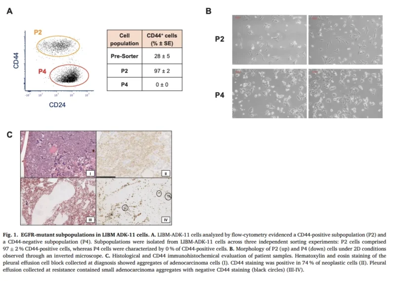
The researchers used CD44 expression to distinguish cellular populations within the LIBM-ADK-11 model. CD44 is a transmembrane glycoprotein involved in cell adhesion, extracellular matrix interactions, migration, and cellular signaling.
In cancer, CD44 has been associated with stemness-related programs, mesenchymal phenotypes, and treatment tolerance. Flow-cytometry analysis revealed that approximately 28% of cells in the unsorted laboratory culture expressed CD44.
The investigators isolated two populations: CD44-positive P2 cells and CD44-negative P4 cells. The sorted P2 population contained approximately 97% CD44-positive cells, whereas CD44 expression was absent in the sorted P4 fraction.
Both populations retained the same EGFR exon 19 driver alteration. The mutation variant allele frequencies were 75.1% in P2 and 81.1% in P4 cells. Despite this shared EGFR-mutant background, the populations demonstrated distinct morphology, transcriptional characteristics, proliferation, and drug sensitivity.
The CD44-positive P2 population exhibited elongated, spindle-shaped morphology and mesenchymal-associated characteristics. The CD44-negative P4 population showed a more epithelial appearance. This finding supports the concept that genetically related tumor cells can occupy functionally distinct states, with important consequences for therapeutic response.
A Nearly 100-Fold Difference in Osimertinib Sensitivity
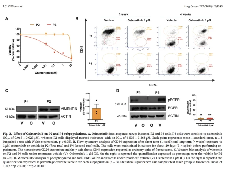
The most striking experimental finding emerged from drug-sensitivity assays. CD44-negative P4 cells remained highly sensitive to osimertinib. Their measured half-maximal inhibitory concentration (IC50) was 0.068 ± 0.023 µM.
In contrast, CD44-positive P2 cells were markedly less sensitive, with an IC50 of 6.535 ± 1.368 µM. This corresponds to an approximately 96-fold difference in measured drug sensitivity between the populations.
Importantly, these measurements were obtained in laboratory assays and should not be interpreted as clinical resistance thresholds. The result demonstrates that retention of a sensitizing EGFR mutation does not necessarily guarantee continued functional dependence on EGFR signaling.
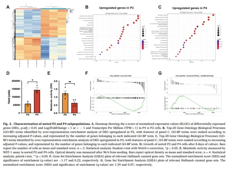
The CD44-positive and CD44-negative states also differed in their growth characteristics. After eight days of culture, P2 cells produced approximately threefold fewer cells than P4 cells. Paradoxically, P2 cells demonstrated higher metabolic activity despite their slower proliferation.
Transcriptomic analysis showed enrichment of glycolysis-associated programs in the resistant population, while cholesterol-associated genes were more prominent in the sensitive population. These findings suggest that treatment tolerance may coexist with reduced proliferative activity and substantial metabolic adaptation.
Such characteristics are consistent with a persister-like phenotype in which cellular survival under drug pressure becomes more important than rapid growth.
EGFR Was Inhibited, Yet Resistant Cells Survived
One of the study’s most clinically relevant observations concerns EGFR phosphorylation. The investigators examined phosphorylated EGFR levels following osimertinib exposure in both populations. Osimertinib reduced EGFR phosphorylation in both the sensitive P4 cells and the resistant P2 cells.
This suggests that resistance in P2 was not simply explained by persistent activation of the receptor that osimertinib was designed to inhibit. Instead, the findings favor an EGFR-independent adaptive mechanism that allows cellular survival despite suppression of proximal EGFR signaling.
The distinction is important. A tumor retaining its original EGFR mutation may contain cells whose survival has become less dependent on the corresponding oncogenic pathway. In such a setting, continued inhibition of EGFR may remain pharmacologically effective at the receptor level while producing an incomplete antitumor response.
However, the study did not establish a single downstream pathway as the definitive cause of resistance. The evidence supports the involvement of broader cellular-state adaptations rather than proving one actionable bypass mechanism.
Mesenchymal Plasticity, AXL, and CD70
Whole-transcriptome analysis identified approximately 4,000 differentially expressed genes between the two populations. The resistant P2 cells were enriched for extracellular matrix organization, matrix remodeling, and mesenchymal-associated programs.
These included transcriptional regulators such as ZEB1, TWIST1, SNAI1, PRRX1, and HMGA2. Several genes associated with treatment resistance were also upregulated, including AXL, CD70, and EMP3.
AXL is a receptor tyrosine kinase previously implicated in EMT-associated resistance and reduced EGFR dependence. ZEB1 is a central transcriptional regulator of mesenchymal differentiation and has also been associated with EGFR inhibitor resistance.
CD70 has emerged as a potential therapeutic target in selected EGFR-TKI-resistant tumor-cell states. The simultaneous enrichment of these programs suggests that the resistant population exhibits a broader biological adaptation rather than a single discrete resistance mechanism.
Nevertheless, increased expression of AXL or CD70 does not establish that either protein is driving resistance in this model. Functional inhibition experiments would be needed to determine whether targeting these pathways restores osimertinib sensitivity.
The observations identify possible vulnerabilities for future research, not validated treatment targets.
A Tumor Cell That Resembles Its Microenvironment
Beyond mesenchymal differentiation, the resistant P2 population demonstrated transcriptional programs associated with stromal and inflammatory biology. The investigators identified expression of genes commonly linked to cancer-associated fibroblast-like phenotypes, including COL1A1, COL3A1, ACTA2, TAGLN, VCAN, and FAP.
Other genes suggested macrophage or tumor-associated macrophage-like mimicry, including CSF1R, SIGLEC15, LGALS1, GAS6, and AXL. The population also expressed inflammatory and antigen-presentation-associated genes, including IL1B, IL32, HLA-DRA, CD74, and CIITA.
Together, these findings suggest that resistant tumor cells may adopt transcriptional characteristics resembling cellular components of the surrounding tumor microenvironment. However, the study does not establish that the tumor cells became functional fibroblasts or macrophages.
These findings describe gene-expression programs within the tumor-cell model. Their biological consequences and interaction with actual immune or stromal cells require further investigation.
The study’s authors interpret this pattern as a broader mesenchymal and inflammatory shift rather than a simple binary transition between epithelial and mesenchymal identities. This distinction may be important as resistance research increasingly recognizes that tumor cells occupy a spectrum of dynamically changing states.
Osimertinib Selects for the Resistant Compartment
The investigators also examined how the two populations changed during prolonged laboratory culture. Without osimertinib exposure, the CD44-positive population gradually declined. The faster-growing CD44-negative cells increasingly dominated the culture.
As this occurred, overall osimertinib sensitivity increased. In low-passage cultures, which contained a larger CD44-positive component, the measured osimertinib IC50 was 5.044 µM. In high-passage cultures, where the CD44-positive fraction had largely disappeared, the IC50 declined to 0.145 µM.
These findings suggest that the relative abundance of distinct cell states can strongly influence the apparent drug sensitivity of the overall tumor model. Under osimertinib exposure, however, the population dynamics changed.
The drug preferentially depleted CD44-negative cells while preserving the CD44-positive compartment. Osimertinib also delayed the spontaneous loss of CD44-positive cells observed during untreated culture. Consequently, treatment acted as a selective pressure favoring the relative persistence of the resistant state.
The CD44-positive population itself was not completely stable. During ordinary culture, some cells lost their CD44-high phenotype, suggesting cellular plasticity or differences in proliferative fitness. The study could not definitively distinguish direct transitions between cellular states from selective expansion of preexisting subpopulations.
These observations nevertheless show how drug exposure and culture conditions can alter the composition of a heterogeneous cancer-cell population.
Three-Dimensional Experiments Reveal Additional Complexity
The investigators extended their experiments beyond conventional two-dimensional culture by evaluating sphere-forming capacity. Interestingly, high-passage cultures demonstrated greater sphere-forming capacity than low-passage cultures, despite having substantially lower CD44 expression.
This suggests that CD44 expression alone does not predict every stemness-associated or three-dimensional growth characteristic. Another important finding was that spheres derived from low- and high-passage cultures showed relatively similar osimertinib sensitivity.
The corresponding IC50 values were 0.061 µM and 0.080 µM. This contrasts with the marked difference observed in conventional adherent cultures. The discrepancy suggests that sensitivity is influenced not only by CD44-defined cellular composition but also by the experimental environment and growth conditions.
It also cautions against assuming that CD44 expression is a universally reliable surrogate for osimertinib resistance. The investigators additionally observed differences in cell-cycle response. In the CD44-positive population, osimertinib increased the G0/G1 fraction while reducing the G2/M fraction, consistent with a shift toward slower cycling.
In CD44-negative cells, treatment reduced the S-phase fraction, consistent with inhibition of proliferation in a more drug-sensitive population. These findings reinforce the concept that distinct cellular states can respond differently to the same therapeutic pressure.
Can Pemetrexed Target Both Populations?
The authors also investigated whether pemetrexed could affect the distinct cellular compartments. In short-term laboratory experiments, pemetrexed partially inhibited both CD44-positive and CD44-negative cells.
Unlike osimertinib, which preferentially depleted the sensitive population, pemetrexed demonstrated activity against both compartments. However, the drug did not completely eliminate the CD44-positive population.
The authors interpret this finding as supporting further investigation of strategies that address multiple cellular states rather than targeting only the EGFR-dependent component. This is biologically consistent with the broader rationale for combining EGFR inhibition with chemotherapy.
Nevertheless, the laboratory result cannot establish clinical benefit from a particular combination or treatment sequence. The study did not test a randomized clinical treatment strategy, and the activity of pemetrexed in a patient-derived cell line should not be equated with clinical outcomes.
The authors also discuss the possibility of investigating PI3K/AKT-related signaling, epigenetic regulation, and alternative treatment schedules. These remain research hypotheses. The findings do not support changing osimertinib dosing, introducing drug holidays, or selecting additional agents on the basis of CD44 expression in routine practice.
A Critical Limitation: The Patient Sample and Cell Model Were Not Identical
An important nuance concerns CD44 expression in the original patient specimens. The diagnostic pleural effusion specimen showed CD44 expression in approximately 74% of tumor cells. However, the pleural effusion specimen collected at osimertinib resistance did not demonstrate CD44-positive tumor cells by immunohistochemistry.
Despite this, CD44-positive cells were present in the derived laboratory culture. The authors suggest several potential explanations, including sampling differences, assay sensitivity, spatial heterogeneity, or adaptation during culture establishment. This discrepancy is especially important when interpreting the biological significance of the findings.
The study demonstrates coexistence of resistant and sensitive states in a cell line derived from clinical resistance, but it does not directly prove that both populations existed in the same proportions within the patient’s progressing tumor.
Furthermore, the model originated from a single patient. Prolonged culture may alter the abundance and characteristics of tumor-cell populations, while the absence of a complete native microenvironment limits the ability to reproduce the full biology of treatment resistance.
Independent patient-derived models, paired clinical samples, and more comprehensive molecular characterization will be essential to determine whether the observed states represent a broadly applicable resistance mechanism.
Clinical Implications: A New Dimension of EGFR Resistance Research
For clinicians treating EGFR-mutant NSCLC, the findings reinforce an increasingly important principle: genetic profiling is essential, but it may not fully capture the biological mechanisms of acquired resistance.
Two cells harboring the same driver alteration may respond differently to treatment because their transcriptional and phenotypic states are different. This may help explain why some tumor populations persist despite apparently effective inhibition of an oncogenic target.
In the future, integrating genomic analysis with cellular-state profiling could improve understanding of resistant disease. Potential approaches include examining EMT-associated transcriptional signatures, metabolic adaptation, CD44-associated cellular states, and emerging therapeutic targets such as AXL or CD70.
However, CD44 is not established as a clinically validated biomarker for selecting therapy after osimertinib progression. The present study does not define a new standard of care. Its importance lies in providing a patient-derived experimental model for understanding why treatment resistance may persist without a newly identified genomic driver.
OncoDaily Takeaway
The new study by Chillico and colleagues highlights the importance of non-genetic heterogeneity in EGFR-mutant NSCLC.
Within a single patient-derived model established after osimertinib resistance, CD44-positive and CD44-negative populations retained the same EGFR exon 19 mutation but demonstrated profoundly different treatment sensitivities.
The CD44-positive population exhibited mesenchymal, inflammatory, and metabolically adapted features and showed nearly 100-fold lower sensitivity to osimertinib in conventional laboratory assays. Crucially, EGFR phosphorylation was suppressed in both populations, suggesting that the resistant phenotype could persist despite effective inhibition of proximal EGFR signaling.
Osimertinib preferentially depleted the sensitive compartment, allowing the resistant state to persist under treatment pressure. Although pemetrexed partially affected both populations, it did not completely eradicate the resistant compartment.
These findings suggest that overcoming acquired resistance may require strategies addressing both genetic drivers and adaptive cellular states. The central message is that retaining an EGFR mutation does not necessarily mean every tumor cell remains equally dependent on EGFR signaling.
The findings are preclinical, derived from a single patient model, and require independent validation. Nevertheless, they provide a compelling framework for investigating phenotypic plasticity, drug-tolerant persister cells, and new therapeutic vulnerabilities in EGFR-mutant lung cancer.
Sources
- Chillico IC, Frascino M, Protopapa G, et al. Coexistence of Osimertinib-Resistant and Osimertinib-Sensitive Cell States in an EGFR-Mutant NSCLC Model Derived at Osimertinib Resistance. Lung Cancer. 2026;222:109680. Published online November 5, 2026. DOI: 10.1016/j.lungcan.2026.109680
- Soria JC, et al. Osimertinib in Untreated EGFR-Mutated Advanced Non–Small-Cell Lung Cancer. New England Journal of Medicine. 2018. DOI: 10.1056/NEJMoa1713137
- Planchard D, et al. Osimertinib With or Without Chemotherapy in EGFR-Mutated Advanced NSCLC. New England Journal of Medicine. 2023. DOI: 10.1056/NEJMoa2306434
- Sharma SV, et al. A Chromatin-Mediated Reversible Drug-Tolerant State in Cancer Cell Subpopulations. Cell. 2010. DOI: 10.1016/j.cell.2010.02.027
- Nilsson MB, et al. CD70 Is a Therapeutic Target Upregulated in EMT-Associated EGFR Tyrosine Kinase Inhibitor Resistance. Cancer Cell. 2023. DOI: 10.1016/j.ccell.2023.01.007