
For decades, cancer was viewed primarily as a disease of malignant cells driven by genetic mutations. Advances in cancer biology have fundamentally changed this perspective. Today, tumors are recognized as highly dynamic ecosystems in which malignant cells constantly communicate with surrounding immune cells, stromal cells, blood vessels, extracellular matrix, and soluble signaling molecules. Together, these components form the tumor microenvironment (TME), a complex network that profoundly influences tumor initiation, progression, metastasis, and response to therapy.
In their landmark review “The Tumor Microenvironment,” Nicole M. Anderson and M. Celeste Simon describe tumors as heterogeneous tissues rather than isolated masses of cancer cells, emphasizing that interactions between malignant cells and the surrounding microenvironment actively drive cancer evolution rather than merely accompany it.
Similarly, Karin E. de Visser and Johanna A. Joyce, in their comprehensive review “The Evolving Tumor Microenvironment: From Cancer Initiation to Metastatic Outgrowth,” highlight that the TME continuously co-evolves with cancer throughout every stage of disease, from the earliest transformed cell to metastatic dissemination, making it one of the central determinants of cancer biology.
What Is the Tumor Microenvironment?
The tumor microenvironment refers to the entire ecosystem surrounding cancer cells.
Rather than existing in isolation, tumor cells are embedded within a highly organized tissue composed of immune cells, stromal cells, vascular networks, extracellular matrix (ECM), cytokines, chemokines, growth factors, metabolites, and even tissue-specific cells such as adipocytes and neurons. Continuous communication among these components determines whether the immune system successfully eliminates the tumor or whether cancer progresses.
Importantly, the TME is not static. It changes throughout tumor development and differs substantially among cancer types, organs, disease stages, and even between individual patients. This dynamic nature explains why tumors with similar genetic alterations can respond very differently to identical therapies.
The Major Components of the Tumor Microenvironment
The TME consists of both cellular and non-cellular elements that continuously influence one another.
Immune Cells
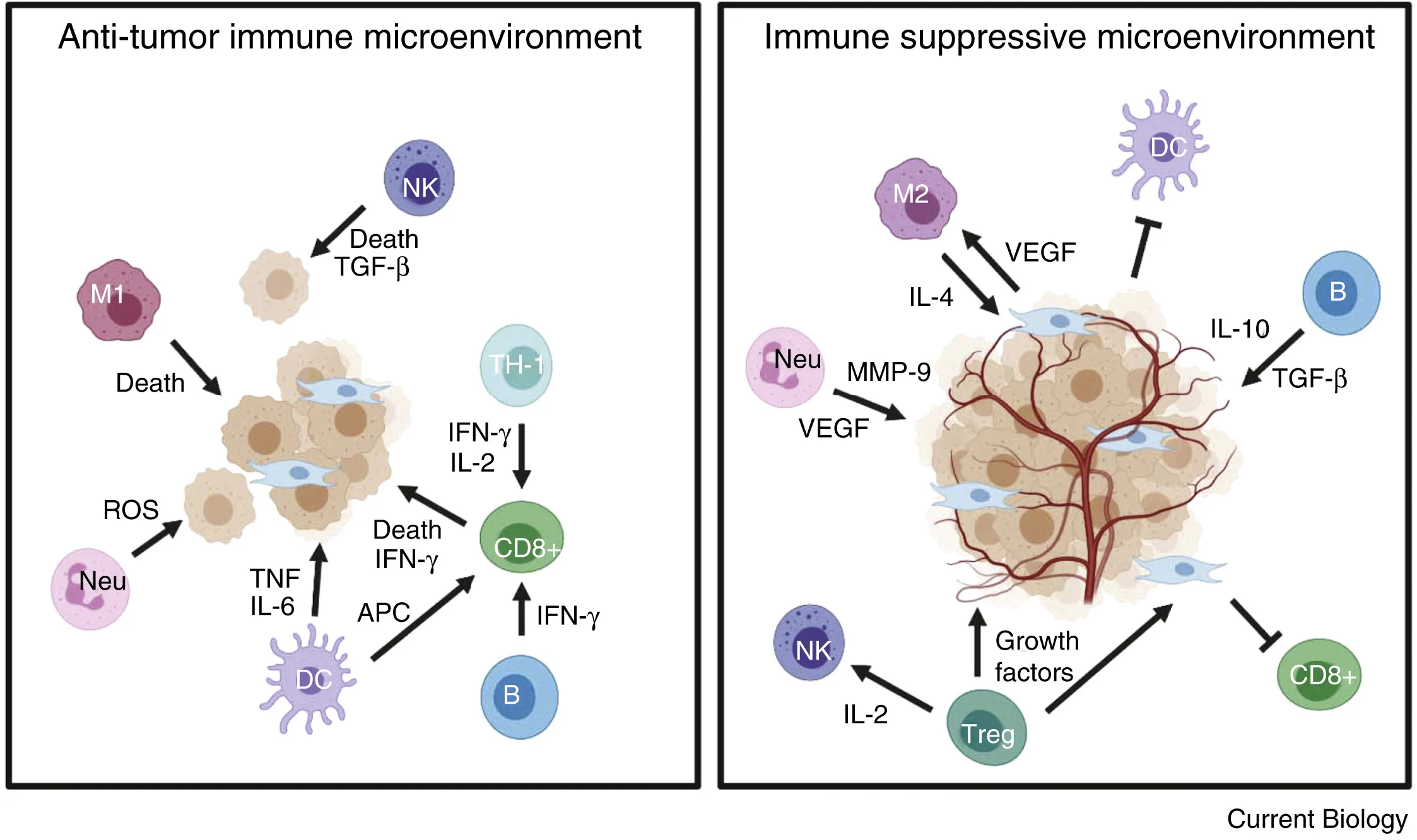
Immune cells represent one of the most important components of the TME. Their role is remarkably complex because the same immune system that initially protects against cancer can later become co-opted to support tumor progression.
Anti-tumor immunity is primarily mediated by:
- CD8⁺ cytotoxic T lymphocytes
- CD4⁺ Th1 helper cells
- Natural killer (NK) cells
- Dendritic cells
- Certain B-cell populations
These cells recognize malignant cells, produce inflammatory cytokines, present tumor antigens, and directly destroy cancer cells.
In contrast, tumors gradually recruit immunosuppressive populations, including:
- Regulatory T cells (Tregs)
- Tumor-associated macrophages (particularly M2-like macrophages)
- Myeloid-derived suppressor cells (MDSCs)
- Regulatory B cells
- Dysfunctional neutrophils
These populations suppress cytotoxic immunity through cytokines such as IL-10 and TGF-β, inhibit T-cell activation, impair NK-cell function, promote angiogenesis, and facilitate immune escape.
Cancer-Associated Fibroblasts (CAFs)
Fibroblasts normally maintain tissue architecture and participate in wound healing. Within tumors, however, they become activated into cancer-associated fibroblasts (CAFs), which actively support cancer progression.
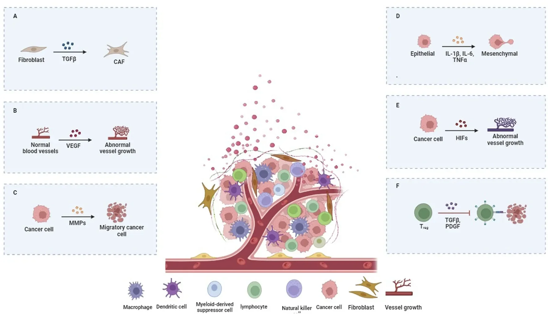
Rather than serving only as structural cells, CAFs remodel the extracellular matrix, secrete growth factors, cytokines, and chemokines, stimulate angiogenesis, promote invasion, and create physical barriers that prevent immune-cell infiltration.
Recent evidence also demonstrates that CAFs contribute substantially to resistance to immune checkpoint inhibitors by establishing an immunosuppressive stromal environment that excludes cytotoxic T cells from tumor nests.
Tumor Vasculature
As tumors grow beyond approximately 1–2 mm, passive diffusion becomes insufficient to supply oxygen and nutrients. Hypoxia activates hypoxia-inducible factors (HIFs), which stimulate production of vascular endothelial growth factor (VEGF) and initiate angiogenesis.
Unlike normal blood vessels, tumor vessels are highly abnormal. They are tortuous, immature, and leaky, resulting in irregular blood flow, poor oxygen delivery, elevated interstitial pressure, and impaired immune-cell trafficking.
These vascular abnormalities not only support metastasis but also reduce delivery of systemic therapies while limiting infiltration of cytotoxic immune cells.
Extracellular Matrix
The extracellular matrix is far more than a structural scaffold.
It consists of collagen, fibronectin, laminins, proteoglycans, and numerous signaling molecules that regulate cell migration, proliferation, and differentiation. During tumor progression, extensive remodeling of the ECM increases tissue stiffness, facilitates invasion, stores cytokines and growth factors, and creates biochemical and mechanical barriers that impair immune-cell penetration.
Matrix remodeling by matrix metalloproteinases (MMPs) further promotes local invasion and metastatic dissemination.
How Tumors Hijack Their Microenvironment
One of the most remarkable features of cancer is its ability to reprogram surrounding normal tissues.
Rather than passively adapting to their environment, tumor cells actively reshape it by secreting cytokines, chemokines, extracellular vesicles, metabolites, and growth factors that recruit supportive stromal and immune cells. This reciprocal communication transforms initially tumor-restrictive tissues into highly permissive environments that support malignant growth.
Cancer cells manipulate macrophages toward pro-tumor phenotypes, recruit Tregs and MDSCs, activate fibroblasts, remodel the ECM, and stimulate angiogenesis. These coordinated changes establish an ecosystem that favors immune evasion, invasion, and metastasis.
Metabolic Reprogramming Inside the TME
The TME is also characterized by profound metabolic alterations.
Rapidly proliferating tumor cells consume enormous quantities of glucose, amino acids, and oxygen, creating intense competition with infiltrating immune cells. As glucose becomes depleted and lactate accumulates, the microenvironment becomes acidic and metabolically hostile.
This metabolic stress impairs the function of cytotoxic T cells and NK cells, reduces cytokine production, inhibits proliferation, and promotes immune exhaustion. Meanwhile, tumor cells adapt through metabolic reprogramming, allowing them to thrive under conditions that suppress anti-tumor immunity.
Hypoxia further amplifies these effects by activating HIF-1α signaling, promoting angiogenesis, increasing PD-L1 expression, and driving additional immunosuppressive mechanisms.
The Tumor Microenvironment and Metastasis
The TME plays a critical role throughout the metastatic cascade.
Cancer-associated fibroblasts remodel the ECM to facilitate local invasion. Tumor-associated macrophages produce proteases and growth factors that promote intravasation into blood vessels. Platelets protect circulating tumor cells from immune attack, while stromal cells at distant organs help establish the pre-metastatic niche that allows disseminated tumor cells to survive and eventually form secondary tumors.
Rather than being driven solely by intrinsic genetic alterations, metastasis is now recognized as a cooperative process orchestrated by continuous interactions between cancer cells and their surrounding microenvironment.
Why the Tumor Microenvironment Matters for Immunotherapy
Modern immunotherapy targets not only cancer cells but also the surrounding immune ecosystem.
Immune checkpoint inhibitors restore exhausted T-cell activity by disrupting inhibitory pathways such as PD-1/PD-L1. At the same time, researchers are developing therapies that target other components of the TME, including macrophages, CAFs, MDSCs, tumor vasculature, and metabolic pathways.
Emerging technologies—including spatial transcriptomics, single-cell sequencing, and multi-omics profiling—are revealing previously unrecognized cellular interactions within tumors. These approaches are helping identify biomarkers of treatment response while enabling increasingly personalized immunotherapeutic strategies.
As emphasized by de Visser and Joyce, understanding how tumors dynamically interact with their microenvironment may become as important as understanding the genetic alterations of cancer cells themselves.
Conclusion
The tumor microenvironment has transformed our understanding of cancer biology. Rather than viewing tumors as isolated collections of malignant cells, modern oncology recognizes cancer as a complex ecosystem in which immune cells, stromal cells, blood vessels, extracellular matrix, and metabolic signals continuously interact to shape disease progression and therapeutic response.
The reviews by Nicole M. Anderson and M. Celeste Simon, Karin E. de Visser and Johanna A. Joyce, Qingjing Wang and colleagues, and Cigir Biray Avci and colleagues collectively demonstrate that successful cancer therapy increasingly depends not only on targeting tumor cells themselves but also on understanding and reprogramming the surrounding microenvironment. As immunotherapy continues to evolve, the TME is likely to remain one of the most important frontiers in precision oncology.