Cancer immunotherapy has traditionally focused on the immune cells that reach a tumor: how many CD8+ T cells are present, whether they recognize cancer antigens, whether they are exhausted and whether inhibitory checkpoints can be removed. Yet the effectiveness of antitumor immunity may depend on something more complex than the presence or absence of individual immune cells.
In some tumors, lymphocytes assemble into highly organized structures containing distinct B-cell and T-cell regions, dendritic cells, follicular networks, specialized blood vessels and sometimes functional germinal centers. These structures, known as tertiary lymphoid structures (TLSs), resemble lymph nodes that have developed directly within or adjacent to diseased tissue. Unlike conventional lymph nodes, they are not anatomically predetermined organs. They emerge in sites of persistent inflammation, including tumors.
Over the past several years, TLSs have evolved from an interesting pathological observation into one of the most compelling areas of tumor immunology. Landmark studies linked their presence to checkpoint inhibitor response across melanoma and other solid tumors. More recent spatial and single-cell studies are beginning to reveal how these structures function, how their maturity matters and why specialized blood vessels may determine whether they form at all.
The question is therefore changing. Rather than simply asking whether a tumor contains immune cells, researchers are beginning to ask whether those cells can be organized into a functional immune ecosystem capable of sustaining antitumor immunity locally. And if some tumors can build such ecosystems naturally, could immunotherapy eventually build them deliberately?
A Lymph Node Where a Lymph Node Should Not Exist
Secondary lymphoid organs such as lymph nodes provide architecture for immune responses. They bring antigen-presenting cells, naïve and memory lymphocytes, B cells and T cells into spatially organized environments where antigen recognition, clonal expansion, affinity maturation and immune-cell differentiation can occur efficiently.
TLSs reproduce parts of this organization inside non-lymphoid tissues.
Their architecture varies substantially. Early lymphoid aggregates may consist of relatively disorganized clusters of T and B cells. More developed TLSs contain segregated T-cell and B-cell zones, follicular dendritic-cell networks and specialized vasculature. Mature structures can develop germinal centers in which B cells proliferate, undergo selection and differentiate toward memory B cells and antibody-producing plasma cells.
This maturation matters because simply counting lymphocytes may miss the biological information encoded in their organization. Two tumors could contain similar numbers of immune cells while having profoundly different immune capabilities if one contains scattered dysfunctional lymphocytes and the other contains a structured environment supporting continuous immune-cell recruitment, antigen presentation and cellular cooperation.
The emerging concept is that antitumor immunity is not only a question of immune-cell abundance. It is also a question of immune architecture.
The 2020 Studies That Changed How We Think About B Cells
For years, checkpoint immunotherapy was largely discussed through the biology of T cells. Then, in January 2020, three independent studies appeared together in Nature and substantially changed the conversation by connecting B cells and TLSs with immunotherapy response.
Beth Helmink, Jennifer Wargo and colleagues reported “B Cells and Tertiary Lymphoid Structures Promote Immunotherapy Response.” In tumors from patients receiving checkpoint blockade, B-cell-associated genes were among the features most strongly enriched in responders. Histological analysis showed that these B cells were frequently organized within TLSs, while single-cell and bulk sequencing demonstrated clonal B-cell expansion and distinct functional B-cell states. Switched memory B cells were also enriched in tumors from responders.
In parallel, Rita Cabrita, Göran Jönsson and colleagues published “Tertiary Lymphoid Structures Improve Immunotherapy and Survival in Melanoma.” Their analysis showed that tumors containing both CD8+ T cells and CD20+ B cells had better survival, and spatial analysis revealed that these populations were organized within TLSs. A TLS-associated gene signature was also linked to outcomes in cohorts receiving checkpoint blockade.
The third study, led by Florent Petitprez and colleagues, extended the observation to soft-tissue sarcoma, where B-cell-rich immune phenotypes were associated with survival and response to checkpoint inhibition. Together, the three studies suggested something that had been underappreciated in the T-cell-dominated model of cancer immunotherapy: successful antitumor immunity may depend on organized collaboration between multiple immune-cell populations rather than T-cell activity alone.
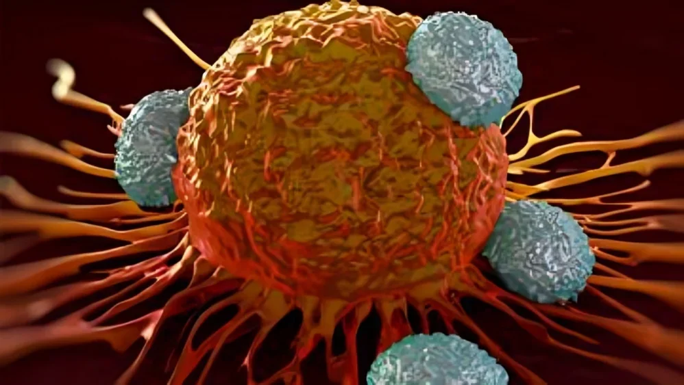
Tertiary lymphoid structures and cancer immunotherapy
What Are B Cells Doing Inside a Tumor?
B cells are often reduced to their best-known function, antibody production. Within tumors, their biology is considerably broader.
B cells can recognize antigen through the B-cell receptor, internalize that antigen and present processed peptides to CD4+ T cells through MHC class II. They can produce cytokines, differentiate into plasma cells, generate tumor-reactive antibodies and participate in the organization of local immune niches. Within germinal center-like TLSs, they can also undergo clonal expansion and maturation.
This creates an environment in which B-cell and T-cell immunity can reinforce one another. T follicular helper-like cells can support B-cell activation and maturation, while B cells and dendritic cells can contribute to antigen presentation and T-cell activation. Plasma cells can generate antibodies with potential antitumor functions, while chemokine networks recruit additional immune populations into the developing structure.
A 2025 Nature Communications study in head and neck squamous cell carcinoma added important mechanistic resolution to this picture. Using single-cell RNA sequencing, antigen-receptor sequencing and spatial transcriptomics, investigators found that mature TLSs were enriched in stem-like T-cell populations and B cells across different stages of maturation. Progenitor-exhausted CD4+ T cells with follicular helper-like characteristics interacted with B cells in germinal-center regions and with DC-LAMP+ dendritic cells, supporting both B-cell maturation and CD8+ T-cell activation.
The TLS therefore begins to look less like a passive collection of immune cells and more like a local immune reaction center.

Why Bring the Immune Response Into the Tumor?
Most conventional adaptive immune responses are initiated in lymph nodes. Dendritic cells capture antigen in peripheral tissues, migrate to draining lymph nodes and present antigen to T cells. Activated lymphocytes then proliferate, enter the circulation and eventually migrate back toward the affected tissue.
Cancer makes this process unusually difficult. Tumor antigens may be poorly presented, tumor vasculature can restrict lymphocyte entry, stromal barriers can prevent infiltration and activated T cells may encounter suppressive signals once they reach the tumor.
TLSs potentially shorten this immunological distance.
When functional lymphoid organization exists inside or immediately adjacent to the tumor, antigen presentation, B-cell maturation and T-cell activation can occur locally. Immune cells no longer necessarily depend entirely on distant lymphoid organs for every stage of the response.
This could be especially important during prolonged cancer evolution. Tumors continually generate and lose antigens under immune and therapeutic pressure. A local immune structure capable of sampling this changing antigenic landscape could theoretically support ongoing immune adaptation rather than relying exclusively on a response initiated much earlier in a distant lymph node.
That possibility helps explain why TLS biology is becoming relevant not only to biomarkers, but to the fundamental question of how durable antitumor immunity is maintained inside human tumors.
High Endothelial Venules May Be the Gateways
Building a local immune ecosystem requires more than organizing cells already present. New immune cells must continuously enter.
This is where high endothelial venules, or HEVs, become particularly important.
HEVs are specialized blood vessels normally found in secondary lymphoid organs. Their endothelial cells express adhesion molecules and chemokines that allow circulating lymphocytes to leave the bloodstream and enter lymphoid tissue. Similar vessels occur within tumor-associated TLSs, creating potential entry routes for lymphocytes directly into the tumor microenvironment.
A 2025 Nature Reviews Immunology review on tumor vasculature emphasized that tumor-associated blood vessels are not merely passive conduits. Abnormal tumor vasculature can actively restrict T-cell homing, whereas HEVs associated with TLSs can create privileged sites supporting lymphocyte entry, antigen presentation and T-cell activation.
Recent spatial studies are now dissecting HEVs at much higher resolution. In nasopharyngeal carcinoma, a 2025 Cell Reports Medicine study integrating spatial transcriptomics with a pan-cancer single-cell atlas identified a population of interferon-responsive HEVs associated with TLS development. These vessels expressed CXCL9 and were linked to recruitment of CXCR3+ CD4+ T cells into TLSs. An HEV/TLS-related score was subsequently associated with anti-PD-1 response across independent cohorts.
Another spatial single-cell analysis in NSCLC identified ID1-high endothelial cells within mature TLS-associated HEVs. These cells displayed adhesion programs consistent with active lymphocyte recruitment, and tumors containing ID1-high HEVs were associated with more durable immunotherapy responses. Interestingly, checkpoint blockade itself appeared capable of remodeling this endothelial state over time, emphasizing that TLS-associated vasculature is dynamic rather than fixed.
The blood vessel, therefore, may be part of the immune response itself.
Mature TLSs May Matter More Than Simply Having TLSs
One major challenge is that the term “TLS-positive” compresses substantial biological diversity.
A small aggregate of lymphocytes is not equivalent to a mature TLS containing organized B-cell follicles, follicular dendritic cells, germinal-center activity, T-cell zones and functional HEVs. Location also matters. TLSs may occur within tumor tissue, at invasive margins or in surrounding stroma, and their biological significance may differ between cancer types.
This is increasingly important as TLSs move toward clinical biomarker development. Histological assessment, immunohistochemistry, transcriptomic signatures and spatial imaging do not necessarily identify exactly the same structures. A tumor classified as TLS-positive by one method might not meet the same definition using another.
The 2026 Nature Cancer article “The Future of Tertiary Lymphoid Structures in Cancer Immunotherapy as Biomarkers and Therapeutic Targets” highlights precisely this transition. TLSs are increasingly being considered not simply as binary structures that are present or absent, but as dynamic immune ecosystems whose cellular composition, maturation and spatial context may determine their clinical significance.
This means that the future biomarker may not simply be TLS yes or no. It may need to capture where the TLS is located, how mature it is, which B-cell and T-cell states it contains, whether germinal-center activity is present and whether functional HEVs are continuously recruiting new lymphocytes.
Clinical Evidence Suggests TLSs May Identify Immunotherapy-Sensitive Tumors
The association between TLSs and checkpoint response has moved beyond retrospective melanoma observations.
Soft-tissue sarcoma provides one of the most striking examples. Checkpoint inhibitors have generally produced modest activity across unselected sarcoma populations, but the phase II PEMBROSARC study included a cohort specifically selected for the presence of TLSs.
Thirty patients with advanced soft-tissue sarcoma and TLS-positive tumors received pembrolizumab with low-dose cyclophosphamide. The 6-month non-progression rate was 40%, and the objective response rate was 30%. In previously reported all-comer cohorts, the corresponding values were 4.9% and 2.4%. Exploratory analyses also linked higher intratumoral plasma-cell abundance with better outcomes.
The study was small and was not a randomized comparison between TLS-positive and TLS-negative patients, so the magnitude of difference should not be interpreted as definitive proof that TLSs themselves caused checkpoint sensitivity. Nevertheless, it provided important prospective evidence that selecting patients according to immune architecture may identify a population with substantially different immunotherapy activity.
More recent studies suggest that even this relationship may be too simple. A 2025 Cancer Discovery analysis of 102 patients with renal cell carcinoma treated with frontline nivolumab found TLS-associated genes enriched in responders, but outcome depended on the broader immune ecosystem. Tumors with high TLS abundance and low levels of a ZNF683+SLAMF7+ exhausted tissue-resident CD8+ population had the most favorable outcomes.
TLSs therefore do not operate independently. Their function depends on the immune states surrounding and inhabiting them.
Can Immunotherapy Create TLSs Rather Than Simply Benefit From Them?
This may be the most important question in the field.
If TLSs merely identify tumors that already possess favorable immune biology, their main clinical value may be as biomarkers. But if functional TLSs can be deliberately generated inside tumors, they become therapeutic targets.
There is growing evidence that treatment can modify TLS biology. Neoadjuvant immunotherapy has been associated with the development or maturation of TLSs in several tumor settings, and recent experimental studies are beginning to identify molecular combinations capable of deliberately inducing these structures.
A particularly interesting 2025 study in Nature Immunology examined simultaneous activation of STING and the lymphotoxin-β receptor (LTβR). These pathways address different components of lymphoid organization. STING activates innate inflammatory programs, while LTβR signaling has an established role in lymphoid tissue organization and stromal biology.
Combined activation produced high endothelial venule development and germinal center-like B-cell responses within tumors, generating functional TLSs in experimental models. The treatment enhanced CD8+ T-cell-dependent tumor suppression and, in a neoadjuvant setting, generated protection against tumor recurrence with long-term survival in mice.
This is still preclinical biology, not evidence that pharmacologically building TLSs will improve outcomes in patients. But it demonstrates something conceptually important: lymphoid architecture inside tumors is experimentally manipulable.
Instead of delivering another cytotoxic signal or blocking another checkpoint, a therapy might eventually attempt to construct the immune infrastructure required for multiple antitumor mechanisms to function together.
Immunogenic Tumor Death May Provide the First Construction Signal
An especially interesting connection has emerged between the way tumor cells die and the way immune structures subsequently organize.
A September 2026 Nature Reviews Immunology Perspective by Rafal Hanc, Robin Demuynck and Dmitri Krysko proposes that immunogenic cell death may act upstream of TLS formation. Rather than viewing tumor-cell killing and lymphoid organization as independent events, the authors suggest that immunogenic death can initiate vascular, stromal and immune remodeling that favors development of tumor-associated TLSs.
The biology is attractive because TLS formation requires several processes to occur together. Tumor antigens must become available, innate immune pathways need to generate inflammatory signals, dendritic cells must participate in antigen presentation, chemokines must recruit lymphocytes, stromal cells must organize the developing structure and local vasculature must permit continued immune-cell entry.
From this perspective, simply killing cancer cells is insufficient. The tumor must die in an immunological context capable of converting destruction into organization.
This could help explain why radiation, certain chemotherapies, local therapies and innate immune agonists sometimes interact productively with checkpoint blockade, while apparently similar tumor destruction in another biological context produces little systemic immunity.
It also creates a new therapeutic question: perhaps some treatments should be evaluated not only by how many cancer cells they kill, but by whether they leave behind an immune microenvironment capable of organizing a sustained response.
Checkpoint Blockade May Remodel Immune Architecture
Checkpoint inhibitors themselves may also influence TLS formation.
A 2026 Nature Communications study examining NSCLC brain metastases found that these lesions contained fewer cytotoxic T cells and fewer TLSs than matched primary tumors, consistent with an immune-excluded environment. In experimental models, combined PD-1 and CTLA-4 blockade increased CD8+ T-cell infiltration and function, expanded T follicular helper-like cells and promoted TLS-like structures, whereas PD-1 monotherapy was less effective.
This observation adds another layer to how combination immunotherapy may work. The benefit of dual checkpoint blockade may not arise solely from releasing two inhibitory receptors on the same T cell. In some contexts, treatment could reorganize the broader immune ecosystem, changing cellular recruitment, helper T-cell states and local lymphoid architecture.
The distinction matters because it suggests that successful combinations could eventually be designed according to the immune structure they are intended to create, rather than simply by stacking active drugs.
But Not Every TLS Is Necessarily Beneficial
The temptation is to interpret TLSs as universally favorable structures. Tumor immunology is rarely that simple.
B cells themselves can adopt both immune-stimulatory and immunosuppressive states. TLSs vary in maturity and composition, and chronic inflammatory environments can generate lymphoid structures that do not necessarily support productive antitumor immunity.
A 2025 Nature Communications study in hepatocellular carcinoma provides an important warning. In fibrotic HCC models, anti-PD-1 treatment or STING agonism increased intratumoral B-cell infiltration and promoted TLS formation, but these changes were accompanied by IL-10 and TIM-1+ regulatory B-cell programs associated with resistance. In that experimental context, inhibiting B cells enhanced the efficacy of STING agonism or checkpoint blockade.
This does not contradict the substantial evidence linking mature TLSs with favorable outcomes in many cancers. Instead, it emphasizes that organization alone is not enough. The functional identity of the cells inside the structure matters.
A therapeutic strategy designed to create TLSs must therefore avoid the assumption that any lymphoid aggregate is beneficial. The objective would be to generate a particular kind of TLS: appropriately located, vascularized, mature and enriched for immune states capable of supporting productive antitumor responses.
B Cells May Even Drive Antitumor Effects We Have Attributed to T Cells
Very recent work is also challenging another assumption: that checkpoint blockade works almost exclusively through T-cell reactivation.
A September 2026 Nature Cancer report highlighted evidence of a local antibody-driven response to anti-PD-1 therapy in which plasma-cell responses contributed to tumor control. The proposed biology involved treatment-associated TLS formation, plasma-cell maturation and local antibody production, including antibodies reactive against HBV-derived antigens associated with tumor cells, with subsequent complement activation.
The finding is context specific and should not be generalized to all checkpoint responses. Nevertheless, it illustrates how organized intratumoral B-cell biology can produce effector mechanisms beyond simply helping CD8+ T cells.
Checkpoint blockade may therefore influence an interconnected immune network involving T cells, B cells, plasma cells, dendritic cells, antibodies, complement, stromal cells and vasculature.
That is a considerably richer model of immunotherapy than simply “releasing the brakes” on exhausted T cells.
Could We Engineer the Architecture Directly?
If functional TLSs become therapeutic objectives, several components of their development could potentially be manipulated.
Chemokines such as CXCL13, CCL19 and CCL21 participate in recruitment and spatial organization of lymphocyte populations. Lymphotoxin signaling contributes to stromal organization and HEV development. Type I and type II interferon programs can alter vascular and immune-cell states. STING activation can provide innate inflammatory signals. Vascular normalization may improve lymphocyte access, while local cytokine delivery or engineered cellular therapies could theoretically provide organizational signals directly within tumors.
The challenge is that these pathways cannot simply be maximally activated everywhere. Systemic manipulation of potent inflammatory or lymphoid-organizing pathways could generate substantial toxicity, while poorly controlled chronic inflammation could create dysfunctional rather than productive immune structures.
Spatial control may therefore become essential. Local delivery systems, engineered immune cells, nanoparticles, intratumoral agents or tumor-restricted cytokine programs could potentially provide the signals needed to construct lymphoid organization where it is useful while limiting systemic exposure.
The ambition is not merely to attract more T cells.
It is to create the cellular, vascular and stromal infrastructure that allows those T cells to continue functioning once they arrive.
Can We Measure TLSs Reliably Enough for Clinical Use?
Before TLSs can become routine biomarkers or therapeutic endpoints, the field needs better standardization.
TLSs can currently be assessed through conventional histopathology, immunohistochemistry, multiplex immunofluorescence, gene-expression signatures, digital pathology and spatial transcriptomic approaches. Each method captures different aspects of the biology.
A transcriptional TLS signature may detect an immune program without showing whether cells are spatially organized into a mature structure. Histology demonstrates architecture but may miss molecular states. A small biopsy can also fail to capture TLSs because these structures are spatially heterogeneous and may cluster at tumor margins rather than within the sampled region.
Maturity adds another complication. Distinguishing an early lymphoid aggregate from a mature germinal-center-containing TLS may ultimately prove more clinically informative than a binary classification.
The 2026 Nature Cancer perspective by Hye Mi Kim, Tejashree Joglekar, Tina Cascone, Tullia Bruno and colleagues argues that these issues must be resolved if TLSs are to move from research observations toward biomarkers and therapeutic targets.
Future trials may therefore need to characterize TLS density, location, maturity, cellular composition and vascular phenotype, rather than recording only whether TLSs are present.
From Biomarker to Therapeutic Organ
The history of TLS research in cancer has progressed through several conceptual stages.
Initially, TLSs were pathological observations associated with chronic inflammation. They then became prognostic features, followed by evidence linking them to checkpoint inhibitor response. Single-cell and spatial technologies revealed that they contain highly organized and functionally distinct immune populations. More recent experimental studies suggest that their formation can be manipulated therapeutically.
The next transition would be the most ambitious: from observing TLSs to deliberately constructing them.
A future immunotherapy strategy might first generate appropriate innate inflammatory signals, then recruit lymphocytes through HEVs, establish chemokine gradients that organize T-cell and B-cell zones, support dendritic-cell antigen presentation and enable B-cell maturation within germinal-center-like structures. Checkpoint blockade or other therapies could then sustain the antitumor cells produced within that environment.
Such a strategy would differ fundamentally from targeting a single immune receptor. It would attempt to engineer an entire immune ecosystem.
Whether that can be achieved safely and reproducibly in human cancers remains unknown.