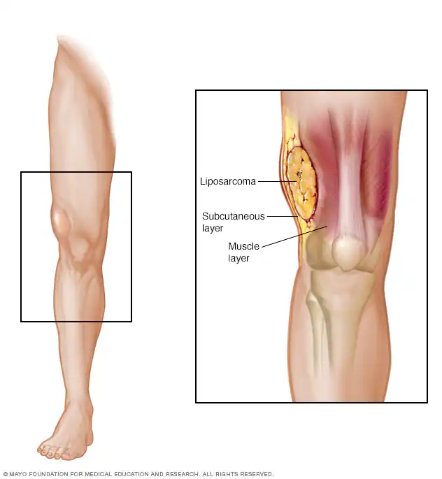
Liposarcoma is a rare malignant tumor that develops in fatty tissues, most commonly in the arms, legs, or abdomen. Unlike a lipoma, which is a benign fatty tumor, liposarcoma is cancerous and can grow aggressively, sometimes spreading to other parts of the body. Early detection is essential, as advanced cases may require complex treatments.

This article will cover the symptoms, causes, types, stages, diagnostic methods, and treatment options for liposarcoma. Understanding its distinction from benign fat tumors, recognizing warning signs, and exploring available treatments can help improve patient outcomes.
What Are the Symptoms of Liposarcoma?
Symptoms vary with tumor location and subtype, leading to delayed diagnoses due to their non-specific nature.
- Limb Tumors: Typically manifest as a painless, firm mass in the thigh or upper arm. As the tumor enlarges, it may cause swelling, numbness, or reduced mobility due to nerve or muscle compression.
- Abdominal Tumors: May remain asymptomatic until substantial growth occurs, leading to abdominal distension, pain, early satiety, or bowel habit changes.
Liposarcoma symptoms vary depending on the subtype, affecting how the disease presents and progresses. Well-differentiated liposarcoma is typically a slow-growing, painless mass, most commonly found in the limbs. Due to its indolent nature, it may go unnoticed for a long time before diagnosis. Myxoid liposarcoma, frequently arising in the thigh, also tends to present as a painless swelling, but it has a higher tendency to metastasize, sometimes spreading to unusual locations such as the retroperitoneum. In contrast, pleomorphic liposarcoma is the most aggressive form, often presenting as a rapidly enlarging tumor that may cause pain and functional impairment, especially if it compresses surrounding nerves or muscles.
Due to their deep-seated nature and non-specific symptoms, liposarcomas often evade early detection. Patients and healthcare providers should maintain vigilance when encountering persistent or enlarging masses, regardless of discomfort, to facilitate timely diagnosis and treatment.
What are the Causes and Risk Factors for Liposarcoma?
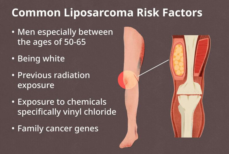
Liposarcoma is primarily driven by genetic mutations, though the exact causes of these alterations remain under investigation. Key risk factors include prior exposure to radiation therapy, certain chemicals like vinyl chloride, and inherited genetic conditions.
- MDM2 Amplification: A significant number of liposarcomas exhibit amplification of the MDM2 gene, which encodes a protein that regulates the tumor suppressor p53. This amplification leads to increased MDM2 protein levels, resulting in the degradation of p53 and promoting tumor growth.
- CDK4 Amplification: Many liposarcomas also show amplification of the CDK4 gene, leading to overexpression of the CDK4 protein, which drives cell cycle progression and contributes to tumor development.
These genetic alterations are particularly prevalent in well-differentiated and dedifferentiated liposarcomas, underscoring their role in the disease’s pathogenesis.

What Are the Types of Liposarcoma?
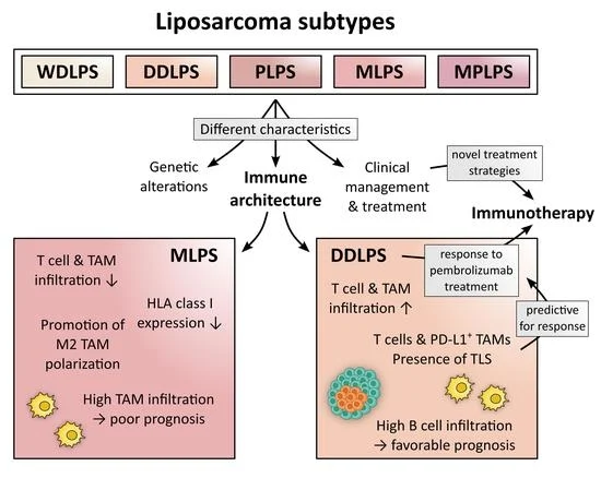
Liposarcoma is classified into four main subtypes based on its cellular characteristics and behavior
- Well-Differentiated Liposarcoma
- Myxoid Liposarcoma
- Pleomorphic Liposarcoma
- Dedifferentiated Liposarcoma
Each subtype varies in aggressiveness, growth pattern, and response to treatment, making accurate diagnosis essential for effective management.

Well-Differentiated Liposarcoma
According to a study by Weiss et al. in The American Journal of Surgical Pathology, well-differentiated liposarcoma is the most common and least aggressive form of liposarcoma. It typically presents as a slow-growing, painless mass, primarily occurring in the limbs and retroperitoneum. According to a study by Lahat et al. in BMC Cancer, the 5- and 10-year survival rates for WDLS are 100% and 82.1%, respectively.
However, it’s important to note that while WDLS has a high survival rate, there is a risk of local recurrence, particularly in cases where complete surgical excision is challenging. Additionally, WDLS has the potential to dedifferentiate into more aggressive forms, underscoring the importance of long-term follow-up. A comprehensive analysis by Dalal et al. in Cancer highlighted that dedifferentiation occurs in approximately 10% of WDLS cases, emphasizing the need for vigilant monitoring post-treatment.
Myxoid Liposarcoma
Myxoid liposarcoma (MLS) is a subtype of liposarcoma that predominantly affects younger adults, with a peak incidence in individuals in their 30s and 40s. It accounts for approximately 30% to 40% of all liposarcoma cases, making it the second most common type. (mdanderson.org)
MLS typically presents as a painless, slow-growing mass, most commonly in the deep soft tissues of the thigh. Despite its indolent presentation, MLS has a propensity for local recurrence and distant metastasis, particularly to unusual sites such as the retroperitoneum and bones.
Prognosis for MLS is generally favorable. A study by Chung et al. in Cancer reported 5-year and 10-year disease-specific survival rates of 93% and 87%, respectively, among patients with localized disease. However, factors such as male gender, age over 45 years, and tumor recurrence were associated with poorer outcomes.
Interestingly, MLS is characterized by a specific chromosomal translocation, t(12;16)(q13;p11), resulting in the FUS-DDIT3 fusion gene. This genetic hallmark is present in approximately 95% of MLS cases and plays a crucial role in tumor development.
Pleomorphic Liposarcoma
Pleomorphic liposarcoma (PLS) is a rare and aggressive subtype of liposarcoma, accounting for approximately 5% to 10% of all cases. It typically presents as a rapidly enlarging, painless mass, most commonly in the extremities. PLS is known for its high metastatic potential, with the lungs being the most frequent site of distant spread. (sarcoma.org.uk)
Treatment of PLS is challenging due to its aggressive nature and tendency for early metastasis. Surgical resection with clear margins is the primary treatment modality; however, achieving complete removal can be difficult, especially in cases with large or anatomically complex tumors. The role of adjuvant therapies, such as radiation and chemotherapy, remains controversial, with limited evidence supporting their efficacy in improving outcomes.
Survival rates for PLS are generally lower compared to other liposarcoma subtypes. According to data from Sarcoma UK, approximately 56% of individuals diagnosed with PLS in England survive for five years or more. Recent studies have highlighted the aggressive behavior of PLS. For instance, a case report published in Journal of Medical Case Reports detailed a PLS of the extremity with a solitary large liver metastasis, underscoring the tumor’s propensity for distant spread.
Dedifferentiated Liposarcoma
Dedifferentiated liposarcoma (DDLPS) is a high-grade malignancy that arises from a well-differentiated liposarcoma (WDLS), transforming into a more aggressive form. This subtype is characterized by a higher propensity for recurrence and metastasis.
According to the American Cancer Society, liposarcoma is among the most common types of soft tissue sarcoma in adults. A study by Lewis et al. in Cancer reported that the 5-year overall survival rate for patients undergoing surgery for a second local recurrence of retroperitoneal sarcoma, which includes DDLPS, was 59% for those who had surgical resection, compared to 18% for those who did not undergo surgery.
How Is Liposarcoma Diagnosed?
Diagnosing liposarcoma involves a combination of imaging techniques and biopsy procedures to accurately determine tumor type, location, and its relationship to surrounding structures.
- Magnetic Resonance Imaging (MRI): MRI utilizes magnetic fields and radio waves to produce detailed images of soft tissues. It is particularly effective in assessing the extent of the tumor and its proximity to vital structures such as major organs and nerves. This information is crucial for surgical planning and determining the feasibility of complete tumor resection.
- Computed Tomography (CT) Scan: CT scans employ X-rays to generate cross-sectional images of the body. They are valuable in evaluating the tumor’s size, location, and involvement with adjacent structures. CT scans are also instrumental in detecting metastasis, particularly in the lungs, which is a common site for liposarcoma spread.
- Core Needle Biopsy: This procedure involves using a hollow needle to extract a tissue sample from the tumor. Imaging guidance, such as CT or ultrasound, is often employed to ensure accurate needle placement, especially for deep-seated tumors. The obtained tissue is then analyzed histologically to confirm the diagnosis and determine the specific liposarcoma subtype.
These diagnostic methods are essential for formulating an effective treatment strategy, as they provide comprehensive information about the tumor’s characteristics and its relationship to surrounding anatomical structures.
Genetic Testing for Liposarcoma
Genetic testing plays a crucial role in accurately diagnosing liposarcoma subtypes and guiding treatment strategies. Molecular analyses can identify specific genetic alterations, such as amplifications of the MDM2 and CDK4 genes, which are characteristic of certain liposarcoma subtypes. These genetic markers are instrumental in distinguishing malignant liposarcomas from benign lipomas.
A study by S. Pilotti et al. in The Journal of Pathology investigated the expression of MDM2 and CDK4 gene products in differentiating well-differentiated liposarcomas from large deep-seated lipomas. The research found that immunohistochemical analysis of these markers could assist in the differential diagnosis, as overexpression of MDM2 and CDK4 is more prevalent in malignant tumors.
Further supporting this, a study by Matthieu Bui Nguyen Binh et al. in The American Journal of Surgical Pathology analyzed 559 soft tissue tumors and concluded that MDM2 and CDK4 immunostainings are valuable adjuncts in diagnosing well-differentiated and dedifferentiated liposarcoma subtypes. The study emphasized the utility of these markers in distinguishing these malignancies from benign adipose tumors.
What Are the Treatment Options for Liposarcoma?
Liposarcoma treatment is tailored based on the tumor subtype and stage, with a combination of surgery, radiation therapy, and chemotherapy being the main approaches. Surgery is the primary treatment, aiming to remove the tumor completely while preserving function. In cases where the tumor is large or near critical structures, radiation therapy may be used before or after surgery to reduce recurrence risk.
Chemotherapy is typically reserved for high-grade or metastatic liposarcomas, such as myxoid, pleomorphic, or dedifferentiated liposarcoma. Certain subtypes, like myxoid liposarcoma, respond better to chemotherapy, particularly anthracycline-based regimens. However, well-differentiated liposarcoma is generally resistant to chemotherapy, making surgery the preferred approach.
Treatment plans are individualized based on tumor aggressiveness, location, and spread, ensuring the best possible outcomes for each patient.
Surgical Options for Liposarcoma
Surgery is the primary treatment for liposarcoma, especially for well-differentiated and dedifferentiated subtypes. The surgical approach is significantly influenced by the tumor’s location and size.
For well-differentiated liposarcomas (WDLPS), which are typically less aggressive, complete surgical resection with clear margins is the standard treatment. However, achieving negative margins can be challenging in anatomically complex areas, such as the retroperitoneum, due to the proximity of vital structures. A study by Annabelle L. Fonseca et al. in The American Surgeon highlighted that tumor size ≤10 cm and location in anatomically difficult areas were predictive of increased risk of recurrence, emphasizing the importance of complete resection in these cases.
In dedifferentiated liposarcomas (DDLPS), which are more aggressive and have a higher propensity for recurrence, wide or radical surgical excision is recommended. A clinico-pathological analysis published in The Journal of Orthopaedic Science indicated that in DDLPS, wide or radical surgery significantly influences local recurrence-free survival, metastasis-free survival, and overall survival, underscoring the necessity for an aggressive surgical approach.
The tumor’s size and location are critical factors in surgical planning. Larger tumors or those situated near major blood vessels, nerves, or organs may require more complex surgical techniques to ensure complete removal while preserving function. In some cases, achieving clear margins may necessitate resecting adjacent structures, which can increase morbidity. Therefore, a multidisciplinary team approach is often employed to balance oncologic control with the preservation of quality of life.
Radiation Therapy for Liposarcoma
Radiation therapy plays a significant role in the management of liposarcoma, particularly in reducing tumor size before surgery and decreasing the risk of recurrence. This approach is especially pertinent for subtypes like myxoid liposarcoma, which exhibit heightened sensitivity to radiation.
Administering radiation therapy before surgery, known as neoadjuvant therapy, aims to shrink tumors, facilitating their removal and potentially allowing for less extensive surgical procedures. This strategy can be beneficial in preserving surrounding tissues and critical structures. However, it’s important to note that the primary intent of preoperative radiotherapy is not always to reduce tumor size; in some cases, the tumor may not visibly shrink, but the treatment can still be effective in controlling the disease.
Myxoid liposarcoma is notably more responsive to radiation compared to other soft tissue sarcomas.. For instance, a study published in the Journal of Radiation Oncology highlighted that preoperative radiotherapy was effective in reducing tumor size in patients with myxoid liposarcoma, thereby facilitating surgical resection.
The impact of radiation therapy on survival and recurrence rates has been a subject of research. A study analyzing data from the Canadian Sarcoma Group found that patients with myxoid liposarcoma who received radiotherapy had a 5-year local recurrence-free survival rate of 97.7%, compared to 89.6% in patients with other soft tissue sarcomas.
Recent Advancement in Liposarcoma
Recent advancements in liposarcoma treatment have focused on targeted therapies and immunotherapies, aiming to improve patient outcomes beyond traditional approaches.
- CDK4 Inhibitors: Abemaciclib, a CDK4/6 inhibitor, has been evaluated in patients with advanced dedifferentiated liposarcoma. A Phase II study reported a median progression-free survival of 30.4 weeks, indicating potential efficacy in this patient population.
- MDM2 Inhibitors: Milademetan, an MDM2 inhibitor, is under investigation in a Phase III trial comparing its efficacy to trabectedin in patients with unresectable or metastatic dedifferentiated liposarcoma. This study aims to determine whether targeting MDM2 can offer therapeutic benefits.
- Checkpoint Inhibitors: A groundbreaking study led by Dr. Christina L. Roland and her team at the University of Texas MD Anderson Cancer Center explored the impact of combining immunotherapy with radiation, followed by surgical removal of the remaining tumor, in patients with soft tissue sarcomas. The results were particularly promising for aggressive subtypes, including undifferentiated pleomorphic sarcoma (UPS) and retroperitoneal dedifferentiated liposarcoma (DDLPS). For patients with UPS, the study found that 90% had less than 15% viable tumor cells remaining after treatment, significantly better than what has historically been observed with radiation alone. Similarly, in patients with resectable DDLPS, the overall survival rate at two years post-treatment was 82%, demonstrating the potential of this combined approach in improving long-term outcomes.
What Are the Stages of Liposarcoma?
Liposarcoma staging is essential for determining treatment strategies and assessing prognosis. The staging system ranges from Stage I (localized) to Stage IV (metastatic), reflecting tumor size, grade, and spread.
- Stage I (Localized): The cancer is confined to its origin, typically low-grade, indicating less aggressive behavior. Surgical removal is often effective.
- Stage II (Locally Advanced): The tumor remains localized but is larger and/or higher grade, suggesting a more aggressive nature. Treatment may involve surgery combined with radiation therapy .
- Stage III (Regional Spread): Cancer has extended to nearby tissues or lymph nodes. A multimodal approach, including surgery, radiation, and possibly chemotherapy, is typically employed.
- Stage IV (Metastatic): Cancer has spread to distant organs, commonly the lungs. Treatment focuses on palliative care to manage symptoms and may include chemotherapy or targeted therapies.
Understanding the stage of liposarcoma is crucial for tailoring treatment plans and providing patients with accurate prognostic information.
After Treatment: What to Expect
Post-treatment recovery for liposarcoma varies depending on the treatment type but commonly includes fatigue, swelling, pain, and limited mobility in the affected area. Surgical recovery may take weeks to months, especially for large tumors or those near critical structures. Radiation therapy can cause localized skin irritation and fatigue, while chemotherapy may lead to nausea, hair loss, and immune suppression.
Monitoring for recurrence is crucial, with regular imaging (MRI or CT scans) every 3–6 months for the first few years. Patients should report any new lumps, pain, or unexplained weight loss to their doctor. To improve quality of life, maintaining a healthy diet, regular exercise, and physical therapy can aid mobility and energy levels. Emotional support, such as joining cancer survivor groups or seeking counseling, helps manage anxiety about recurrence. Early detection of recurrence significantly improves treatment options and outcomes.
How Is Liposarcoma Prognosis Determined?
The prognosis of liposarcoma is influenced by several factors, including tumor subtype, size, location, and the presence of metastasis.
- Tumor Subtype and Grade: Low-grade tumors, such as well-differentiated liposarcomas, tend to grow slowly and have a better prognosis. In contrast, high-grade subtypes like pleomorphic and dedifferentiated liposarcomas are more aggressive and associated with poorer outcomes. According to the American Cancer Society, low-grade tumors are slower to spread and often have a better outlook than higher-grade tumors.
- Tumor Size and Location: Larger tumors and those located in challenging areas, such as the retroperitoneum, can complicate surgical removal and are linked to higher recurrence rates. The American Cancer Society notes that the location where the tumor started (arm, leg, or retroperitoneum) can affect the outlook.
- Metastasis: The spread of cancer to distant organs significantly worsens the prognosis. The American Cancer Society reports that the 5-year relative survival rate for soft tissue sarcomas with distant spread is 15%.
- Advancements in Treatment: Recent developments in targeted therapies and immunotherapies have shown promise in improving outcomes for liposarcoma patients. For instance, the American Cancer Society highlights ongoing research into new treatment combinations, such as surgery, radiation, and chemotherapy, as well as novel approaches like immunotherapy.
Can Liposarcoma Be Prevented?
Preventive measures for liposarcoma focus on early detection and minimizing exposure to potential risk factors. Regular medical checkups are essential, as they can help identify unusual lumps or growths early on. While no specific screening tests are recommended for individuals without a family history of sarcoma or other risk factors, promptly reporting any unexplained lumps or symptoms to a healthcare provider is crucial.
Avoiding exposure to known risk factors is also important. Although the exact cause of liposarcoma is not well understood, certain factors may increase the risk, including prior radiation therapy and exposure to specific chemicals.
Monitoring for genetic predispositions is vital, especially for individuals with a family history of cancer. Some genetic syndromes can increase the risk of developing soft tissue sarcomas, including liposarcoma. Identifying these genetic factors can lead to increased surveillance and early detection.Early detection of liposarcoma significantly improves treatment outcomes. Detecting the disease at an early stage allows for more effective treatment options and can reduce the risk of metastasis.
You Can Watch More on OncoDaily Youtube TV
Written by Toma Oganezova, MD
FAQ
What is Liposarcoma?
Liposarcoma is a rare cancer that forms in fat cells, most commonly in the arms, legs, or abdomen. Unlike benign lipomas, liposarcomas are malignant and can spread.
What Are the Symptoms of Liposarcoma?
Common symptoms include a painless lump, swelling, numbness, or restricted movement. In advanced cases, pain and weight loss may occur.
How is Liposarcoma Different from a Lipoma?
A lipoma is a benign, soft, and movable fatty tumor, while a liposarcoma is malignant, firm, and can grow aggressively, sometimes invading nearby tissues.
What Are the Causes and Risk Factors for Liposarcoma?
The exact cause is unknown, but genetic mutations, radiation exposure, and certain inherited conditions may increase risk.
What Are the Types of Liposarcoma?
The main types include well-differentiated, dedifferentiated, myxoid, pleomorphic, and mixed-type liposarcoma, each with varying growth rates and treatment responses.
How is Liposarcoma Diagnosed?
Diagnosis involves imaging tests (MRI, CT scans) and a biopsy to confirm malignancy and determine the specific subtype.
What Are the Stages of Liposarcoma?
Liposarcomas are staged from I to IV, based on tumor size, location, spread to lymph nodes, and metastasis.
What Are the Treatment Options for Liposarcoma?
Surgery is the primary treatment, often combined with radiation or chemotherapy for aggressive or advanced cases.
What is the Survival Rate for Liposarcoma?
The survival rate depends on type and stage. Localized liposarcoma has a 5-year survival rate of 75–90%, while advanced cases have lower survival rates.
Can Liposarcoma Be Prevented?
There are no known preventive measures, but early detection and monitoring of suspicious lumps improve outcomes.


