
Stage I triple-negative breast cancer presents a clinical paradox. TNBC is generally considered an aggressive breast cancer subtype, associated with a substantial risk of early recurrence. Yet when the disease is detected at stage I, particularly in small node-negative tumors, outcomes can be excellent.
The difficulty is that stage I does not necessarily mean biologically uniform disease. Some tumors may carry a sufficiently favorable immune phenotype that their recurrence risk is already very low. Others may display highly proliferative biology and remain at meaningful risk despite apparently favorable anatomical staging.
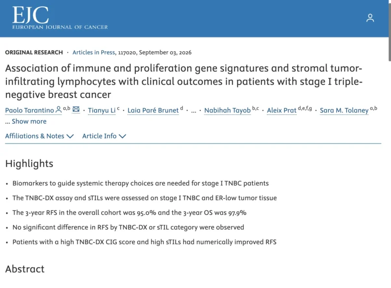
A new study published online in the European Journal of Cancer on September 3, 2026, explores whether immune gene expression, proliferation signatures and stromal tumor-infiltrating lymphocytes (sTILs) could help separate these biologically different groups. The results are intriguing, but appropriately preliminary.
Overall outcomes were excellent. High immune activity appeared associated with favorable prognosis, while a high proliferation signature showed the opposite numerical trend. However, neither TNBC-DX nor sTIL category significantly separated recurrence-free survival in this cohort.
That distinction is essential. The study supports further development of biological risk stratification in stage I TNBC. It does not yet provide a biomarker for safely omitting systemic therapy.
Stage I TNBC May Be Where Better Risk Stratification Matters Most
Approximately one-third of patients with TNBC are diagnosed with stage I disease, according to the study authors. Yet biomarkers capable of reliably distinguishing higher- and lower-risk tumors within this group remain an important unmet need. This is particularly relevant because treatment decisions in small node-negative TNBC often involve balancing two competing risks.
On one side is undertreatment of a biologically aggressive tumor capable of early distant recurrence. On the other is overtreatment of a patient whose baseline recurrence risk may already be extremely low. Traditional clinicopathologic variables, tumor size, nodal status, grade and patient factors, remain central to treatment decisions.
But TNBC is biologically heterogeneous. Two anatomically similar stage I tumors may have very different immune environments and proliferative programs. The long-term objective is therefore not simply to identify which patients have stage I disease. It is to identify which stage I disease actually behaves as high-risk TNBC.
The Study Evaluated Both Tumor Immunity and Proliferation
The retrospective cohort included 253 consecutive patients treated at Dana-Farber/Brigham Cancer Center between 2016 and 2021. Patients underwent upfront breast surgery and received standard-of-care postoperative management.
The study defined TNBC as:
- ER <10%
- PR <1%
- HER2-negative disease.
That definition is worth noting because it includes ER-low tumors, not exclusively ER-zero cancers. The investigators evaluated two complementary biological approaches. The TNBC-DX assay measured a Core Immune Gene, or CIG, signature together with a proliferation signature.
Separately, stromal tumor-infiltrating lymphocytes were centrally reviewed. The hypothesis was conceptually attractive, a tumor with strong immune activation might carry a more favorable prognosis, while one dominated by proliferation might represent a more biologically aggressive stage I phenotype.
Overall Outcomes Were Excellent
The most striking result may actually be how well the cohort performed overall.
Among the 253 patients:
- 88.9% had ductal tumors
- 73.1% had high-grade disease
- 65.2% received adjuvant chemotherapy.
Despite the aggressive histologic characteristics traditionally associated with TNBC, there were only 18 recurrence events.
At three years:
- RFS was 95.0%
- 95% CI, 92.1%–98.1%
- OS was 97.9%
- 95% CI, 95.9%–100%.
These figures provide important context for everything that follows. When recurrence events are this uncommon, demonstrating statistically robust differences between biomarker-defined subgroups becomes difficult.
The excellent overall prognosis is clinically reassuring. But statistically, it also means that the study has relatively little information with which to distinguish one biomarker subgroup from another.
High Immune Gene Activity Produced a Striking Numerical Signal
Among the 117 patients evaluable for TNBC-DX, the assay did not significantly differentiate recurrence-free survival overall. Yet the highest quartile of the Core Immune Gene signature produced a noteworthy observation.
Among 29 patients with the highest CIG scores, the three-year RFS was:
- 100%
- with a reported 95% CI of 100%–100%.
This is biologically plausible.
TNBC is one of the breast cancer subtypes in which immune infiltration and immune activation have repeatedly been associated with more favorable outcomes. A highly immune-active tumor may represent a cancer that is more effectively recognized and controlled by the host immune system.
But the small sample size is critical. Twenty-nine patients with no recurrence events is an encouraging observation. It is not sufficient evidence to conclude that a high CIG score identifies patients who can safely avoid chemotherapy.
That requires a very different level of validation.
sTILs Pointed in the Same Direction
The stromal TIL analysis produced a similar pattern. Among 123 patients with evaluable sTILs, there was again no statistically significant difference in RFS according to sTIL category.
However, patients with high sTILs, defined as >20%, had a three-year RFS of:
- 97.0%
- 95% CI, 90%–100%.
The direction of the signal is consistent with the TNBC-DX immune signature. That concordance is perhaps more interesting than either biomarker considered in isolation. A gene-expression measure of immune activity and a conventional histopathologic assessment of stromal lymphocytes appear to point toward the same biological phenotype, an immune-enriched stage I TNBC associated with very favorable outcomes.
The challenge is transforming that biological observation into a sufficiently reliable clinical tool.
Proliferation Told the Opposite Story
The TNBC-DX proliferation signature produced a contrasting numerical pattern. Patients with a high proliferation score had a three-year RFS of approximately:
- 83%
- 95% CI, 68%–100%.
The confidence interval is wide, reflecting the small number of events and patients. Still, the direction of the association is biologically coherent. A highly proliferative tumor may carry greater metastatic potential even when its anatomical stage is favorable.
This creates an appealing conceptual model:
- high immune activity → more favorable biology
versus
- high proliferation → potentially less favorable biology.
The investigators therefore conclude that immune and proliferative characteristics showed opposing prognostic trends and suggest that integrating the two may ultimately improve risk stratification.
The Most Interesting Future Strategy May Be Integration, Not a Single Biomarker
This is probably the study’s most important translational message. sTILs alone may not be enough. An immune gene signature alone may not be enough. A proliferation score alone may not be enough. But their combination could potentially identify biologically distinct risk groups.
Conceptually, a tumor with:
- strong immune activation + low proliferation
- could represent a particularly favorable stage I phenotype
Conversely:
- weak immune activation + high proliferation
could define a population in whom apparently favorable stage does not fully capture the underlying biological risk. That type of integrated model may ultimately be more useful than treating all stage I TNBC as one prognostic category. But the present study does not validate such a clinical classifier.
It provides a rationale for building one.
Could This Eventually Support Chemotherapy De-Escalation?
This is the question that will naturally attract the most attention.
If some stage I TNBCs have an extremely favorable immune phenotype, could systemic therapy safely be reduced, or perhaps omitted in carefully selected patients? The possibility is clinically important because stage I disease includes patients for whom the absolute benefit of chemotherapy may already be relatively small.
But the present dataset cannot answer that question. There are several reasons. First, 65.2% of the study population received adjuvant chemotherapy. Therefore, excellent outcomes among biomarker-defined groups cannot automatically be interpreted as their untreated natural history.
A patient with high TILs who remains recurrence-free after chemotherapy does not prove that the same patient would have remained recurrence-free without chemotherapy. Second, only 18 recurrences occurred across the entire cohort. That sharply limits statistical power and makes subgroup estimates unstable.
Third, the investigators themselves report no significant RFS difference by TNBC-DX or sTIL category. The study should therefore stimulate prospective de-escalation research, not justify de-escalation in routine practice.
Excellent Prognosis Can Make Biomarker Development Harder
There is an interesting methodological paradox here. Stage I TNBC is precisely the population in which better prognostic biomarkers could be most useful. But it is also one of the hardest settings in which to validate them.
Why?
Because if three-year RFS is already 95%, very large numbers of patients and prolonged follow-up are required to demonstrate that one subgroup does meaningfully better or worse than another.
This is particularly important when the ultimate clinical purpose is treatment de-escalation.
To justify omitting chemotherapy, investigators need a biomarker with an exceptionally high negative predictive value and confidence that excellent prognosis is preserved without the therapy being removed. A suggestive retrospective association is not sufficient. The evidentiary bar appropriately needs to be high.
ER-Low Disease Adds Another Layer of Complexity
The cohort definition also deserves attention. The investigators included tumors with ER expression below 10%, PR below 1%, and HER2-negative status.
Thus, some ER-low tumors were analyzed together with conventional TNBC. This reflects a clinically relevant contemporary issue. ER-low/HER2-negative tumors can share biological and clinical characteristics with TNBC, but they do not necessarily represent an identical disease entity in every context.
Any future clinical implementation of TNBC-DX or immune-based risk stratification will need to determine whether biomarker performance is consistent across:
- ER-zero TNBC
- ER-low tumors
- different tumor sizes
- younger versus older patients
- treated versus untreated populations.
The present abstract does not provide sufficiently detailed subgroup results to answer those questions.
What the Study Does, and Does Not Show
The central conclusion should remain restrained. The study shows that stage I TNBC has an excellent overall short-term prognosis, with three-year RFS of 95% and OS of nearly 98%. It also provides biologically coherent signals:
- higher immune activity appears favorable
- higher sTILs appear favorable
- high proliferation appears less favorable
But none of the evaluated biomarker categories produced a statistically significant difference in RFS. Therefore, this is hypothesis-generating evidence, not a clinically validated treatment-selection tool.
That difference becomes especially important when discussing de-escalation. A prognostic biomarker may tell us who does well.
A predictive biomarker must tell us who can safely receive a different treatment strategy. The current study addresses the first question.
The second remains unanswered.
The Bottom Line
This European Journal of Cancer analysis reinforces an increasingly important concept: Stage I TNBC should not automatically be considered one biological risk group.
Among 253 patients, three-year outcomes were excellent:
- RFS: 95.0%
- OS: 97.9%
High immune activity appeared particularly favorable. The highest CIG-score quartile had a three-year RFS of 100%, while patients with sTILs >20% had a three-year RFS of 97%. In contrast, a high TNBC-DX proliferation score was associated numerically with a lower three-year RFS of 83%.
Yet neither TNBC-DX nor sTIL category significantly differentiated RFS in the study. That is the key clinical caution. These biomarkers may eventually help distinguish patients who require treatment escalation from those who could safely receive less therapy.
But today, they remain promising tools for risk stratification rather than validated tools for selecting or omitting adjuvant therapy. The future of stage I TNBC may therefore move beyond tumor size alone.
The challenge is proving that biological risk can be measured accurately enough to change treatment without compromising cure.
Reference
- Tarantino P, Li T, Paré Brunet L, et al. Association of immune and proliferation gene signatures and stromal tumor-infiltrating lymphocytes with clinical outcomes in patients with stage I triple-negative breast cancer. European Journal of Cancer. 2026;117020. Published online September 3, 2026. doi:10.1016/j.ejca.2026.117020.