





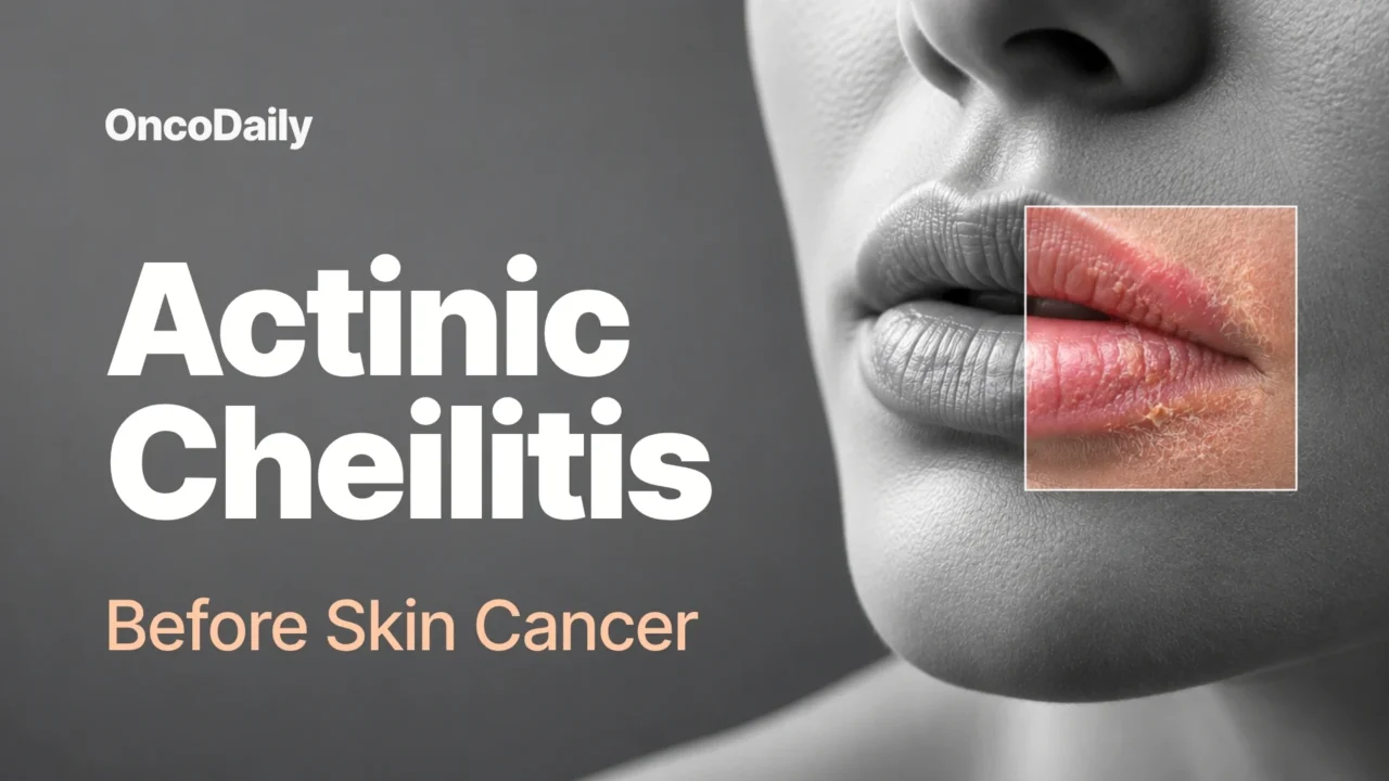
Actinic cheilitis (AC) is a sun-related, potentially malignant disorder of the lip, most often affecting the lower vermilion. It develops primarily as a result of chronic and cumulative ultraviolet (UV) exposure and is often considered the lip counterpart of actinic keratosis. The condition may begin with persistent dryness, loss of elasticity, scaling, erythema, and blurring of the vermilion border. With time, some lesions may develop fissures, erosions, hyperkeratosis, crusting, or white patches.
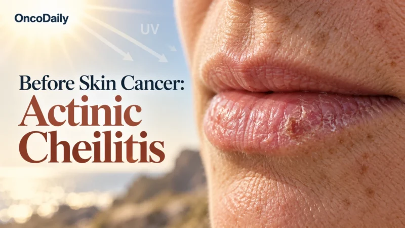
Experts generally classify actinic cheilitis as a potentially malignant disorder rather than established invasive cancer, although terminology remains debated. Approximately 95% of squamous cell carcinomas of the lip have been reported to occur on a background of actinic cheilitis. Importantly, this does not mean that 95% of actinic cheilitis lesions will become cancer. High-quality prospective studies directly measuring the malignant-transformation risk of AC remain limited.
Is Actinic Cheilitis Cancer or Precancer?
Actinic cheilitis is most commonly classified as a potentially malignant disorder rather than invasive cancer. Some authors consider severe or dysplastic forms of AC to represent an intraepithelial neoplastic process or even squamous cell carcinoma in situ, but there is no universal agreement on this terminology.
AC is primarily a clinical diagnosis, and the histopathological findings can vary. Hyperkeratosis, inflammation, solar elastosis, keratinocyte atypia, and epithelial dysplasia may be present, but dysplasia is not found in every lesion and is not required for the clinical diagnosis. What clearly distinguishes invasive squamous cell carcinoma is invasion beyond the epithelium, with malignant cells breaching the basement membrane and extending into underlying tissue.
Is Actinic Cheilitis the Same as Squamous Cell Carcinoma?
No. Actinic cheilitis and invasive squamous cell carcinoma (SCC) are not the same condition. AC represents chronic UV-related damage with potentially malignant epithelial changes. SCC is an invasive cancer capable of destroying surrounding tissue and, in some cases, spreading to regional lymph nodes or distant sites. The two are closely related because invasive SCC can arise in an area affected by actinic cheilitis. Clinically, however, the distinction may not always be obvious. Hypertrophic, ulcerated, indurated, or heterogeneous AC can resemble invasive SCC.
Histopathology helps determine whether atypical cells have breached the basement membrane. However, a limited biopsy may not represent the entire lesion, particularly when the affected area is diffuse or heterogeneous. Additional targeted biopsies or excision may therefore be needed when clinical suspicion remains high.
What Does Actinic Cheilitis Look Like?
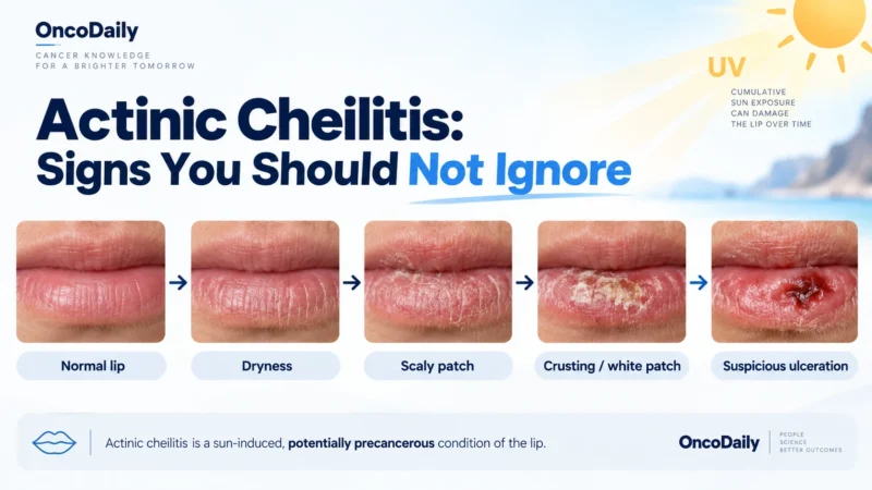
Actinic cheilitis usually affects the lower lip, which receives greater direct exposure to sunlight than the upper lip. Early changes may include persistent dryness, roughness, reduced elasticity, scaling, erythema, and gradual loss of the normally sharp border between the lip and surrounding skin. The lesion may appear as a well-defined or poorly defined dry, scaly patch or plaque. Other findings can include atrophy, edema, desquamation, mottled coloration, and areas of hyperkeratosis.
In more advanced or clinically suspicious cases, the lip may develop fissures, erosions, ulceration, bleeding, induration, leukoplakic or white areas, crusting, or hyperkeratotic projections. These findings do not by themselves prove that cancer has developed, but they should prompt appropriate medical assessment. The extent of AC varies. Some people have a small focal lesion, while others develop changes involving much of the lower lip.
Actinic Cheilitis vs Other Lip Conditions
Actinic cheilitis can resemble several other inflammatory and dermatological conditions. Angular cheilitis typically affects the corners of the mouth and is commonly associated with irritation, inflammation, or infection. Contact or eczematous cheilitis may cause itching, scaling, and more diffuse or bilateral lip involvement and can be associated with allergens or irritants. Lichen planus may produce characteristic reticular white striae and can involve the oral mucosa, while discoid lupus may cause areas of central atrophy with surrounding scale. Actinic cheilitis is distinguished by its close association with chronic cumulative UV exposure, its preference for the lower vermilion, and the presence of surrounding actinic damage.
What Causes Actinic Cheilitis?
The strongest and most consistently demonstrated risk factor for actinic cheilitis is chronic cumulative ultraviolet exposure. Risk increases in people who spend substantial amounts of time outdoors, particularly over many years. A 2021 meta-analysis found associations between AC and several characteristics, including low skin phototype, age over 50, high cumulative sun exposure, high daily sun exposure, male sex, alcohol use, and smoking.
These findings describe associations, however, and do not prove that every factor independently causes actinic cheilitis. Smoking may contribute to lip carcinogenesis or progression toward squamous cell carcinoma, but its causal role in the initial development of AC is less firmly established than the role of UV radiation. Human papillomavirus has also been investigated as a possible cofactor in lip carcinogenesis, but its specific contribution to actinic cheilitis remains less clearly established than cumulative UV exposure. Geographic factors can also influence risk. People living closer to the equator or at higher altitudes may experience greater UV intensity and cumulative exposure.
Who Is Most at Risk of Actinic Cheilitis?
Actinic cheilitis is most frequently reported among fair-skinned people with lower skin phototypes who have accumulated substantial sun exposure over many years. Risk increases with age. In a 2021 meta-analysis, people older than 50 had approximately three times the odds of AC compared with younger individuals. Outdoor workers and people who spend many hours outside recreationally are particularly vulnerable because of their cumulative UV exposure. Men have historically been affected more frequently than women, a pattern that may partly reflect differences in occupational sun exposure.
Immunosuppressed patients may also warrant greater clinical vigilance because of their increased vulnerability to UV-related malignancies. Overall, the typical high-risk profile is an older, fair-skinned individual with substantial long-term sun exposure. Rodriguez-Archilla A, et al. “Risk factors for actinic cheilitis: A meta-analysis.” 2021.
Can Actinic Cheilitis Become Invasive Skin Cancer?
Yes. Actinic cheilitis can be associated with or progress to invasive squamous cell carcinoma of the lip, but the exact probability of malignant transformation remains uncertain. A systematic review of observational studies estimated malignant transformation at approximately 3.07%. Other reviews and case series have reported estimates ranging from roughly 3% to 16.9%, while older literature has cited rates as high as 10%–30%.
These numbers should not be treated as directly interchangeable. The studies used different diagnostic definitions, patient populations, follow-up periods, treatment histories, and methods of identifying malignant transformation. For this reason, there is no single percentage that can reliably predict the risk for an individual untreated lesion. Transformation, when it occurs, may develop over years. Published reports have described intervals ranging from approximately 1 to 30 years, and the timing cannot be predicted for an individual patient.
The frequently cited observation that approximately 95% of lip SCCs occur on a background of AC describes the close association between the two conditions. It does not represent the probability that a person with AC will develop cancer. Squamous cell carcinoma of the lip also has clinically important metastatic potential and may carry a greater metastatic risk than many cutaneous SCCs at other sites. Individual risk depends on factors including tumor size, depth, differentiation, perineural invasion, lymph-node involvement, immune status, and other tumor characteristics.
In literature cited by expert consensus, patients who already had lip carcinoma together with actinic changes were reported to have an approximately 2.5-fold greater risk of developing a second primary lip cancer compared with patients without AC. This finding applies to a specific high-risk population and should not be generalized to everyone with actinic cheilitis. M.H. Trager et al. “Actinic cheilitis: a systematic review of treatment options.” 2021. Vasilovici A, et al. “Actinic cheilitis – From risk factors to therapy.” Front Med (Lausanne). 2022.
Warning Signs That Should Not Be Ignored
Certain changes should prompt medical assessment because they may indicate more advanced dysplasia or invasive SCC. These include persistent erosions or ulceration, a new nodule, induration, bleeding, persistent white or red patches, marked hyperkeratosis, crusting, friability, heterogeneous appearance, and progressive blurring of the vermilion border. These findings are warning signs rather than a diagnosis of cancer. Biopsy may be necessary when suspicious changes develop. Seoane Lestón JM, et al. “Assembling a consensus on actinic cheilitis: A Delphi study.” 2021.
How Is Actinic Cheilitis Diagnosed?
Actinic cheilitis is often suspected clinically through careful inspection and palpation of the lips. Typical findings can include dryness, scaling, atrophy, erythema, mottling, hyperkeratosis, loss of elasticity, and blurring of the vermilion border.
When Is a Biopsy Needed?
Biopsy is particularly important when a lesion is persistent, atypical, heterogeneous, recurrent, resistant to treatment, ulcerated, nodular, indurated, or bleeding. A single biopsy may not accurately represent a broad or multifocal lesion. When clinical concern remains high, multiple targeted biopsies or excision may be required because a limited sample can miss an area of invasion. Histopathological assessment can determine the degree of epithelial atypia or dysplasia and, crucially, whether invasive carcinoma is present.
In selected mild and clinically typical lesions, treatment may sometimes begin after clinical assessment without waiting for biopsy, but this decision should be made by an appropriately qualified clinician with suitable follow-up.
Can Dermoscopy or Confocal Microscopy Help?
Dermoscopy may provide additional clues, including white structureless areas, scaling, a white halo along the vermilion border, and telangiectatic or tortuous vessels. Reflectance confocal microscopy can provide non-invasive microscopic imaging and may demonstrate keratinocyte atypia or an abnormal honeycomb pattern. Both techniques can help clinicians identify suspicious areas, particularly in specialist settings. However, neither dermoscopy nor confocal microscopy replaces histopathology when invasive SCC is suspected.
How Is Actinic Cheilitis Treated?
Treatment should be individualized according to the extent of the lesion, clinical appearance, histological findings, suspected invasion, cosmetic considerations, comorbidities, previous treatment, and patient preference.
Approaches reported in the literature include vermilionectomy, CO₂ or Er laser ablation, cryotherapy, electrosurgery, photodynamic therapy, topical 5-fluorouracil, and imiquimod. There is no single treatment that is appropriate for every patient. Available systematic reviews generally report higher clearance and lower recurrence with vermilionectomy or laser ablation than with some topical, chemical, or photodynamic treatments. However, the available studies are often small, nonrandomized, heterogeneous, and difficult to compare directly. Moderate or severe dysplasia is more commonly treated with surgical approaches, while selected mild or nondysplastic lesions may be considered for nonsurgical treatment under specialist supervision.
Vermilionectomy
Vermilionectomy removes the affected vermilion and superficial lip tissue. One important advantage is that it provides a relatively large specimen for histopathological examination, potentially identifying dysplasia or an invasive focus that may have been missed by a smaller incisional biopsy. Reported complications can include swelling, bruising, pain, altered sensation, bleeding, infection, delayed healing, and temporary difficulty eating. These complications do not occur in every patient.
Laser Treatment
CO₂ and Er laser ablation can provide high clearance rates and favorable cosmetic results in appropriately selected patients. Possible adverse effects include pain, edema, bleeding, delayed healing, changes in sensation or pigmentation, and scarring, although scarring may be limited in many patients. Laser ablation does not provide the same full tissue specimen for pathological examination as vermilionectomy, which may influence treatment choice when invasive disease is suspected.
Topical Treatments
Topical 5-fluorouracil can produce substantial clinical improvement in some patients. Local pain, irritation, inflammation, difficulty eating, and difficulty speaking may occur, and recurrence has been reported. Imiquimod may also achieve clinical and histological improvement in selected lesions but commonly causes a local inflammatory reaction that can include swelling, pain, ulceration, and crusting. Historical studies have also evaluated other topical agents, including ingenol mebutate. However, its regulatory status and clinical use have changed because of safety concerns, and it should not be presented as a routine contemporary treatment without region-specific regulatory confirmation.
Photodynamic Therapy
Photodynamic therapy (PDT) may provide clinical improvement and favorable cosmetic outcomes in selected patients. However, recurrence can be higher than with vermilionectomy or laser ablation, and the evidence is limited by small studies and inconsistent treatment protocols. Limited studies suggest that combining laser therapy with PDT may reduce recurrence in selected cases, but this approach has not been established as a universal standard of care. Regardless of the treatment chosen, regular follow-up and ongoing UV protection remain important.
Can Actinic Cheilitis Come Back After Treatment?
Actinic cheilitis can recur after treatment, and recurrence rates vary considerably according to the treatment modality, lesion characteristics, follow-up period, and study design. Vermilionectomy and laser ablation generally show relatively low recurrence rates in published reviews, while some topical treatments and photodynamic approaches have reported higher recurrence. Persistent UV damage can also affect surrounding areas of the lip even after the original lesion has been successfully treated. For this reason, patients require ongoing follow-up. Any recurrent or changing lesion—particularly one that becomes ulcerated, indurated, nodular, or prone to bleeding—should be reassessed.
When Should You See a Doctor?
Persistent lip changes should not automatically be assumed to be ordinary chapped lips. A medical assessment is appropriate when dryness, scaling, cracking, burning, pain, altered texture, discoloration, or loss of the normal vermilion border persists for several weeks without healing. More urgent evaluation is appropriate when there is persistent ulceration, erosion, a new lump or nodule, induration, recurrent bleeding, marked crusting, or a persistent white or red patch. These signs do not necessarily mean cancer is present, but they warrant assessment because advanced dysplasia or invasive SCC may look similar. People with substantial lifelong sun exposure, fair skin, older age, immunosuppression, or other risk factors should be especially attentive to persistent lip changes.
Can Actinic Cheilitis Be Prevented?
Because chronic cumulative UV exposure is the primary established driver of actinic cheilitis, prevention focuses on protecting the lips from ultraviolet radiation throughout life. A 2021 meta-analysis found that reported use of sunscreen creams and caps or hats was not statistically associated with a lower risk of AC. This does not prove that sunscreen or protective clothing are ineffective.
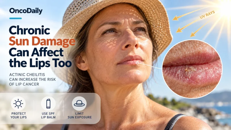
The finding may reflect inconsistent use, poor measurement of sun exposure, inadequate coverage of the lips, or other limitations of the observational studies. Practical preventive measures include using a broad-spectrum lip balm with SPF 30 or higher and reapplying it after eating, drinking, swimming, or prolonged outdoor activity. A wide-brimmed hat can provide more complete facial and lip shade than relying on a cap alone. Seeking shade and limiting intense midday UV exposure can further reduce cumulative sun exposure.
Avoiding tobacco is also advisable, particularly because of its broader association with lip and oral carcinogenesis. Limiting alcohol can form part of broader oral-cancer risk reduction. Ordinary facial sunscreen should not be assumed to adequately protect the lips unless it is specifically formulated and labeled for lip use. Finally, prevention does not replace surveillance. Any persistent or changing lip lesion should be medically assessed, particularly when ulceration, induration, bleeding, nodularity, or other suspicious features develop. Rodriguez-Archilla A, et al. “Risk factors for actinic cheilitis: A meta-analysis.” 2021.
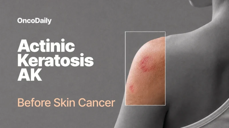
You Can Also Read Actinic Keratosis (AK) : The Precancerous Skin Lesion You Shouldn’t Ignore by OncoDaily
Written by Written by Lily Tumanyan MD
FAQ
What is actinic cheilitis?
A potentially malignant disorder caused by chronic sun exposure, mainly affecting the lower lip and characterized by dryness, scaling, atrophy, and blurred borders. Can it be prevented? Mainly by lifelong lip protection from the sun, avoiding tobacco and alcohol, and having persistent lip changes checked early.
Can it turn into cancer?
Yes. Up to 95% of lip squamous cell carcinomas develop on preexisting actinic cheilitis, with transformation taking years.
Who is most at risk?
People over 50 with fair skin, high cumulative sun exposure, outdoor occupations, immunosuppression, or tobacco and alcohol use.
What are the symptoms?
Often none, but dryness, cracking, burning, pain, mottled red and white patches, fissures, and blurred lip borders can occur.
How is it diagnosed?
Mainly by clinical inspection and palpation, supported by dermoscopy and reflectance confocal microscopy; biopsy confirms the diagnosis.
What treatments are available?
Surgical options (vermilionectomy, CO2 or Er:YAG laser ablation) work best; topical treatments (imiquimod, 5-fluorouracil, diclofenac) and photodynamic therapy are alternatives.
Which treatment is most effective?
Surgery: CO2 laser ablation and vermilionectomy give the most favourable outcomes with the fewest recurrences.
Can it come back after treatment?
Yes. Recurrence is lowest after surgery but higher with topical treatments and chemical peels, so periodic follow-up is needed.