Queensland Spatial Biology Centre shared a post on X about a recent paper by Habib Sadeghirad et al. titled “Spatial dynamics of tertiary lymphoid aggregates in head and neck cancer: insights into immunotherapy response” published in Journal of Translational Medicine.
Authors: Habib Sadeghirad, James Monkman, Chin Wee Tan, Ning Liu, Joseph Yunis, Meg L. Donovan, Afshin Moradi, Niyati Jhaveri, Chris Perry, Mark N. Adams, Ken O’Byrne, Majid E. Warkiani, Rahul Ladwa, Brett G.M. Hughes and Arutha Kulasinghe.
“Excited to share the tweetorial for our latest publication on spatial profiling and phenotyping of tertiary lymphoid aggregates in head and neck cancer!
This work was led by the talented PhD student Habib Sadeghirad (not on Twitter). We are grateful to the amazing team that included James Monkman (UQ), Chin Wee Tan (WEHI), Arutha Kulasinghe (UQ), Joseph Yunis (UQ), Meg Donovan (QSBC).
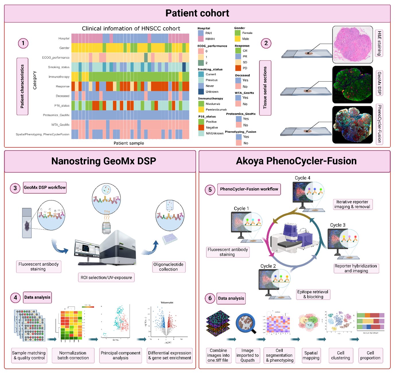
Our goal was to identify potential predictive biomarkers of response to immunotherapy. In our study, we profiled whole tissue samples in serial sections from patients with head and neck squamous cell carcinoma (HNSCC) before their immune checkpoint inhibitor therapies.
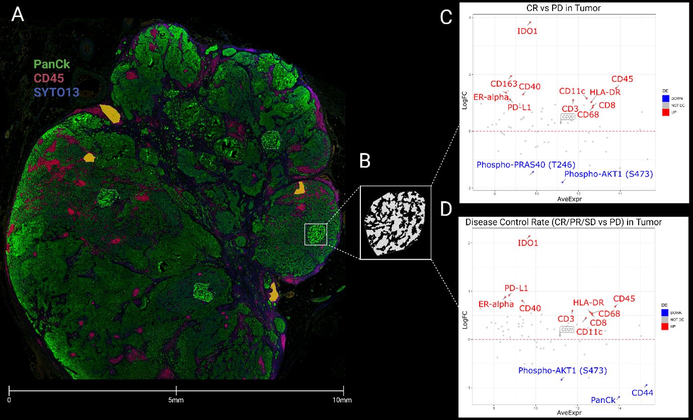
Using Nanostring GeoMx DSP, we found that antigen presentation biomarkers (CD40, CD11c, CD68, CD163, HLA-DR) were highly expressed in responders vs. non-responders. Responders also had a ‘hot tumor’ phenotype in their TMEs.
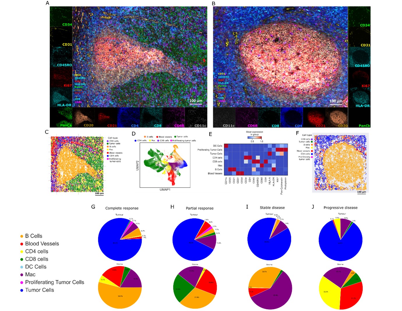
By applying whole transcriptome spatial analysis of TLS and germinal center (GC)-like structures, we observed a significant increase in genes related to immune modulation, diverse immune cell recruitment, and a potent interferon response within TLS compared to normal GCs.
By employing single-cell spatial phenotyping of TLS and GC structures, we characterized the spatial organization of immune cells within the tumor microenvironment of HNSCC tumors. CD8 T cell and blood vessel markers were enriched in TLSs compared to normal GCs.
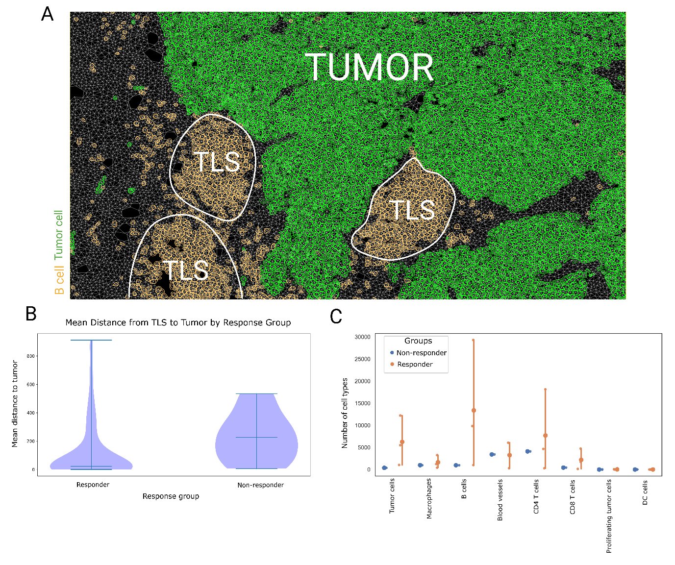
We further investigated the association of TLS distance to tumor cells and patient response to immunotherapy. We found that the proximity of TLSs to tumor cells could be a critical indicator of ICI response in head and neck cancer patients.
Last but not least, massive thanks to The Passe and Williams Foundation and collaborating clinicians who supported this work.”


