Down syndrome has one of the most unusual cancer profiles in medicine. Trisomy 21 is present in every cell, but its effect on cancer risk isn’t consistent. Children with Down syndrome are at much higher risk of leukemia, while adults have lower rates of many solid tumors.
A Swedish population-based study of nearly 10,000 individuals with Down syndrome quantified the contrast. Childhood risk of acute lymphoblastic leukemia (ALL) was more than 20-fold higher than in matched controls. For acute myeloid leukemia (AML), the increase was starker still: before age 5, risk was nearly 500-fold higher. In adulthood, overall solid tumor incidence ran roughly half that of the general population, with lower rates of breast, prostate, lung, and colorectal cancer.
So trisomy 21 isn’t a general cancer-promoting state. Its effects depend on cell type, developmental timing, and which additional mutations follow, a pattern visible earliest and most clearly in the blood.
Leukemia Risk Begins in Utero
The increased leukemia risk associated with Down syndrome begins before birth. During fetal development, when the liver is the primary site of hematopoiesis, Down syndrome is associated with expansion of hematopoietic stem cell populations, along with increased megakaryocyte-erythroid progenitors. Trisomy 21 alone does not cause leukemia, but it reshapes the progenitor pool into one that is unusually susceptible to a second hit.
The second hit is an acquired mutation in GATA1 transcription factor. Its N-terminus gets truncated, producing a short protein. These are present in essentially all TAM (transient abnormal myelopoiesis: clonal proliferation of GATA1-mutated blasts) and ML-DS cases.
In a prospective cohort of 200 Down syndrome newborns, Roberts and colleagues found circulating blasts in 195 of them, nearly all, regardless of GATA1 status. Most had neither leukemia nor transient abnormal myelopoiesis.
TAM: A Preleukemia That Can Resolve on Its Own
TAM occurs only in newborns with Down syndrome or mosaic trisomy 21, identified clinically in around 10% of Down syndrome neonates. Molecular studies suggest these clones are far more common. Most cases resolve spontaneously in infancy, without leukemia-directed therapy, even after the blast count has climbed.
Resolution does not equal harmlessness. Severe TAM can cause hepatic dysfunction, effusions, extreme leukocytosis, and other life-threatening complications, some infants require low-dose cytarabine. In a minority of children small numbers of clonal cells persist after apparent remission and, years later, acquire the additional mutations needed to become ML-DS.
In the Oxford-Imperial Down Syndrome Cohort Study, conventional testing identified GATA1 mutations in 17 of 200 newborns. Targeted next-generation sequencing of the remaining samples found GATA1-mutant clones in another 18 infants who had shown no clinical or hematologic evidence of TAM, a phenomenon the investigators termed silent TAM.
Progression to ML-DS
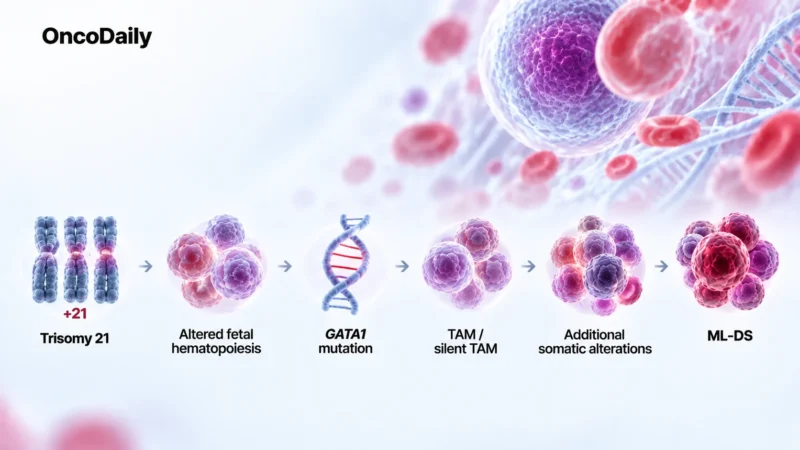
ML-DS develops before age 5, often with megakaryoblastic differentiation, through a multistep sequence:
- trisomy 21
- altered fetal hematopoiesis
- GATA1 mutation
- TAM/silent TAM
- additional somatic alterations
- ML-DS
Genomic studies of ML-DS have identified recurrent alterations in cohesin components, epigenetic regulators, and signaling pathways, the events that separate overt leukemia from its precursor state. Few cancers offer this degree of visibility into their own evolution.
Unusual Sensitivity to Chemotherapy
Children with the disease generally do better than children with non-Down syndrome acute megakaryoblastic leukemia: the leukemic blasts are particularly sensitive to cytarabine.
In the Children’s Oncology Group AAML0431 trial, 204 patients achieved a 5-year event-free survival of 89.9% and overall survival of 93.0%. The regimen reduced cumulative daunorubicin exposure by 25% and moved high-dose cytarabine from later intensification to the second induction cycle. Pediatric protocols have reduced chemotherapy intensity while maintaining high cure rates, a case where predisposition and prognosis clearly diverge.
DS-ALL: A Different Pathway
Down syndrome raises ALL risk roughly 20-fold, with most cases being B-cell precursor ALL. One of its most characteristic molecular features is CRLF2 dysregulation, present in half of cases and frequently accompanied by JAK-STAT pathway hyperactivation. Not a single “Down syndrome leukemia gene,” but the pattern points to chromosome 21 dosage reshaping the signaling context.
The evolutionary sequence here is less defined. CRLF2 rearrangements can arise early and remain stable from diagnosis to relapse, when JAK-STAT and RAS pathway lesions are more changeable, one study of paired diagnosis-relapse samples found signaling mutations frequently replaced at relapse, arguing against any single targetable JAK-STAT abnormality.
Greater Treatment Risks
These children have historically faced both greater treatment toxicity, infection risk in particular, and worse leukemia outcomes than children with ALL without Down syndrome, so simply intensifying chemotherapy carries different risks than it would in an average-risk child.
Immunotherapy has become an alternative: blinatumomab, inotuzumab ozogamicin and CD19-directed CAR T-cell therapy are the options. A 2025 review of DS-ALL flagged these approaches as ways to reduce reliance on conventional cytotoxic therapy, but their optimal use in this population is still under study.
Why Does the Same Chromosome Seem to Protect Against Solid Tumors?
Epidemiological studies show lower rates of many solid tumors in Down syndrome, including breast, prostate, lung, colorectal cancers, and melanoma. Testicular cancer risk increased though, an indicator that trisomy 21’s relationship to cancer cannot be reduced to “higher” or “lower” risk. The direction depends on tissue.
Proposed mechanisms include chromosome 21 genes that influence angiogenesis, proliferation, apoptosis, immune function, and tumor suppression. DSCR1/RCAN1 and ETS2 have drawn particular interest in experimental models, along with differences in hormonal exposure and metabolism. No single mechanism accounts for the full pattern, and the “protection” is not uniform, some cancers show no reduction at all.
Can Preleukemia Be Stopped Before It Progresses?
A genetically defined preleukemic clone, detectable shortly after birth and sometimes present years before ML-DS develops, makes GATA1-mutant clones an obvious candidate for risk prediction. But their sheer frequency is the obstacle, sensitive sequencing finds far more GATA1-mutant clones than the number of children who will ever develop ML-DS.
TAM therapy already shows the limit of this: cytarabine controls severe, life-threatening neonatal disease, but has not been shown to reliably prevent later ML-DS. It might be more reasonable to monitor clone size, persistence, and additional alterations for signs of progression
What Makes Down Syndrome Different From Other Leukemia Predisposition Syndromes?
Fanconi anemia predisposes to MDS and AML through defective DNA repair combined with bone marrow failure. Shwachman-Diamond syndrome follows a related path, pairing chronic marrow dysfunction with clonal evolution toward myeloid malignancy. GATA2 deficiency presents differently, cytopenias and immunodeficiency alongside MDS/AML risk, while germline RUNX1 and CEBPA variants raise myeloid malignancy risk without the marrow failure.
Down syndrome stands apart because several steps toward leukemia can be observed from birth and offers an unusually clear model of how genetic predisposition and acquired mutations interact over time. What separates a dormant clone from a progressing one is unresolved, and that, not new genes, is what stands between molecular monitoring and real prediction.
You can also read about
FAQ
Why does Down syndrome increase leukemia risk but lower the risk of many solid tumors?
Trisomy 21 does not affect every tissue in the same way. In developing blood cells, extra chromosome 21 dosage alters hematopoiesis and creates conditions that favor specific leukemias. In other tissues, chromosome 21 genes may influence processes such as angiogenesis, proliferation, immune function, and tumor suppression in ways that can reduce the risk of some solid cancers.
Are babies with Down syndrome routinely checked for leukemia?
Newborns with Down syndrome typically have a complete blood count and blood film assessment because transient abnormal myelopoiesis (TAM) can appear shortly after birth. This is important because TAM may be clinically silent or, at the other extreme, cause serious complications requiring treatment.
Does finding a GATA1 mutation mean a child will develop leukemia?
No. GATA1-mutant clones can be detected in newborns who never develop myeloid leukemia of Down syndrome (ML-DS). The mutation is an important early step, but progression usually requires additional genetic changes. This makes predicting which clones will eventually become leukemia difficult.
Can transient abnormal myelopoiesis come back after it disappears?
TAM itself usually resolves during infancy, but disappearance of circulating blasts does not necessarily mean every abnormal clone has been eliminated. In some children, residual GATA1-mutant cells may persist and later acquire additional alterations that contribute to ML-DS.
Could immunotherapy make ALL treatment safer for children with Down syndrome?
Potentially. Blinatumomab, inotuzumab ozogamicin, and CD19-directed CAR T-cell therapy offer ways to treat B-cell ALL without relying entirely on conventional chemotherapy. Their optimal timing and ability to reduce treatment-related toxicity specifically in DS-ALL remain important areas of study.
Which cancers are not reduced in people with Down syndrome?
The lower solid-tumor risk is not universal. Testicular cancer is a notable exception, with an increased risk reported in people with Down syndrome, while the incidence of several other cancers may not differ substantially. This is one reason describing trisomy 21 as broadly “protective against cancer” is misleading.
Could newborn sequencing eventually predict ML-DS before it develops?
Sequencing can already detect very small GATA1-mutant clones, including silent TAM. The harder problem is determining which clones matter. Future prediction will likely require more than detecting GATA1, potentially combining clone size and persistence with additional somatic alterations and other biological markers of progression.