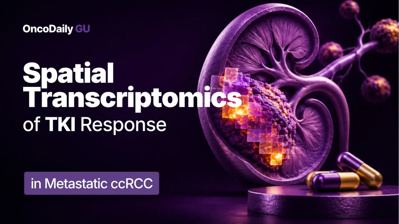
Despite advances in the treatment of metastatic clear cell renal cell carcinoma (ccRCC), predicting which patients will respond to tyrosine kinase inhibitors (TKIs) remains challenging. Although immune checkpoint inhibitor-based combinations have become standard first-line treatment options, VEGFR-targeted therapies continue to play an important role in the management of advanced disease.
Responses to VEGFR inhibition vary considerably between patients, and the mechanisms underlying treatment resistance are not fully understood. While angiogenesis has traditionally been the main focus of TKI therapy, growing evidence suggests that interactions between tumor cells, immune cells, and the surrounding microenvironment may also influence treatment outcomes.
A study published on November 10, 2026, in Signal Transduction and Targeted Therapy, titled “Spatial transcriptomics tumor microenvironment determinants of tyrosine kinase inhibitor response in renal cell carcinoma: results from a phase 2 clinical trial,” investigated the molecular characteristics associated with sunitinib response and resistance in metastatic ccRCC.
The study was conducted by Elshad Hasanov, Mostafa I. H. Ali, Shriya Deshmukh, Jose A. Ovando-Ricardez, Truong Lam, Anh Hoang, Xian-De Liu, Xuemei Wang, Christine B. Peterson, Lianchun Xiao, Surena F. Matin, Kanishka Sircar, Nizar M. Tannir, Merve Hasanov, and Eric Jonasch.
Study Design and Patient Characteristics
This single-arm phase II trial (NCT00715442) enrolled 50 patients with previously untreated, biopsy-proven metastatic ccRCC between June 2008 and October 2011. Of these, 46 patients were included in the clinical efficacy analysis after four patients with non-clear cell histology identified on surgical specimens were excluded.
Patients received sunitinib 50 mg daily on a four-week-on, two-week-off schedule. Following the initial treatment cycle and restaging, eligible patients underwent cytoreductive nephrectomy during the second cycle. Those with stable or responding metastatic disease could resume sunitinib after surgery and continue treatment until disease progression or unacceptable toxicity.
The study evaluated time to progression (TTP) and sunitinib-related toxicity. Overall survival (OS) and objective response rate (ORR) were secondary efficacy endpoints, while progression-free survival (PFS) was assessed post hoc.
To better understand the biological mechanisms associated with treatment response, investigators also performed an exploratory spatial transcriptomic analysis of available nephrectomy specimens. Using whole-transcriptome digital spatial profiling (DSP), they examined gene expression patterns across tumor, myeloid, endothelial, and CD8+ T-cell compartments.
Clinical Outcomes
At a median follow-up of 118 months, median TTP was 8.2 months (95% CI: 6.1–19.3), while median PFS was 7.8 months (95% CI: 5.7–19.2). Median OS reached 36.3 months (95% CI: 19.7–70.5), with an estimated five-year OS rate of 36.1% (95% CI: 24.4–53.3). The ORR was 37% (95% CI: 23.1–50.9), including two complete responses and 15 partial responses. Of the 50 patients who started sunitinib, 41 underwent nephrectomy, while nine discontinued the study before surgery because of disease progression or unacceptable toxicity. Following nephrectomy, 32 patients resumed sunitinib treatment.
Regarding safety, the most frequently reported sunitinib-related adverse events included diarrhea, fatigue, mucositis, rash, and hand-foot syndrome. Grade 3 hand-foot syndrome occurred in 14% of patients, while grade 3 fatigue was reported in 8%. Hematologic toxicities included grade 3–4 thrombocytopenia and neutropenia. Two patients experienced grade 4 thromboembolic events that resulted in treatment discontinuation. No grade 5 sunitinib-related toxicities were reported.
Spatial Transcriptomic Analysis
Exploratory biomarker analysis was conducted using available nephrectomy tissue from 23 patients, including 11 responders who achieved a complete or partial response and 12 non-responders whose best response was stable disease. Spatial transcriptomic profiling revealed differences in immune regulation, angiogenic signaling, and metabolic pathways between the two groups, suggesting that sensitivity to sunitinib may depend on several interacting components of the tumor microenvironment.
Myeloid Cell Signatures and Treatment Resistance
One of the main findings involved differences in myeloid cell transcriptional programs. Responders demonstrated enrichment of pro-inflammatory and immune-activating pathways, including interferon- and chemokine-associated signaling. They also showed higher expression of C1QC+ tumor-associated macrophage signatures, which have been associated with antigen presentation and inflammatory immune responses.
In contrast, non-responders exhibited increased expression of SPP1+ tumor-associated macrophage signatures, alongside immunosuppressive macrophage-related programs and elevated expression of CD274, the gene encoding PD-L1.
The investigators also identified predicted differences in communication between myeloid and endothelial cells. Ligand–receptor analysis predicted more interactions involving collagen-related ligands and endothelial integrin ITGA11 in non-responders, suggesting a potential contribution of extracellular matrix remodeling and angiogenic signaling to resistance. These findings indicate that myeloid cell programs may influence sunitinib response through both immune regulation and interactions with the tumor vasculature.
CD8+ T-Cell Exhaustion and Immune Signaling
Differences were also observed in the CD8+ T-cell compartment. Non-responders demonstrated stronger T-cell exhaustion signatures, with increased expression of inhibitory checkpoint-associated genes, including PDCD1 (PD-1), HAVCR2, and LAG3. They also showed enrichment of pathways related to autophagy, cell adhesion, and complement signaling.
By comparison, responders showed increased TNF expression and enrichment of pro-inflammatory MAPK/ERK-associated pathways, suggesting a more immune-active environment.
Ligand–receptor analysis additionally identified predicted SPP1–CD44 interactions involving CD8+ T cells in non-responders. These interactions may link macrophage-associated signaling with T-cell dysfunction and extracellular matrix remodeling, providing another potential explanation for resistance to VEGFR-targeted treatment.
Angiogenesis and Metabolic Adaptation
The endothelial and tumor compartments provided further insight into the molecular differences between response groups. In non-responders, endothelial cells showed enrichment of pathways associated with extracellular matrix remodeling, vasculogenesis, epithelial–mesenchymal transition, and oxidative phosphorylation. Enrichment of pathways involving genes such as MMP2, MMP14, and NRP1, together with predicted collagen–ITGA11 interactions, suggested possible mechanisms of angiogenic escape that may extend beyond VEGF-dependent signaling.
In contrast, responders demonstrated enrichment of metabolic pathways involving amino acid and fatty acid catabolism in the endothelial compartment. The tumor-background compartment also showed differences in lipid-related gene expression. Responders had increased expression of APOE, APOC1, PLIN2, and ANGPTL4, genes associated with lipid processing and adaptation to hypoxic conditions. Non-responders, meanwhile, showed increased expression of PDCL3, a VEGFR-associated chaperone protein that may contribute to adaptive resistance.
These findings suggest that metabolic adaptation, particularly lipid metabolism, may be associated with sunitinib sensitivity, alongside differences in angiogenic and immune signaling.
Machine Learning Analysis and External Evaluation
To determine whether the transcriptional features identified through spatial profiling were associated with outcomes in an independent population, investigators developed machine learning-based survival models using data from the phase III JAVELIN Renal 101 trial. The analysis incorporated gene expression features derived from the tumor, endothelial, myeloid, and CD8+ T-cell compartments and evaluated their association with OS and PFS.
In the sunitinib-treated cohort, the best-performing model for OS demonstrated a concordance index (C-index) of 0.72 in the training dataset and 0.67 in the independent test dataset. The model also separated patients into high- and low-risk groups with different survival outcomes.
In the avelumab plus axitinib cohort, the OS model achieved a C-index of 0.72 in training and 0.56 in the independent test dataset, compared with 0.67 in the sunitinib test dataset. This difference suggests that transcriptional features associated with outcomes during sunitinib treatment may have limited generalizability to TKI–immunotherapy combinations, where additional immune-related mechanisms can influence treatment response.
Although the findings support the potential value of spatial transcriptomic profiling for outcome prediction, the derived signatures require further validation before clinical application.
Takeaway
This phase II study provides insight into the molecular mechanisms associated with sunitinib response and resistance in metastatic ccRCC, highlighting the importance of the tumor microenvironment beyond angiogenic signaling alone.
Non-responding tumors demonstrated greater enrichment of immunosuppressive myeloid programs, CD8+ T-cell exhaustion signatures, and pathways associated with extracellular matrix remodeling and angiogenic escape. In contrast, responding tumors showed more prominent immune-activating programs and distinct lipid metabolism-related signatures.
However, the exploratory biomarker analysis included only 23 patients with available nephrectomy specimens collected during sunitinib treatment, excluding those who discontinued therapy before surgery because of early progression or unacceptable toxicity. The findings therefore require confirmation in larger cohorts, particularly in the context of contemporary immunotherapy-based combinations.
While sunitinib monotherapy is no longer the preferred first-line treatment for metastatic ccRCC, this research may help inform future biomarker development and studies investigating strategies to overcome TKI resistance by targeting multiple components of the tumor microenvironment.
The full article is available in Signal Transduction and Targeted Therapy.