
Bone is one of the most common destinations for metastatic prostate cancer, but this pattern is far from random.
For a prostate cancer cell to establish itself in bone, it must do more than simply travel through the bloodstream. It must find the right signals, survive within a highly specialized marrow environment, and interact with cells that normally regulate bone formation and breakdown.
These interactions help explain why the skeleton becomes such a distinctive site of prostate cancer progression and why bone metastases behave differently from disease in other organs.
Why Does Prostate Cancer Commonly Spread to Bone?
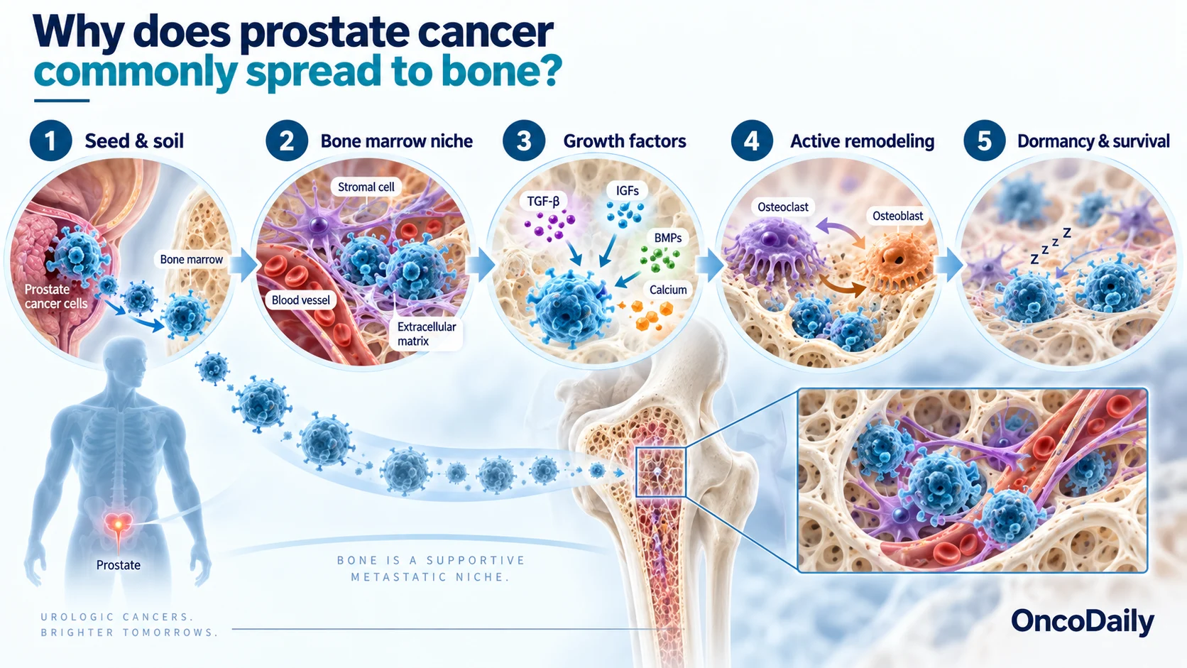
Prostate cancer has a strong tendency to metastasize to bone because the bone marrow provides a particularly favorable environment for disseminated tumor cells to survive and eventually grow.
This relationship is often described through the “seed and soil” hypothesis: prostate cancer cells represent the “seed,” while the bone microenvironment provides a receptive “soil.” Bone marrow contains growth factors, stromal cells, extracellular matrix proteins, and specialized cellular niches that can support metastatic cells after they leave the primary tumor. (Jiang, 2025; Wong et al., 2019).
Bone is also biologically active throughout life. Continuous remodeling releases signaling molecules from the bone matrix, while interactions among marrow cells, blood vessels, and bone-forming and bone-resorbing cells create conditions that can support tumor-cell survival.
Some prostate cancer cells that reach bone may remain dormant for years before forming clinically detectable metastases. This combination of a supportive marrow niche, active remodeling, and long-term tumor-cell survival helps explain the strong preference of prostate cancer for the skeleton. (Jiang, 2025; Wong et al., 2019).
How Do Prostate Cancer Cells Reach and Colonize Bone?
Reaching bone requires prostate cancer cells to complete several stages of the metastatic process.
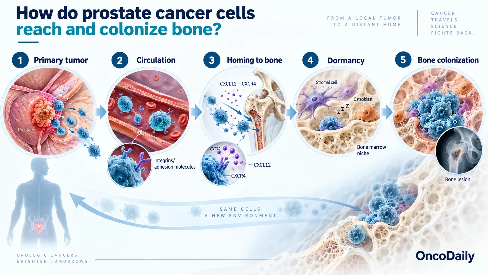
Cells must first detach from the primary tumor, invade surrounding tissue, and enter the circulation. Changes associated with epithelial–mesenchymal transition (EMT) can increase tumor-cell mobility and invasive capacity, while enzymes that remodel the extracellular matrix help cells cross tissue and vascular barriers. (Jiang, 2025; Wong et al., 2019).
Once in the bloodstream, only a small proportion of circulating tumor cells survive long enough to reach distant organs. Homing to bone is influenced by chemokine signaling, particularly the CXCL12–CXCR4 axis. Bone marrow stromal and endothelial cells produce CXCL12, while many prostate cancer cells express CXCR4, helping direct them toward the marrow. Integrins, CD44, and other adhesion molecules then help tumor cells attach to marrow endothelium and enter the surrounding tissue. (Jiang, 2025; Rahim et al., 2014).
After entering the marrow, disseminated tumor cells may either proliferate or enter dormancy. Signals from osteoblasts, stromal cells, and the perivascular niche can keep some cells quiescent for prolonged periods, while later changes in inflammation, bone remodeling, or local signaling may allow them to reactivate. (Yu-Lee et al., 2018; Singh et al., 2021).
Successful colonization therefore involves more than simply reaching bone. Tumor cells must adapt to the marrow environment, establish supportive interactions with surrounding cells, and eventually acquire the conditions needed for sustained metastatic growth. (Jiang, 2025).
What Makes the Bone Microenvironment Favor Prostate Cancer Growth?
Bone is not an inert structure. It is a metabolically active tissue containing osteoblasts, osteoclasts, stromal cells, endothelial cells, immune cells, and a mineralized extracellular matrix rich in signaling molecules. Together, these components create a highly interactive metastatic niche. (Jiang, 2025; Prigol et al., 2023).
The bone matrix stores factors including TGF-β, IGFs, BMPs, and calcium. During bone remodeling, some of these molecules are released and can promote prostate cancer-cell survival, proliferation, and adaptation to the metastatic environment. Tumor cells can then alter bone remodeling further, creating feedback between cancer growth and the surrounding tissue. (Wong et al., 2019; Jiang, 2025).
The marrow also contains specialized niches that can protect disseminated tumor cells. Signals from osteoblast-lineage cells and stromal cells may induce dormancy, allowing cancer cells to persist without forming an obvious lesion. Changes in these signals may later favor reactivation and metastatic outgrowth. (Yu-Lee et al., 2018; Singh et al., 2021).
Immune regulation adds another layer. Myeloid cells, macrophages, regulatory T cells, and other components of the marrow immune environment can create conditions that limit effective antitumor immunity and support metastatic progression. (Jiang, 2025; Prigol et al., 2023).
The bone microenvironment therefore does more than provide a physical location for metastasis. It actively influences whether prostate cancer cells survive, remain dormant, or progress into established metastatic disease.

What Is the Role of Osteoblasts and Osteoclasts in Prostate Cancer Bone Metastases?
Normal bone is continuously remodeled through the coordinated actions of osteoblasts, which form bone, and osteoclasts, which resorb it. Prostate cancer disrupts this balance and uses both cell types to support metastatic growth.
Osteoclast activity breaks down mineralized bone and releases matrix-bound factors such as TGF-β and IGFs. These molecules can stimulate tumor-cell survival and proliferation, while prostate cancer cells release signals that further influence osteoclast formation and activity. This reciprocal interaction contributes to the vicious cycle of bone metastasis. (Wong et al., 2019; Jiang, 2025).
Osteoblasts are especially important in prostate cancer because skeletal metastases are typically osteoblastic or scleroticon imaging. Tumor-derived signals such as endothelin-1, BMPs, and Wnt-related factors stimulate abnormal osteoblast activity and new bone formation. Despite their dense appearance, these lesions contain disorganized bone and usually retain an active osteoclastic component. (Wong et al., 2019; Jiang, 2025).
Osteoblasts may also influence whether disseminated cancer cells remain dormant. Osteoblast-derived signals including TGF-β2 and GDF10, together with pathways involving BMP7 and GAS6/AXL, have been implicated in maintaining prostate cancer-cell quiescence within bone. Disruption of these signals may contribute to later reactivation. (Yu-Lee et al., 2018; Yumoto et al., 2016).
Prostate cancer bone metastasis is therefore not simply an osteoblastic process. It reflects a dynamic interaction among tumor cells, osteoblasts, and osteoclasts in which both bone formation and bone resorption contribute to disease progression.

What Symptoms and Complications Can Bone Metastases Cause in Prostate Cancer?
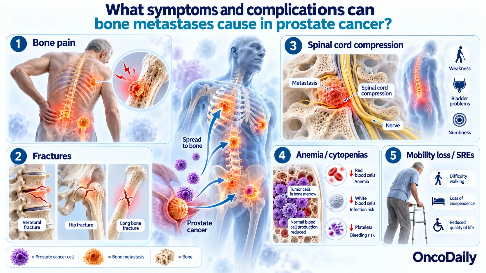
Bone metastases are a major source of morbidity in advanced prostate cancer, particularly through pain, structural bone damage, neurologic complications, and impairment of normal marrow function.
Bone pain is one of the most common symptoms. It may present as a persistent ache and become increasingly severe as metastatic lesions progress. Abnormal bone remodeling, microfractures, periosteal stretching, nerve sensitization, and inflammatory signaling within the metastatic site can all contribute to pain. (Smith et al., 2022; Baldessari et al., 2023).
Metastatic involvement can also weaken bone and cause pathologic fractures, particularly in the vertebrae, pelvis, and long bones. These events may severely affect mobility, independence, and quality of life. (Tsuzuki et al., 2016; Baldessari et al., 2023).
Vertebral metastases can result in metastatic spinal cord compression, which may present with worsening back pain, weakness, sensory changes, difficulty walking, or bowel and bladder dysfunction. This is an oncologic emergency because delayed treatment can result in irreversible neurologic injury. (Tsuzuki et al., 2016).
Extensive bone and marrow involvement can also contribute to anemia, thrombocytopenia, and other cytopenias. Disturbances in calcium metabolism may occur as well, although hypercalcemia is less characteristic of the predominantly osteoblastic pattern seen in prostate cancer than in many osteolytic malignancies. (Baldessari et al., 2023).
Many of these complications are described clinically as skeletal-related events (SREs), including pathologic fracture, spinal cord compression, and the need for radiation or surgery to bone. Preventing these events is an important part of managing metastatic prostate cancer. (Tsuzuki et al., 2016).
How Are Prostate Cancer Bone Metastases Detected and Treated?
Detection combines the clinical picture, PSA kinetics, disease setting, and imaging. PSMA PET/CT has become increasingly important for staging and restaging prostate cancer because it can detect metastatic disease more accurately than conventional imaging in many clinical settings. Bone scintigraphy, CT, and MRI remain useful, while MRI is particularly important when marrow disease or spinal cord compression is suspected. (Fendler et al., 2023).
Treatment depends on whether the disease is metastatic hormone-sensitive or castration-resistant, as well as metastatic burden, symptoms, previous therapies, molecular characteristics, and patient fitness. Androgen deprivation therapy (ADT) remains the foundation of systemic treatment, but in metastatic hormone-sensitive disease it is generally combined with an androgen-receptor pathway inhibitor and, in selected patients, chemotherapy rather than used alone. (Tilki et al., 2026).
For men with metastatic castration-resistant prostate cancer (mCRPC) and bone metastases, denosumab or zoledronic acid can reduce skeletal-related complications. Because these therapies can cause hypocalcemia and osteonecrosis of the jaw, appropriate calcium and vitamin D management, dental assessment, and relevant laboratory monitoring are important. (Fizazi et al., 2011; Tilki et al., 2026).
Local therapy remains valuable for symptomatic or structurally threatening lesions. External-beam radiotherapy can provide effective relief from bone pain, while surgery may be required for impending or established fractures, spinal instability, or selected cases of spinal cord compression. (Tilki et al., 2026).
Other treatments depend on the clinical setting. Radium-223 remains an option for appropriately selected patients with symptomatic bone-predominant mCRPC without visceral metastases, while PSMA-targeted radioligand therapy and molecularly targeted treatments may be used according to PSMA expression, genomic findings, prior therapy, and current treatment indications. (Scarpato et al., 2026; Tilki et al., 2026).
Management of bone-metastatic prostate cancer therefore requires two parallel strategies: controlling the underlying cancer and protecting the skeleton from pain, fracture, neurologic complications, and loss of function.

FAQ
Why does prostate cancer commonly spread to bone?
Bone marrow provides a supportive environment rich in growth factors, stromal cells, and signaling molecules that help prostate cancer cells survive and grow.
What are the most common symptoms of prostate cancer bone metastases?
Persistent bone pain is common, especially in the spine, pelvis, hips, and ribs. Fractures and neurologic symptoms can also occur.
Are prostate cancer bone metastases osteoblastic or osteolytic?
They are usually predominantly osteoblastic, producing dense sclerotic lesions, although osteoclast-mediated bone resorption also remains active.
How are prostate cancer bone metastases detected?
Imaging may include PSMA PET/CT, bone scintigraphy, CT, and MRI, depending on the clinical situation and available resources.
Can prostate cancer bone metastases be treated?
Yes. Treatment may include systemic prostate cancer therapy, bone-targeted agents, radiotherapy, radiopharmaceuticals, and surgery for selected complications.