
PI-RADS 3, 4, and 5 are categories used in prostate MRI reports to describe the likelihood of clinically significant prostate cancer. A PI-RADS 3 score indicates an uncertain finding, while PI-RADS 4 and 5 suggest progressively higher levels of suspicion.
Receiving an abnormal prostate MRI result can be concerning, but a high PI-RADS score does not automatically mean that cancer is present. The score helps doctors decide whether further testing, particularly a prostate biopsy, is needed. Understanding the differences between PI-RADS categories can help patients interpret their MRI results and discuss the next steps with their urologist.
What Is PI-RADS and How Is It Used in Prostate MRI?
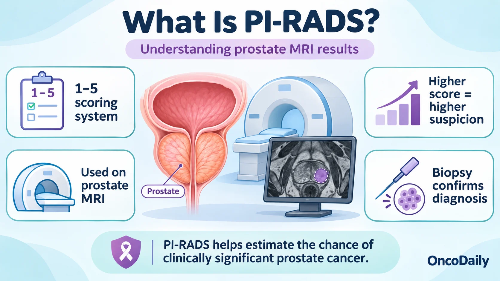
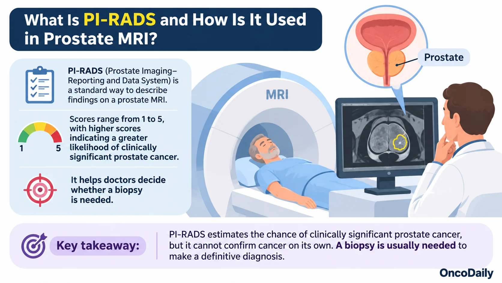
PI-RADS (Prostate Imaging–Reporting and Data System) is a standardized scoring system used to assess suspicious areas detected on prostate magnetic resonance imaging (MRI). Scores range from 1 to 5, with higher numbers indicating a greater likelihood of clinically significant prostate cancer.
Prostate MRI is commonly recommended when a patient has an elevated prostate-specific antigen (PSA) level, an abnormal digital rectal examination, or other findings that raise suspicion of prostate cancer.
The examination uses detailed images to identify abnormalities within the prostate. Multiparametric MRI (mpMRI) combines several imaging techniques, including anatomical imaging, diffusion-weighted imaging, and contrast-enhanced sequences, to evaluate prostate tissue.
Radiologists assess features such as the location, size, and appearance of suspicious lesions before assigning a PI-RADS score. The system helps standardize reporting and guides decisions about whether a biopsy is necessary.
An important distinction is that PI-RADS evaluates the likelihood of clinically significant prostate cancer, generally referring to disease with a Gleason score of 3+4 or higher (Grade Group 2 or above). These cancers are more likely to require treatment or careful clinical management than low-grade tumors.
However, MRI findings alone cannot establish a diagnosis. Even highly suspicious lesions require histopathological confirmation, usually through a prostate biopsy (Steiger and Thoeny, 2016; Ponsiglione et al., 2026).
What Does PI-RADS 3 Mean on a Prostate MRI?
PI-RADS 3 indicates an equivocal MRI finding, meaning that clinically significant prostate cancer cannot be confidently confirmed or excluded based on imaging alone.
These lesions are often described as borderline because their appearance may overlap with non-cancerous prostate conditions, including inflammation, benign prostatic hyperplasia, and tissue changes related to previous procedures.
Not every patient with PI-RADS 3 requires an immediate biopsy. The decision depends on additional risk factors, particularly PSA density, previous biopsy results, family history, and the appearance of the lesion.
PSA density is calculated by dividing the PSA blood level by prostate volume. It helps distinguish whether an elevated PSA may be related to an enlarged prostate or whether further investigation for cancer is necessary.
A 2025 retrospective study involving 671 patients with 981 PI-RADS 3 lesions found clinically significant prostate cancer in 15.8% of biopsied lesions. PSA density was one of the strongest predictors of cancer detection, supporting its role in deciding which patients require biopsy (Franz et al., 2025).
For patients with a PI-RADS 3 result and otherwise low clinical risk, doctors may recommend continued PSA monitoring and repeat MRI rather than immediate biopsy. However, patients with elevated PSA density or other concerning findings may benefit from targeted biopsy.

What Does PI-RADS 4 Mean on a Prostate MRI?
PI-RADS 4 indicates a high likelihood of clinically significant prostate cancer. It means that the suspicious area has imaging characteristics strongly associated with malignancy, although cancer is not yet confirmed.
These findings are more concerning than PI-RADS 3 lesions because they demonstrate clearer abnormalities on MRI, such as restricted water diffusion or changes in prostate tissue structure.
Patients with PI-RADS 4 are generally advised to undergo a prostate biopsy to determine whether cancer is present.
An MRI-targeted biopsy allows doctors to collect tissue samples directly from the suspicious area. Depending on the clinical situation, additional systematic biopsies may be performed to examine other parts of the prostate.
However, not every PI-RADS 4 lesion is malignant. Prostatitis, benign nodules, and other tissue changes can occasionally resemble cancer on MRI, leading to false-positive findings.
The likelihood of cancer also varies according to PSA density, age, lesion location, and previous biopsy history.
A PI-RADS 4 result therefore indicates a need for further investigation, not an automatic cancer diagnosis. If biopsy confirms cancer, the pathology report determines its Grade Group, which helps guide subsequent management (National Cancer Institute, 2026; Ponsiglione et al., 2026).
What Does PI-RADS 5 Mean on a Prostate MRI?
PI-RADS 5 is the highest suspicion category on prostate MRI and indicates that clinically significant prostate cancer is highly likely.
These lesions generally demonstrate pronounced MRI abnormalities. Under PI-RADS assessment criteria, a lesion may receive a score of 5 because of its suspicious imaging characteristics combined with a size of at least 1.5 cm, or because there is evidence of definite extension beyond the prostate.
The score indicates a higher probability of clinically significant cancer than PI-RADS 4, but it does not reveal the cancer’s Gleason score or determine its complete stage.
Patients with PI-RADS 5 findings generally require a timely urological assessment and targeted prostate biopsy.
If cancer is confirmed, the next steps depend on the biopsy results, PSA level, clinical stage, and overall health. Depending on these findings, additional imaging may be necessary to assess whether cancer extends beyond the prostate.
Importantly, PI-RADS 5 does not automatically mean advanced or metastatic prostate cancer. Some patients have disease confined to the prostate that can be treated with curative intent.
Although the probability of malignancy is high, a biopsy remains necessary to confirm the diagnosis and identify the tumor’s histological characteristics (Steiger and Thoeny, 2016; Gottumukkala and Lee, 2021).
What Is the Difference Between PI-RADS 3, 4, and 5
The main difference between PI-RADS 3, 4, and 5 is the level of suspicion for clinically significant prostate cancer. As the score increases, the likelihood that the suspicious area contains clinically significant disease generally increases.
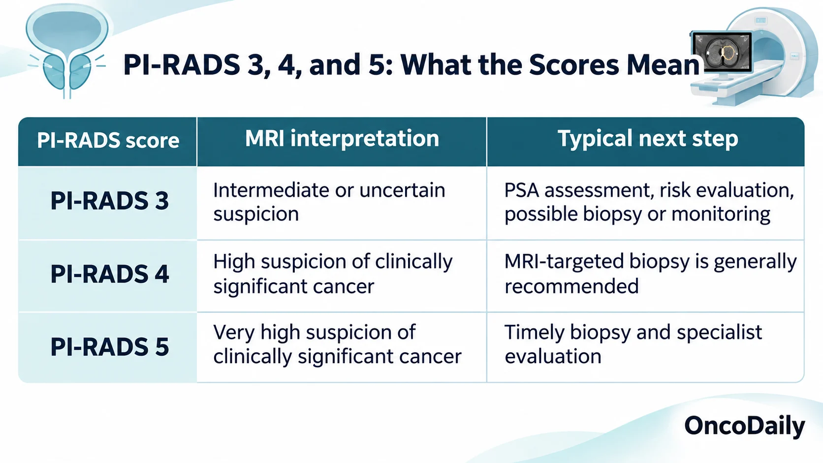
Although PI-RADS scores indicate different levels of risk, they are not direct measurements of tumor aggressiveness. A PI-RADS 5 lesion is more suspicious on imaging than a PI-RADS 3 lesion, but the score does not establish the tumor’s grade.
The distinction between PI-RADS and the Gleason grading system is particularly important. PI-RADS describes MRI findings before tissue diagnosis, while Gleason scores and Grade Groups are determined by examining cancer cells under a microscope.
Both assessments may contribute to treatment planning, but they provide different information about the disease.
Does a High PI-RADS Score Always Mean Prostate Cancer?
No. Even a PI-RADS 4 or 5 result does not confirm prostate cancer without a biopsy. Although higher PI-RADS scores are associated with a greater probability of clinically significant disease, MRI cannot always distinguish malignant lesions from benign prostate conditions.
Inflammation, particularly prostatitis, can produce MRI changes similar to those seen in prostate cancer. Benign prostatic hyperplasia and scar tissue may also affect the appearance of the prostate.
The accuracy of PI-RADS interpretation depends partly on image quality, lesion location, and radiologist experience. Some abnormalities are more difficult to classify, particularly those in the transition zone of the prostate.
A 2026 review published in Cancer Imaging highlighted that PI-RADS remains an important tool for prostate cancer detection but has limitations related to reader variability and the classification of equivocal lesions.
The authors emphasized the importance of combining MRI findings with PSA density and other clinical factors to improve diagnostic decisions (Ponsiglione et al., 2026).
It is also possible for prostate cancer to be present despite a low or negative PI-RADS result. Therefore, patients with persistently elevated PSA or other concerning risk factors may still require further evaluation even when MRI findings are reassuring.
What Happens After a PI-RADS 3, 4, or 5 Result?
The next step after a prostate MRI depends on the PI-RADS score and the patient’s overall risk of clinically significant prostate cancer.
For PI-RADS 3, doctors often review PSA levels, PSA density, family history, and previous biopsy findings before deciding whether a biopsy is necessary. In selected low-risk patients, repeat MRI and PSA monitoring may be appropriate.
PI-RADS 4 and 5 findings generally lead to a recommendation for prostate biopsy because of their higher likelihood of clinically significant disease.
During an MRI-targeted biopsy, tissue samples are collected from suspicious areas identified on imaging. This may be performed using MRI-ultrasound fusion guidance or other targeting techniques. Systematic sampling may also be recommended, depending on the patient’s circumstances.
If the biopsy identifies prostate cancer, the tissue is examined to determine the Gleason score and Grade Group. These results, together with PSA and clinical staging, help doctors decide whether active surveillance, surgery, radiation therapy, or another approach is appropriate.
If the biopsy is negative despite a highly suspicious MRI result, further evaluation may still be necessary. Depending on the level of concern, doctors may recommend imaging review, repeat PSA testing, or another biopsy.
Research also supports individualized follow-up for patients with uncertain MRI findings. In a 2024 retrospective cohort study, Önder and colleagues examined the long-term prognostic value of PI-RADS assessments, highlighting the importance of continued risk evaluation rather than relying solely on a single MRI result (Önder et al., 2024).
For patients, the most important point is that PI-RADS scores guide the diagnostic process but do not determine treatment on their own. Decisions should be made after reviewing MRI findings together with PSA results, biopsy findings when available, and individual clinical risk factors.
FAQ
Does PI-RADS 4 mean you have prostate cancer?
No. PI-RADS 4 indicates a high suspicion of clinically significant prostate cancer, but a biopsy is needed to confirm the diagnosis.
Can PI-RADS 5 be benign?
Yes. Although PI-RADS 5 is highly suspicious for prostate cancer, inflammation and other benign conditions can occasionally produce similar MRI findings.
Does PI-RADS 3 require a biopsy?
Not always. The decision depends on PSA levels, PSA density, family history, and other risk factors. Some patients may undergo monitoring instead.
What is the difference between PI-RADS 4 and 5?
PI-RADS 4 indicates a high likelihood of clinically significant prostate cancer, while PI-RADS 5 indicates a very high likelihood. Neither score confirms cancer.
Can prostate cancer be present with a normal MRI?
Yes. Prostate MRI can miss some cancers, including clinically significant tumors. Doctors consider PSA levels and other risk factors when deciding whether further testing is necessary.