
Key takeaways
- Intratumoral heterogeneity complicates diagnostic accuracy, molecular profiling, and personalized treatment selection.
- VHL, PBRM1, BAP1, and SETD2 alterations contribute to the biological diversity and clinical behavior of clear cell RCC.
- Sarcomatoid differentiation is associated with aggressive disease but also greater sensitivity to immune checkpoint inhibition.
- The abandonment of papillary RCC type 1 and type 2 classification reflects the molecular diversity of these tumors.
- Rare molecularly defined subtypes, such as FH-deficient and ALK-rearranged RCC, carry important hereditary or therapeutic implications.
Eva Compérat, Professor of Pathology at the Medical University of Vienna, specializing in urological pathology, examined the growing molecular and histological complexity of renal cell carcinoma (RCC) during her presentation at VIRO 2026 – Global Voices in Renal Oncology.
Understanding kidney cancer increasingly requires moving beyond conventional histological classification. Tumors with similar microscopic appearances may have different molecular drivers, clinical outcomes, and responses to systemic therapies. Conversely, considerable genetic diversity can exist within a single tumor.
These differences have important implications for diagnostic accuracy, prognostic assessment, and the development of personalized treatment strategies.
The Changing Classification of Renal Tumors
The 2022 World Health Organization (WHO) classification of renal tumors marked an important transition toward integrating molecular characteristics with conventional histopathology.
Several previously grouped tumors are now recognized as distinct disease entities, while the introduction of molecularly defined renal carcinomas reflects a more detailed understanding of their underlying biology.
According to Compérat, work is already underway on the next edition of the WHO classification, which is expected to further refine existing entities and incorporate additional molecular information.
“It will become more molecular. There will be new names and so on. But one thing is sure, the clear cell carcinoma will remain the same, and it will be called the same.”
Although clear cell RCC remains a well-established diagnostic entity, its underlying molecular landscape is far from uniform.
One of the central challenges for contemporary renal pathology is understanding how a tumor’s genotype contributes to its morphology and, ultimately, its clinical behavior.
The Molecular Architecture of Clear Cell RCC
Clear cell renal cell carcinoma (ccRCC), the most common histological subtype of kidney cancer, is strongly associated with alterations involving the von Hippel-Lindau (VHL) gene.
Under normal oxygen conditions, the VHL protein contributes to the degradation of hypoxia-inducible factors (HIFs). Inactivation of VHL disrupts this regulatory mechanism, leading to the accumulation of HIF proteins and the activation of genes involved in angiogenesis, cellular metabolism, and tumor growth.
This mechanism helps explain one of the characteristic histological features of clear cell RCC: its extensive vascular network.
Using CD31 immunohistochemical staining, Compérat illustrated the abundance of blood vessels within these tumors. Their abnormal vascular architecture also contributes to intratumoral hemorrhage.
The biological dependence of clear cell RCC on angiogenic pathways provided the rationale for developing antiangiogenic treatments, including vascular endothelial growth factor receptor (VEGFR) tyrosine kinase inhibitors such as sunitinib.
A more direct approach to targeting this molecular pathway has subsequently emerged through HIF-2α inhibition.
However, VHL inactivation represents only one component of clear cell RCC biology.
Intratumoral Heterogeneity: When One Tumor Contains Multiple Molecular Profiles
A particularly challenging characteristic of clear cell RCC is the extent of its intratumoral heterogeneity.
Different regions of the same tumor may contain distinct genetic alterations, cellular populations, and biological characteristics.
“If you take one millimeter, let’s say, on the left part of the tumor, it will not at all be like the tumor on the right side of the tumor.”
This spatial heterogeneity has implications for tissue sampling, molecular profiling, and treatment selection.
A single biopsy may not fully capture the molecular diversity of a tumor, particularly when clinically relevant alterations are present only in specific subclonal populations.
Among the most important genes implicated in clear cell RCC are four tumor suppressor genes located on the short arm of chromosome 3:
- VHL: A central regulator of the cellular response to hypoxia and angiogenesis.
- PBRM1: Involved in chromatin remodeling and associated with distinct tumor evolutionary patterns.
- BAP1: Frequently associated with higher-grade tumors, aggressive biological behavior, and unfavorable prognosis.
- SETD2: Involved in histone methylation, chromatin regulation, and genomic stability.
Compérat discussed the contrasting biological characteristics of PBRM1- and BAP1-altered tumors. These alterations are frequently mutually exclusive, although exceptions exist, and their prognostic significance depends on the broader molecular and clinical context.
While certain PBRM1-altered tumors may demonstrate more favorable outcomes, BAP1 alterations have repeatedly been associated with aggressive disease.
The prognostic role of SETD2 is more complex and may depend on accompanying genetic alterations and tumor evolution.
Importantly, none of these alterations should be interpreted in isolation as a definitive predictor of treatment response.
Beyond Chromosome 3p
Although loss of chromosome 3p is a defining genomic characteristic of clear cell RCC, additional chromosomal alterations contribute to its biological diversity.
Compérat reviewed genomic studies reporting 3p loss in up to 93% of clear cell RCC cases, alongside recurrent alterations involving other chromosomes.
These include gains of chromosome 5q and chromosome 7, as well as losses involving chromosomes 9p and 14q.
Loss of 14q has been associated with unfavorable clinical outcomes, while the accumulation of additional genomic abnormalities may contribute to tumor progression and metastatic potential.
Understanding kidney cancer therefore requires examining not only its initiating molecular events but also the subsequent genetic changes that shape disease evolution.
Sarcomatoid Differentiation: Aggressive Biology and Immunotherapy Sensitivity
Sarcomatoid differentiation represents one of the most aggressive morphological features encountered in renal cell carcinoma.
Rather than constituting a separate RCC subtype, it reflects dedifferentiation that can occur across different histological entities, including clear cell, papillary, and chromophobe RCC.
In renal carcinomas for which the WHO/ISUP grading system is applicable, sarcomatoid differentiation is classified as grade 4.
Its presence is associated with aggressive clinical behavior, an increased risk of metastasis, and historically poor survival outcomes.
The molecular mechanisms underlying sarcomatoid transformation remain incompletely understood. Several processes and genetic alterations have been implicated, including epithelial-mesenchymal transition, TP53 and BAP1 mutations, alterations involving CDKN2A and CDKN2B, and dysregulation of the NF2-Hippo signaling pathway.
However, the aggressive nature of these tumors is accompanied by a particularly interesting immunological profile.
A 2021 study published in Nature Communications investigated the molecular characteristics of sarcomatoid and rhabdoid RCC across clinical trial and real-world cohorts.
The study identified an immune-inflamed tumor microenvironment characterized by increased immune cell infiltration, activation of immune-related pathways, and greater PD-L1 expression.
These findings help explain why renal tumors with sarcomatoid or rhabdoid differentiation can demonstrate substantial sensitivity to immune checkpoint inhibition despite their unfavorable underlying biology.
Compérat illustrated this phenomenon using histological images showing strong PD-L1 staining in a clear cell RCC with sarcomatoid differentiation, in contrast to a non-sarcomatoid tumor without detectable staining.
Nevertheless, PD-L1 expression alone is not an established requirement for selecting immunotherapy in RCC.
Why the Type 1 and Type 2 Papillary RCC Classification Was Abandoned
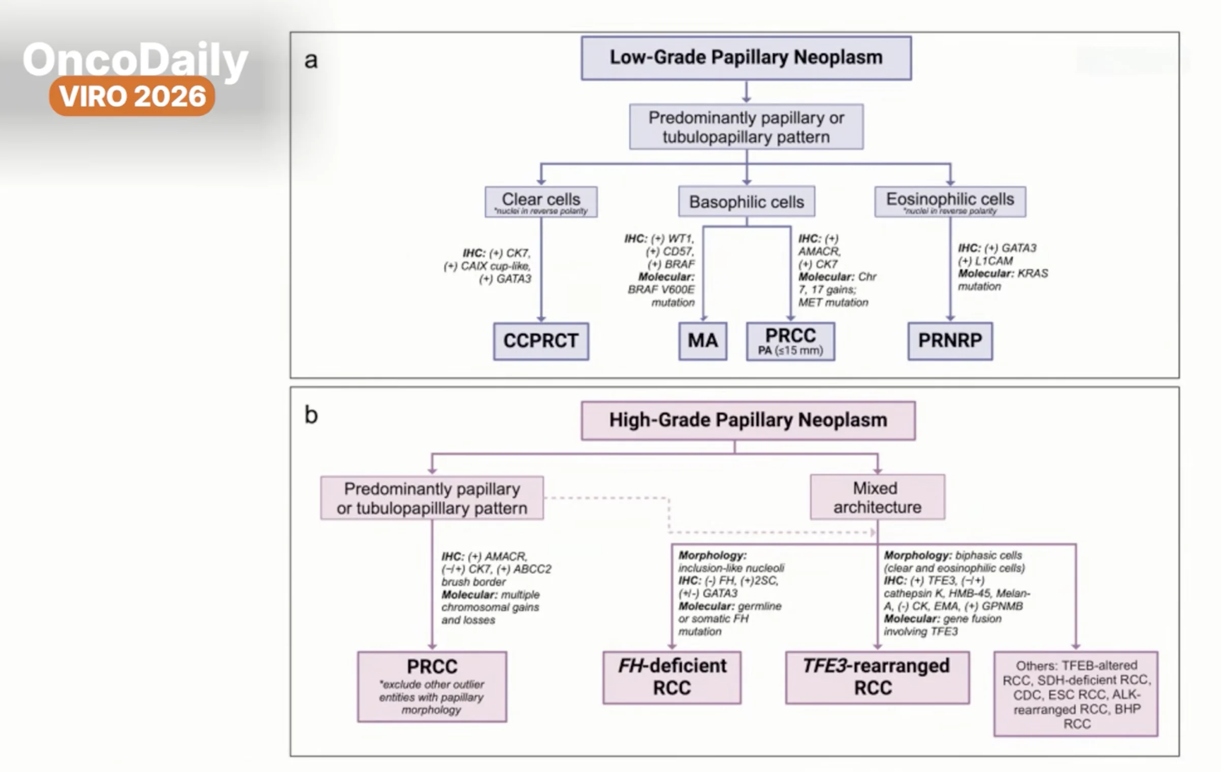
One of the most consequential changes introduced by the 2022 WHO classification was the abandonment of the traditional subdivision of papillary RCC into type 1 and type 2 tumors.
The previous classification grouped tumors according to their morphological characteristics. However, molecular investigations revealed that many tumors historically classified as type 2 papillary RCC represented biologically distinct diseases.
“We have sorted out very many others in between from the type 2. And so the type 2 has been abandoned.”
Several renal tumor entities that may exhibit papillary architecture are now recognized separately, including:
- Fumarate hydratase (FH)-deficient RCC
- TFE3-rearranged RCC
- TFEB-altered RCC
- ALK-rearranged RCC
- Acquired cystic disease-associated RCC
The distinction is clinically important because these tumors may differ considerably in their prognosis, hereditary implications, and potential therapeutic vulnerabilities.
Papillary architecture, therefore, does not necessarily establish a diagnosis of papillary RCC.
In high-grade tumors with overlapping morphological features, immunohistochemical and molecular investigations can be particularly valuable in establishing a more precise diagnosis.
FH-Deficient RCC: How Molecular Classification Revealed a High-Risk Subgroup
An important example of the limitations of the former papillary RCC classification emerged from The Cancer Genome Atlas (TCGA) analysis published in The New England Journal of Medicine in 2016.
The study performed comprehensive molecular characterization of 161 primary papillary renal cell carcinomas and identified four major molecular clusters with different clinical outcomes.
The clusters included:
- C1: Predominantly composed of classical, historically type 1 papillary tumors.
- C2a: Enriched for former type 2 tumors, with relatively favorable outcomes.
- C2b: Associated with more advanced disease and less favorable survival.
- C2c: Characterized by a CpG island methylator phenotype (CIMP) and particularly poor survival.
The C2c subgroup comprised only nine tumors. However, five of these carried germline or somatic alterations in the FH gene.
This observation helped demonstrate that the unfavorable outcomes of certain tumors historically classified as type 2 papillary RCC were related to distinct underlying molecular mechanisms.
FH deficiency disrupts normal cellular metabolism, leading to fumarate accumulation and extensive metabolic and epigenetic changes that contribute to tumor development.
Today, FH-deficient RCC is recognized as a distinct molecularly defined entity.
Its identification is important not only because of its potentially aggressive clinical course but also because some cases arise in the context of hereditary leiomyomatosis and renal cell cancer syndrome.
Not all FH-deficient tumors are hereditary, however. Somatic alterations can also result in FH deficiency, making appropriate genetic evaluation essential.
MET Alterations in Papillary RCC: Searching for a Predictive Biomarker
The MET signaling pathway represents another important area of investigation in papillary RCC.
Molecular alterations involving MET may include activating mutations in the kinase domain, gene amplification, and chromosome 7 gains.
Compérat referred to research investigating whether these alterations could identify patients more likely to benefit from targeted therapies.
In a biomarker-based phase II study evaluating the selective MET inhibitor savolitinib in 109 patients with advanced papillary RCC, objective responses were observed in 18% of patients with MET-driven tumors, compared with no responses in patients with MET-independent disease.
Median progression-free survival was 6.2 months and 1.4 months, respectively.
Although these findings support the biological relevance of MET signaling, different MET alterations may not confer the same degree of therapeutic sensitivity.
The identification of a molecular alteration does not necessarily establish that it is a sufficiently reliable predictive biomarker for routine clinical practice.
“Finding a biomarker is something which seems to be extremely complicated in kidney cancer.”
ALK-Rearranged RCC: A Rare Diagnosis With Therapeutic Implications
Among the molecularly defined renal carcinomas, ALK-rearranged RCC provides a particularly interesting example of how diagnostic precision may create new treatment opportunities.
These tumors are exceptionally rare and can exhibit substantial morphological variability, making them difficult to recognize through conventional histopathology alone.
Compérat suggested that their incidence may be underestimated because they do not share a single, easily identifiable microscopic appearance.
The clinical relevance of identifying these tumors lies in their potential sensitivity to anaplastic lymphoma kinase (ALK) inhibitors.
Published case reports have documented meaningful and sometimes durable responses to ALK-targeted agents, including alectinib, in patients with advanced ALK-rearranged RCC.
Although the available clinical evidence remains limited and does not establish a standardized treatment approach, these observations demonstrate the potential value of identifying rare but therapeutically actionable molecular alterations.
For selected patients, establishing the correct molecular diagnosis may fundamentally change the available treatment options.
When Greater Diagnostic Precision Changes Clinical Management
The increasing complexity of renal tumor classification is not simply a question of nomenclature.
Distinguishing an indolent oncocytic lesion from an aggressive renal carcinoma, recognizing sarcomatoid differentiation, identifying a potentially hereditary tumor, or detecting an actionable gene rearrangement can have substantial consequences for patient management.
Compérat noted that kidney tumors are among the lesions for which she most frequently receives requests for a second pathological opinion.
Even as molecular technologies become more sophisticated, diagnostic challenges remain, particularly among rare renal tumors, emerging oncocytic entities, and tumors with overlapping histological characteristics.
“There are really very big differences among the different RCC groups, but also within the RCC groups.”
The integration of morphology, immunohistochemistry, molecular profiling, and clinical information will become increasingly important as renal oncology moves toward more individualized treatment strategies.
For some patients, the most consequential advance in kidney cancer care may begin not with a new drug, but with a more precise understanding of the tumor being treated.
Written by Eliz Baloyan, MD, Features Writer and Editor at OncoDaily and CancerWorld