
A study published in Nature reveals that hormone receptor-positive breast tumors can harbor epigenetically distinct basal-like cancer cells associated with aggressive biological characteristics. By linking these hidden cellular populations to circulating nucleosome signatures, researchers also demonstrate the potential of a blood-based assay to identify breast cancer and estimate recurrence risk. The findings offer a new perspective on intratumoral heterogeneity, although prospective validation is required before clinical application.
Breast cancer classification has traditionally relied on a combination of histopathology, hormone receptor status, HER2 expression, and molecular characteristics. These features guide systemic treatment selection and provide essential information about prognosis.
Hormone receptor-positive tumors are generally considered biologically distinct from basal-like breast cancers, which substantially overlap with the triple-negative subtype. Yet patients with apparently similar luminal tumors can experience markedly different clinical outcomes.
Some remain disease-free for decades following endocrine therapy, whereas others experience early or late recurrence despite favorable initial characteristics. A study published on November 11, 2026, in Nature suggests that part of this variability may arise from epigenetically distinct tumor-cell populations hidden within cancers classified as luminal.
The research, Epigenetic Heterogeneity in Breast Tumours Informs Diagnosis and Prognosis, was led by Noa Furth, Christina Riemenschneider, Yishai Arieli, Guy Ron, Efrat Shema, and an international group of investigators.
Using single-cell epigenetic profiling, DNA methylation analysis, proteomics, and a novel blood-based platform, the researchers investigated how differences in chromatin organization influence tumor-cell identity.
Their results identified a recurrent basal-like population within hormone receptor-positive tumors, characterized by reduced estrogen signaling, distinct histone modifications, and increased expression of aggressive biological programs.
The team then investigated whether these epigenetic signatures could be detected in blood and whether they were associated with future disease recurrence. The findings connect two important areas of breast cancer research: the biology of intratumoral cellular plasticity and the development of molecular biomarkers for early detection and prognosis.
Luminal Breast Cancer Is Not a Uniform Cellular Population
The conventional molecular classification of breast cancer divides tumors into luminal A, luminal B, HER2-enriched, and basal-like categories. These groups reflect broad differences in gene expression and biological behavior.
However, the classification of an entire tumor does not necessarily describe every malignant cell within it. Single-cell studies have increasingly demonstrated that tumors contain multiple cellular populations with different transcriptional, metabolic, and signaling programs.
Some populations may be more proliferative, while others may be relatively dormant, adapted to stress, or less dependent on the dominant molecular pathway. In hormone receptor-positive breast cancer, such heterogeneity may be clinically important because endocrine therapy primarily targets estrogen-dependent tumor biology.
The new study investigated whether chromatin-associated changes could help explain how different cellular identities coexist within the same tumor. Unlike genomic mutations, epigenetic changes can modify gene regulation without altering the underlying DNA sequence.
These changes include DNA methylation and chemical modifications of histone proteins around which DNA is organized. Together, these processes influence which genes remain accessible for transcription and which become repressed.
The investigators hypothesized that distinct epigenetic patterns could support the coexistence of luminal and basal-like cell states within hormone receptor-positive malignancies.
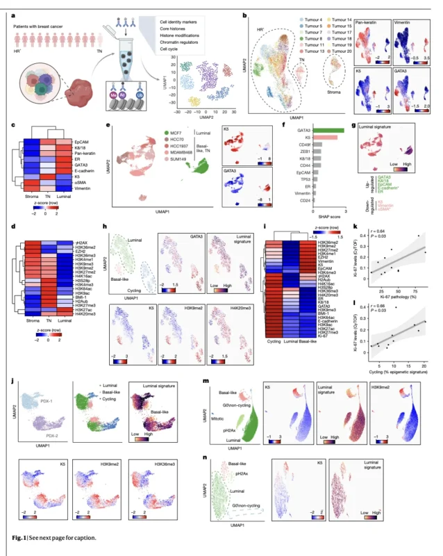
Profiling More Than One Million Cells
The research began with freshly resected breast tumors obtained from patients who had not received systemic treatment before surgery. The primary single-cell analysis included 12 human tumors: 11 hormone receptor-positive tumors and one basal-like triple-negative tumor used for comparison.
Using cytometry by time-of-flight, or CyTOF, the researchers analyzed approximately 1.16 million cells across these tumors. The experimental panel measured proteins associated with cellular identity, cell-cycle activity, chromatin regulation, and multiple histone modifications.
Rather than examining only one molecular marker at a time, the platform enabled simultaneous evaluation of complex epigenetic patterns at single-cell resolution. The investigators identified three recurring malignant-cell states within luminal tumors.
The first population retained typical luminal characteristics, including estrogen receptor-associated signaling and expression of luminal identity proteins. The second represented cycling cells with increased proliferative activity and characteristic chromatin modifications.
The third, and most unexpected, consisted of cells displaying basal-like features within tumors classified clinically as luminal. These basal-like cells expressed higher levels of cytokeratin 5, or K5, while showing reduced expression of luminal markers such as GATA3 and estrogen receptor.
The population was identified across different tumors and reproduced in patient-derived xenograft models and experimental breast cancer cell systems.Importantly, these were basal-like cellular states within luminal tumors, not evidence that the tumors should be reclassified as triple-negative breast cancer.
The Hidden Basal-Like Population Shows Aggressive Biological Features
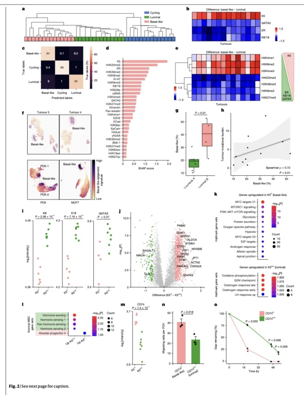
The researchers next investigated whether the basal-like cells differed functionally from their luminal counterparts. Multiple observations suggested that they possessed biological characteristics associated with more aggressive behavior.
Basal-like cells demonstrated reduced hormone receptor signaling and enrichment of pathways associated with epithelial–mesenchymal transition. They also expressed programs involving MYC, mTOR, PI3K–AKT signaling, glycolysis, and cellular adaptation.
The researchers identified increased expression of CD74 in basal-like populations. In laboratory experiments, cells selected for higher CD74 expression demonstrated greater migratory activity than cells with lower expression.
These experiments provided functional support for a relationship between the basal-like phenotype and increased cellular migration. The study also found that the proportion of basal-like cells was greater in luminal B tumors than in luminal A tumors within the small tissue cohort.
Higher basal-like fractions correlated with tumor mutational burden, and tumors carrying TP53 mutations tended to contain more of these cells. Together, these observations linked the basal-like state to molecular and pathological characteristics associated with greater tumor aggressiveness.
However, they did not establish that these cells independently cause metastasis or clinical treatment resistance. The proposed contribution of these states to disease recurrence remains a biologically supported hypothesis requiring additional functional and clinical validation.
Histone Modifications Reveal the Epigenetic Basis of Cellular Identity
A central objective of the study was identifying the chromatin features distinguishing basal-like cells from luminal cells. The investigators found consistent differences in multiple histone modifications. Basal-like cells exhibited increased H3K9me2, H3K4me1, and H3K4me3.
In contrast, luminal cells showed greater enrichment of modifications including H4K20me3, H3K36me3, H3K9me3, and H3K27me3. These patterns indicated that cellular identity was associated with coordinated differences in chromatin organization.
H3K4me1 is commonly linked to enhancer-associated chromatin, whereas H3K4me3 is associated with active regulatory regions near transcription initiation sites. Their distribution can therefore provide information about the gene regulatory programs active within a cell.
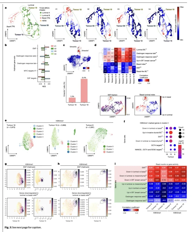
Using single-nucleus CUT&Tag, the researchers mapped selected histone modifications across the genome. This approach confirmed that basal-like populations showed distinct H3K4-associated patterns near genes involved in basal differentiation and epithelial–mesenchymal transition.
The researchers also examined the repressive modification H3K9me2. Their findings suggested increased H3K9me2-associated regulation of estrogen-related programs in the basal-like population. These complementary analyses provided evidence that the cellular differences were not simply the result of changes in cell-cycle activity.
They reflected broader alterations in chromatin states associated with luminal or basal-like identity.
Can Cancer Cells Change Identity Without Acquiring New Mutations?
The study also raises an important question about tumor evolution. Classically, cancer heterogeneity is often understood as a consequence of genetically distinct subclones acquiring different mutations. However, genetically similar or identical cells can also adopt different phenotypic states through changes in gene regulation.
The investigators observed that single-cell-derived MCF7 clones could regenerate cellular diversity resembling that of the parental population. Similarly, independently grown xenograft tumors derived from the same donor demonstrated comparable distributions of distinct cellular states.
These observations support the possibility that luminal and basal-like populations exist within a dynamic, potentially interconvertible system. In such a model, cells could shift between states without necessarily acquiring a new driver mutation.
This mechanism may be relevant to treatment resistance. A tumor containing cells with reduced dependence on estrogen signaling could theoretically retain a population capable of surviving endocrine treatment pressure.
However, the investigators did not directly demonstrate that the identified basal-like population causes endocrine resistance in patients. Nor did the study evaluate a new epigenetic therapy capable of preventing these cellular transitions. The findings identify a potential biological mechanism that future research can investigate.
DNA Methylation Supports the Same Biological Pattern
To determine whether the observed cellular states were reflected in other epigenetic features, the researchers performed DNA methylation analyses. They integrated whole-genome methylation data from selected luminal tumors with publicly available datasets, including The Cancer Genome Atlas.
The analysis identified distinct methylation patterns associated with luminal differentiation, basal-like identity, and epithelial–mesenchymal transition. Additional experiments comparing sorted K5-high and K5-low populations demonstrated differentially methylated regulatory regions.
In basal-like cells, genes associated with estrogen response were more frequently hypermethylated, whereas genes linked to mesenchymal and oncogenic programs showed different regulatory patterns. The basal-like population also exhibited greater epigenetic variability or noise.
Such variability may reflect a more flexible regulatory state, potentially allowing tumor cells to adapt to changing environmental conditions. Nevertheless, measuring epigenetic noise does not directly prove that these cells are more resistant to a particular therapy. The clinical implications of these patterns remain to be established.
From Tumor Tissue to Blood: The EPINUC Approach
One of the most translational aspects of the study involved applying the tumor epigenetic findings to a blood-based diagnostic platform. The investigators used a technology called EPINUC, short for epigenetics of plasma-isolated nucleosomes.
Nucleosomes are structures composed of DNA wrapped around histone proteins. When cells die, nucleosomes can be released into the circulation. Some of these circulating structures retain epigenetic features reflecting their tissue of origin and biological state.
The EPINUC system captures plasma-isolated nucleosomes and uses single-molecule imaging to quantify histone modifications and their combinations. Unlike conventional circulating tumor DNA approaches focused primarily on sequence alterations, EPINUC examines the chromatin-associated features carried by circulating nucleosomes.
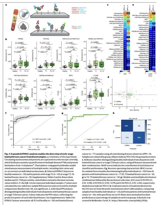
For breast cancer, the team expanded its existing platform to include 29 parameters. These included several histone modifications and breast cancer-associated proteins, including K5, L1CAM, and MUC1. The initial expanded analysis included plasma from 77 women with luminal breast cancer and 30 healthy women.
The researchers identified differences in circulating histone modifications between the groups. Several features were detectable even among patients with small T1 tumors, supporting the possibility that the assay could identify molecular changes associated with relatively low tumor burden.
Blood-Based Cancer Detection Reached an AUC of 0.95
The researchers trained machine-learning classifiers to distinguish plasma samples from patients with breast cancer and healthy controls. Using the earlier 20-parameter EPINUC panel, the diagnostic model achieved an area under the receiver operating characteristic curve, or AUC, of 0.85.
Expanding the panel to 29 parameters improved the reported AUC to 0.95. The researchers also reported encouraging performance among patients with T1 tumors. In a separately processed cohort of samples collected later, the previously trained model achieved an AUC of 0.98 following batch correction.
Across multiple centers, the investigators pooled 188 luminal breast cancer samples and trained a model on 75% of the data. The remaining 25% were used for evaluation, producing an AUC of 0.96. These findings suggest that circulating epigenetic patterns can distinguish breast cancer samples from healthy controls in the studied retrospective datasets.
However, an AUC describes overall discrimination across possible test thresholds. It is not equivalent to sensitivity, specificity, or positive predictive value at a clinically selected threshold.
Most importantly, the study did not establish how EPINUC would perform as a population screening test, particularly among asymptomatic women with benign breast abnormalities or other medical conditions.
The reported performance therefore should not be interpreted as proof that EPINUC can replace mammography or detect breast cancer before it becomes visible on imaging.
Could EPINUC Detect Breast Cancer in Dense Breasts?
Breast density can reduce the sensitivity of conventional mammographic screening. The investigators explored whether EPINUC measurements were associated with breast density categories. Within the study dataset, assay characteristics and performance did not show substantial differences across BI-RADS breast density groups.
This finding is interesting because blood-based molecular testing does not depend on the anatomical visualization of a lesion within breast tissue. Nevertheless, the analysis does not establish EPINUC as a validated alternative screening strategy for women with dense breasts.
A clinical screening study would need to evaluate performance in the intended population, the number of false-positive results, downstream diagnostic procedures, and whether the test identifies clinically meaningful cancers.
The present findings provide a rationale for such research rather than evidence sufficient for screening implementation.
Predicting Recurrence Before Surgery
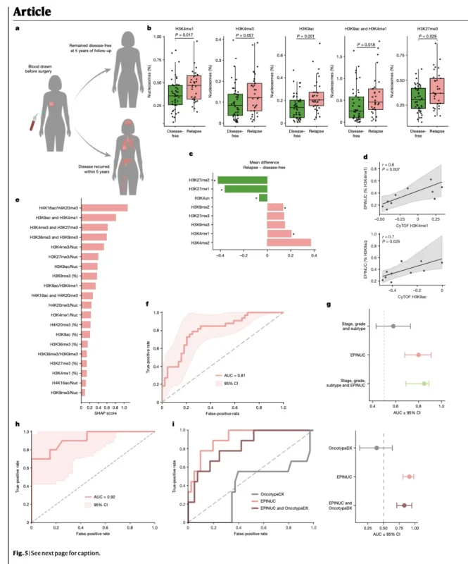
The researchers then investigated whether circulating epigenetic patterns could provide prognostic information. The initial MAYO-1 cohort included 77 women with luminal breast cancer who had preoperative plasma samples and at least five years of clinical follow-up.
Of these, 44 remained disease-free, while 33 experienced recurrence. Several epigenetic features differed between patients who subsequently relapsed and those who remained disease-free. Notably, H3K4me1 and H3K4me3 were elevated in the plasma of patients who later experienced recurrence.
These marks were also associated with the basal-like populations identified in tumor tissue. Additional recurrence-associated features included H3K27me3, H3K9ac, and nucleosomes carrying combinations of H3K9ac and H3K4me1.
The findings created a potential biological connection between aggressive tumor-cell states and detectable circulating epigenetic signatures. A machine-learning model developed from the EPINUC data achieved a recurrence-discrimination AUC of 0.81, with a 95% confidence interval of 0.71–0.90.
The investigators reported that these signatures provided prognostic information beyond conventional clinical variables in the analyzed cohort. However, the cohort was relatively small and enriched for patients with recurrence.
Its recurrence proportion should not be interpreted as the expected recurrence rate in an unselected population of patients with luminal breast cancer.
Independent Recurrence Analysis: AUC of 0.92
The researchers further evaluated recurrence prediction in an independent cohort, MAYO-2. This cohort included 30 patients, comprising 20 who remained disease-free and 10 who experienced recurrence.
The reported recurrence-discrimination AUC was 0.92, with a 95% confidence interval of 0.79–1.00. This result provides preliminary evidence that the prognostic signal may be reproducible. However, the confidence interval was relatively wide, and the number of recurrence events was small.
The findings therefore cannot establish the test’s definitive clinical accuracy. Prospective evaluation in larger, more representative populations will be necessary to determine whether EPINUC can reliably improve recurrence prediction.
How Does EPINUC Compare With Oncotype DX?
The investigators also explored whether EPINUC could provide additional information relative to Oncotype DX.
Oncotype DX is an established tissue-based genomic assay used in selected patients with HR-positive/HER2-negative early breast cancer to estimate recurrence risk and inform chemotherapy decisions.
It is important to distinguish these purposes. The new EPINUC approach measures circulating epigenetic features and was evaluated here for associations with five-year recurrence. In a subset of 49 patients with available Oncotype DX scores, including nine who experienced recurrence, EPINUC retained discriminatory ability.
Oncotype DX scores did not differ significantly between the recurrence and disease-free groups within this small selected sample. Combining both assays did not improve performance in the reported analysis.
However, these results do not establish that EPINUC is superior to Oncotype DX. The comparison was exploratory, involved few recurrence events, and did not reproduce the full clinical context in which Oncotype DX has been validated.
Furthermore, the study did not demonstrate that EPINUC predicts chemotherapy benefit. The two assays therefore cannot be considered interchangeable. Before EPINUC can influence adjuvant treatment decisions, it will need prospective validation demonstrating clinically meaningful information beyond established risk-assessment tools.
A Biological Link Between Tumor and Plasma Epigenetics
The researchers also examined whether circulating histone modifications reflected the epigenetic patterns observed in tumor tissue. Using paired tumor and blood samples, they identified positive correlations for several histone modifications.
H3K4me1 and H3K9ac showed particularly notable associations between tumor-level and circulating measurements. These findings strengthen the biological plausibility of the liquid biopsy approach. However, circulating nucleosomes originate from multiple tissues and cellular compartments.
Their composition can be influenced by normal tissue turnover, tumor-associated stromal cells, inflammatory responses, and systemic biological processes. Consequently, EPINUC does not directly isolate or count the basal-like cancer cells identified within tumors.
Instead, it measures a composite circulating epigenetic signal that may reflect tumor biology together with other physiological contributions. This distinction is important when interpreting what the blood assay can reveal.
What Are the Main Limitations?
The study presents substantial technical innovation, but several limitations must be considered. The original single-cell tumor cohort was relatively small, involving 12 tumors, predominantly hormone receptor-positive disease.
Although the researchers used several complementary platforms and external datasets, broad generalizability across breast cancer subtypes remains uncertain. The diagnostic analyses were retrospective case-control comparisons, not prospective screening studies.
Healthy controls do not fully represent the diagnostic complexity of routine clinical practice. The recurrence analyses involved limited numbers of events and require evaluation in larger independent cohorts.
The study also did not demonstrate that adapting treatment according to EPINUC results improves patient outcomes. Additionally, the connection between the basal-like state and endocrine treatment resistance remains mechanistic and inferential rather than clinically proven.
Finally, EPINUC relies on specialized single-molecule measurements and computational analysis. Analytical standardization, reproducibility, turnaround time, accessibility, cost-effectiveness, and clinical utility will all require evaluation.
OncoDaily Takeaway
The new Nature study provides important evidence that hormone receptor-positive breast cancers can contain epigenetically distinct basal-like cell populations with aggressive biological characteristics. These populations show reduced luminal signaling, different histone modification patterns, and greater expression of programs associated with cellular migration and phenotypic plasticity.
The research also demonstrates that some of these epigenetic features can be reflected in circulating nucleosomes. Using the EPINUC platform, investigators reported an AUC of 0.95 for breast cancer detection in the initial expanded-panel analysis and AUCs of 0.81 and 0.92 for recurrence discrimination in two retrospective cohorts.
These are promising findings, but they do not establish clinical readiness. The broader implication is that a tumor’s epigenetic state may contain prognostic information not fully captured by its conventional molecular classification.
Future prospective studies must determine whether circulating epigenetic measurements can reliably improve early detection, recurrence-risk assessment, or treatment selection. Until then, EPINUC remains an investigational technology, and its potential role alongside conventional pathology, imaging, and established genomic assays requires further validation.
Sources
- Furth N, Riemenschneider C, Arieli Y, et al. Epigenetic Heterogeneity in Breast Tumours Informs Diagnosis and Prognosis. Nature. Published online November 11, 2026. DOI: 10.1038/s41586-026-11230-y
- Fedyuk V, et al. Multiplexed, Single-Molecule, Epigenetic Analysis of Plasma-Isolated Nucleosomes for Cancer Diagnostics. Nature Biotechnology. 2023;41:212–221. DOI: 10.1038/s41587-022-01447-3
- Regner MJ, et al. Defining the Regulatory Logic of Breast Cancer Using Single-Cell Epigenetic and Transcriptome Profiling. Cell Genomics. 2025;5:100765. DOI: 10.1016/j.xgen.2025.100765
- Rueda OM, et al. Dynamics of Breast-Cancer Relapse Reveal Late-Recurring ER-Positive Genomic Subgroups. Nature. 2019;567:399–404. DOI: 10.1038/s41586-019-1007-8